. 2012;7(7):e41087.
doi: 10.1371/journal.pone.0041087. Epub 2012 Jul 20.

C-terminal substitution of HBV core proteins with those from DHBV reveals that arginine-rich 167RRRSQSPRR175 domain is critical for HBV replication

Affiliations

C-terminal substitution of HBV core proteins with those from DHBV reveals that arginine-rich 167RRRSQSPRR175 domain is critical for HBV replication

Jaesung Jung et al. PLoS One. 2012.

Abstract

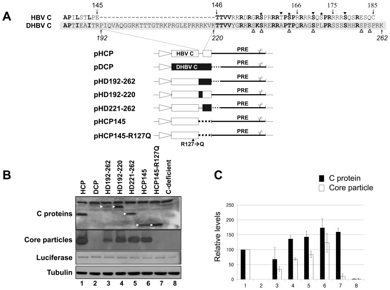
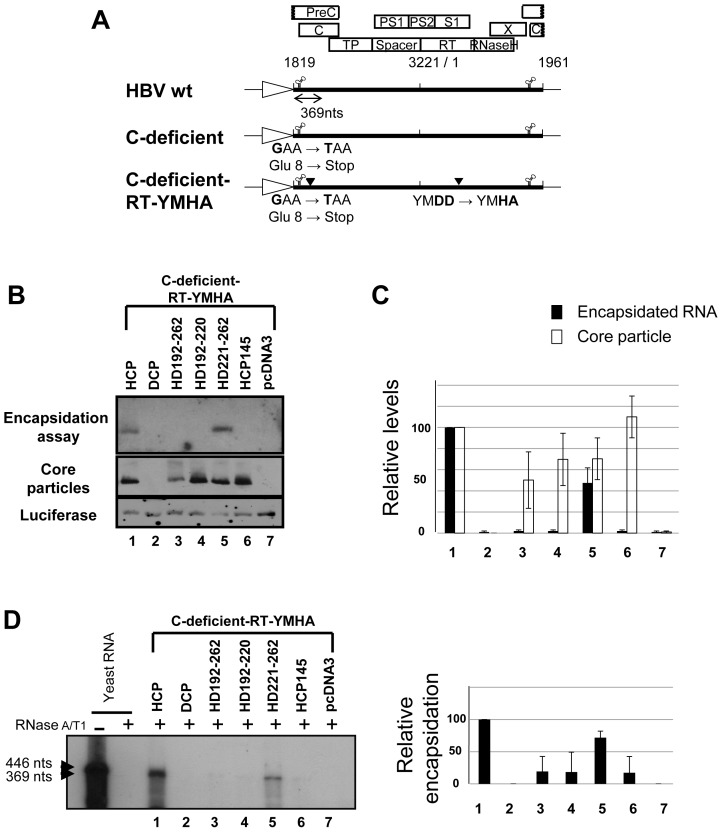
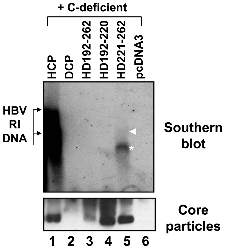
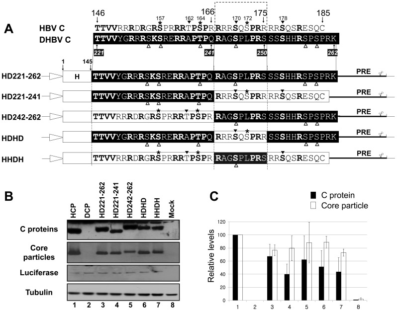
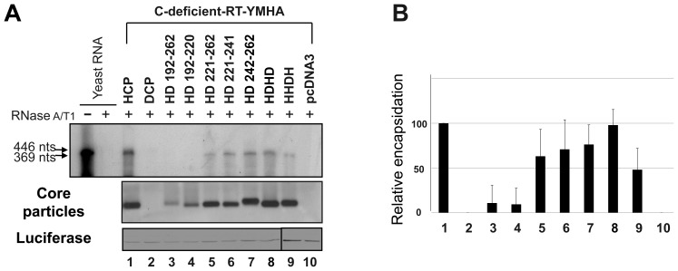
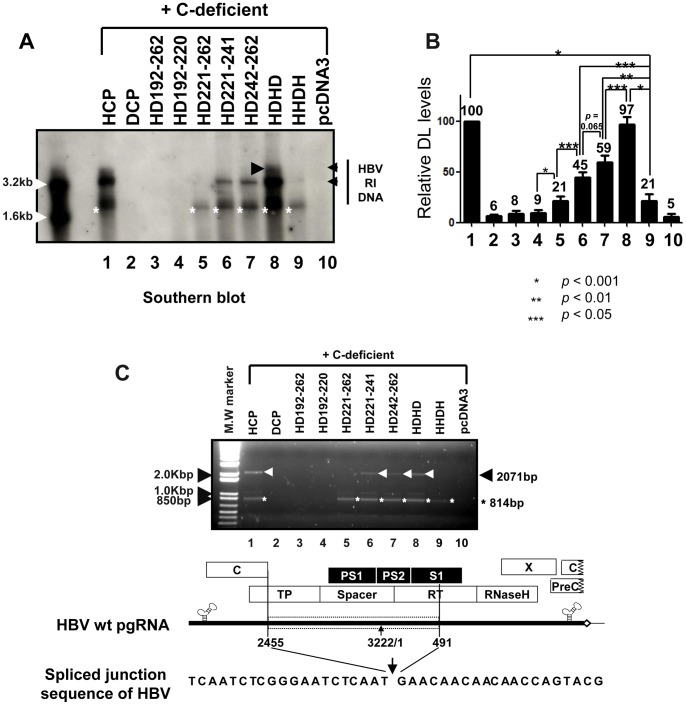
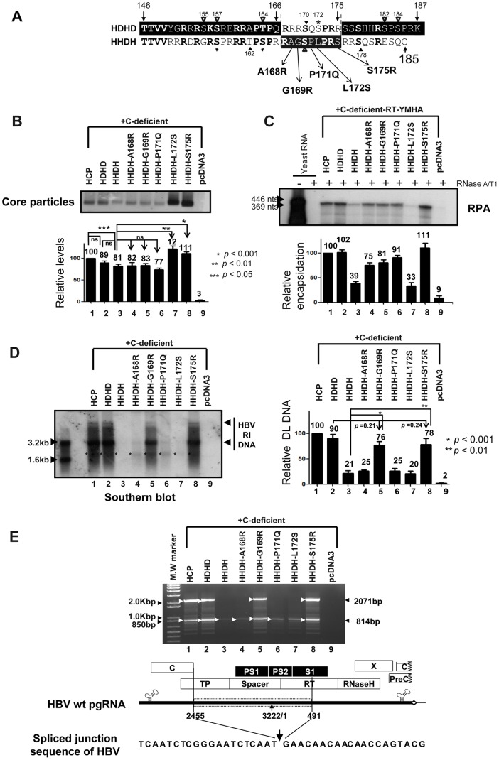
To investigate the contributions of carboxyl-terminal nucleic acid binding domain of HBV core (C) protein for hepatitis B virus (HBV) replication, chimeric HBV C proteins were generated by substituting varying lengths of the carboxyl-terminus of duck hepatitis B virus (DHBV) C protein for the corresponding regions of HBV C protein. All chimeric C proteins formed core particles. A chimeric C protein with 221-262 amino acids of DHBV C protein, in place of 146-185 amino acids of the HBV C protein, supported HBV pregenomic RNA (pgRNA) encapsidation and DNA synthesis: 40% amino acid sequence identity or 45% homology in the nucleic-acid binding domain of HBV C protein was sufficient for pgRNA encapsidation and DNA synthesis, although we predominantly detected spliced DNA. A chimeric C protein with 221-241 and 251-262 amino acids of DHBV C, in place of HBV C 146-166 and 176-185 amino acids, respectively, could rescue full-length DNA synthesis. However, a reciprocal C chimera with 242-250 of DHBV C ((242)RAGSPLPRS(250)) introduced in place of 167-175 of HBV C ((167)RRRSQSPRR(175)) significantly decreased pgRNA encapsidation and DNA synthesis, and full-length DNA was not detected, demonstrating that the arginine-rich (167)RRRSQSPRR(175) domain may be critical for efficient viral replication. Five amino acids differing between viral species (underlined above) were tested for replication rescue; R169 and R175 were found to be important.


Conflict of interest statement

Competing Interests: The authors have declared that no competing interests exist.

Figures

Figure 1
Figure 1. C protein expression and core particle assembly by chimeric C protein variants.
(A) Schematic diagrams of HBV, DHBV, and chimeric C protein variant constructs aligned with amino acid sequences of HBV and DHBV C protein carboxyl-terminal domains. Amino acids in bold are identical or homologous. SRPK and PKA phosphorylation sites of HBV are marked with asterisks and arrowheads, respectively. Phosphorylation sites of DHBV , are marked with open arrowheads. Amino acid sequences of the HBV and DHBV C proteins are presented as open and closed boxes, respectively. The cytomegalovirus immediate early (CMV IE) promoter is represented as an open arrow. PRE, post-transcriptional regulatory element. (B) Identification of C protein and core particles by chimeric C protein variants. To examine expression of C protein variants, lysates from HuH7 cells transfected with a pHCP, pDCP, pHD192–262, pHD192–220, pHD221–262, pHCP145, pHCP145–R127Q, or C-deficient mutant were electrophoresed on 12% SDS-PAGE gels and protein levels visualized by Western blotting using polyclonal rabbit anti-HBc antibody (top panel). C protein variants (arrowheads) with expected molecular weights are indicated. The C-deficient mutant lacks C protein due to the introduction of a stop codon at Glu 8 in the C ORF. The pHCP and the C-deficient mutant constituted positive and negative controls, respectively. Transfection experiments were repeated four times. To detect core particles formed by C protein variants from native agarose gels, isolated core particles were transferred to PVDF membranes and incubated with polyclonal rabbit anti-HBc antibody (second panel). The Renilla luciferase expression plasmid phRL-CMV was co-transfected into HuH7 cells as a transfection control (third panel). Luciferase and α-tubulin (bottom panel) levels were determined by Western blotting using polyclonal rabbit anti-luciferase and monoclonal mouse anti-tubulin antibodies as transfection and loading controls, respectively. HRP-conjugated secondary antibody and enhanced chemiluminescence were used to visualize C, α-tubulin, and luciferase proteins and core particles. (C) Relative levels of C protein expression and core particle assembly by chimeric C protein variants. Relative levels of C proteins, core particles, and luciferase were measured with the Fujifilm Image Gauge V4.0 program. Relative levels of C protein variant expression and core particle assembly were compared to normalized transfection efficiencies. The data represent the mean ± standard deviation (SD) from four independent experiments.
Figure 2
Figure 2. HBV
pgRNA encapsidation in core particles with chimeric C protein variants. (A) Schematic diagram of HBV wt , C-deficient mutant, and C-deficient-RT-YMHA mutant. The C-deficient-RT-YMHA mutant is RT- and C-protein deficient due to mutation of the YMDD motif to YMHA, in addition to the presence of a stop codon at Glu 8 in the C ORF. The positions of point mutations are indicated as closed arrowheads. Four ORFs of HBV are shown at the top as open boxes. The CMV promoter is denoted by an open arrow. (B) Encapsidation assay to detect HBV nucleic acid in situ from disrupted core particles. To examine encapsidation by chimeric C protein variants, the C-deficient-RT-YMHA mutant was co-transfected into HuH7 cells with the pHCP, pDCP, pHD192–262, pHD192–220, pHD221–262, or pHCP145. HBV wt C protein from pHCP served as a positive control. Isolated core particles were electrophoresed through a 1% native agarose gel and transferred to nylon membrane. A 32P-labeled HBV DNA probe was hybridized to HBV nucleic acids in core particles after disruption of the particles in situ. Core particles were also detected as described for Figure 1B. (C) Relative levels of RNA encapsidation and core particle assembly by chimeric C protein variants. Relative levels of encapsidated RNA and core particles were measured with the Fujifilm Image Gauge V4.0 program. Relative levels of encapsidated RNA and core particles were compared with normalized transfection efficiencies (n = 3). (D) RNase protection assay (RPA) to detect encapsidated pgRNA. In vitro transcribed radiolabeled antisense RNA probe (446 nt) was hybridized overnight at 50°C with pgRNA from isolated core particles. Following RNase digestion, the protected pgRNA (369 nt), nt 1819–2187 of the HBV sequence, was run on a 5% polyacrylamide–8 M urea gel and visualized by autoradiography. Relative levels of encapsidated pgRNA were measured with the Fujifilm Image Gauge V4.0 program. Transfection experiments were repeated three times. The Renilla luciferase expression plasmid phRL-CMV was used as a transfection control and pcDNA3.1 was used to equalize the total amount of DNA transfected. The data represent the mean ± SD from three independent experiments.
Figure 3
Figure 3. HBV DNA synthesis in core particles with chimeric C protein variants.
To examine HBV DNA synthesis in core particles with chimeric C variants, the C-deficient mutant and the pHCP, pDCP, pHD192–262, pHD192–220, or pHD221–262 were co-transfected into HuH7 cells. HBV DNA was extracted from isolated core particles and Southern blot analysis performed. Briefly, HBV DNA was separated, transferred to nylon membranes, hybridized with a random-primed 32P-labeled HBV specific probe, and subjected to autoradiography. Transfection experiments were repeated more than three times. The HBV replicative intermediate (RI) DNA is marked. Core particle formation (bottom panel) was determined as described for Figure 1B.
Figure 4
Figure 4. Expression and core particle assembly of additional C protein variants.
(A) Aligned amino acid sequences of carboxyl-terminal domains of HBV and DHBV C proteins and schematic diagrams of additional chimeric C protein variant constructs. Amino acids in bold are identical or homologous. SRPK and PKA phosphorylation sites in the HBV genome are marked with asterisks and arrowheads, respectively. DHBV phosphorylation sites , are marked with open arrowheads. The amino acid sequences of HBV and DHBV C protein are presented as open and closed boxes, respectively. The cytomegalovirus immediate early (CMV IE) promoter is represented as an open arrow. PRE, post-transcriptional regulatory element. (B) Expression of chimeric C proteins and core particle assembly by additional chimeric C protein variants. To examine C protein expression by HBV variants with chimeric C sequence, Western blotting was performed on lysates from HuH7 cells and HuH7 cells transfected with pHCP, pDCP, pHD221–262, pHD221–241, pHD242–262, pHDHD, or pHHDH variants, as described for Figure 1B (top panel). Core particle formation by C protein variants was detected as described for Figure 1B (second panel). Transfection experiments were repeated four times. As the respective transfection and loading controls, luciferase (third panel) and α-tubulin (bottom panel) levels were determined as described for Figure 1B. (C) Relative levels of C protein expression and core particle assembly by additional chimeric C protein variants. Relative levels of C proteins, core particles, and luciferase were measured with the Fujifilm Image Gauge V4.0 program. Relative levels of C protein variant expression and core particle assembly were compared with normalized transfection efficiencies. The data represent the mean ± SD from four independent experiments.
Figure 5
Figure 5. pgRNA encapsidation in core particles by additional C protein variants.
(A) RPA to detect encapsidated pgRNA. To detect the pgRNA encapsidated by chimeric C protein variants, the C-deficient-RT-YMHA mutant and the C protein chimeras pHCP, pDCP, pHD192–262, pHD192–220, pHD221–262, pHD221–241, pHD242–262, pHDHD, or pHHDH were co-transfected into HuH7 cells. RPA (top panel) was performed as described for Figure 2D. Core particle formation (second panel) and luciferase levels (bottom panel) were determined as described for Figure 1B. Transfection experiments were repeated three times. (B) Relative levels of encapsidated pgRNA and core particle assembly by additional chimeric C protein variants. Relative levels of encapsidated pgRNA, core particles, and luciferase were measured with the Fujifilm Image Gauge V4.0 program. Relative levels of encapsidated pgRNA and core particle assembly were compared to normalized transfection efficiencies. The data represent the mean ± SD from three independent experiments.
Figure 6
Figure 6. Full-length HBV DNA synthesis in core particles with additional chimeric C protein variants.
(A) To examine synthesis of HBV DNA in core particles with chimeric C variants, the C-deficient mutant and the C protein chimeras pHCP, pDCP, pHD192–262, pHD192–220, pHD221–262, pHD221–241, pHD242–262, pHDHD, or pHHDH were co-transfected into HuH7 cells. HBV DNA was extracted from isolated core particles and Southern blot analysis performed as described for Figure 3. Transfection experiments were repeated five times. The HBV replicative intermediate (RI) DNA is marked. Core particle formation (bottom panel) was determined as described for Figure 1B. (B) Relative levels of HBV double-stranded linear (DL) DNA from isolated core particles were measured with the Fujifilm Image Gauge V4.0 program and compared after normalization to transfection efficiencies (top right panel). The data represent the mean ± SD from five independent experiments. * p<0.001, ** p<0.01, *** p<0.05 (n = 5). (C) PCR and sequence alignment of the spliced junction of DNAs from isolated core particles. HBV DNA was extracted from isolated core particles and PCR was performed. The 814 base-pair (bp) DNA that was 1257 nt smaller than 2,071 bp of full-length HBV DNA and full-length HBV DNA were amplified (arrowheads).
Figure 7
Figure 7. HBV core particle formation, pgRNA encapsidation, and HBV DNA synthesis by C variants.
(A) Aligned amino acid sequences of HDHD and HHDH and amino acid substitutions in the HHDH-derived C variants HHDH-A168R, HHDH-G169R, HHDH-P170Q, HHDH-L172S, and HHDH-S175R. Amino acids in bold are identical or homologous. SRPK and PKA phosphorylation sites of HBV are marked with asterisks and arrowheads, respectively. Phosphorylation sites of DHBV , are marked with open arrowheads. Amino acid sequences of HBV and DHBV C proteins are presented as black and white letters, respectively, on contrasting background. (B-D) HBV core particle formation, pgRNA encapsidation, and HBV DNA synthesis by C variants. To examine HBV core particle formation (B), pgRNA encapsidation (C), and HBV DNA synthesis (D) in core particles with the C-deficient or C-deficient-RT-YMHA mutants and pHCP or the C protein chimeras, pHDHD, pHHDH, HHDH-A168R, HHDH-G169R, HHDH-P170Q, HHDH-L172S, or HHDH-S175R, were co-transfected into HuH7 cells. pcDNA3.1 was used to equalize the amount of DNA transfected. (B) Core particle formation and luciferase levels (data not shown) were determined as described for Figure 1B. The data represent the mean ± SD (n = 5). * p<0.001, ** p<0.01, and * p<0.05 (n = 5). (C) To examine pgRNA encapsidation, RPA was performed as described for Figure 2D. The data represent the mean ± SD from four independent experiments. (D) HBV DNA was extracted from isolated core particles and Southern blot analysis performed as described for Figure 3. The HBV replicative intermediate (RI) DNA is marked. DNAs from spliced RNAs are indicated by asterisks. Relative levels of core particles and encapsidated pgRNA and HBV DL DNA from isolated core particles were measured with the Fujifilm Image Gauge V4.0 program and compared after normalization to transfection efficiencies. The data represent the mean ± SD from five independent experiments. * p<0.001 HHDH vs HHDH-G169R, ** p<0.01 HHDH vs HHDH-S175R, p = 0.21 HDHD vs HHDH-G169R, or p = 0.24 HDHD vs HHDH-S175R (n = 5). (E) PCR and sequence alignment of the spliced junction. HBV DNA was extracted from isolated core particles and PCR was performed as described for Figure 6C. The 814 base-pair (bp) DNA that was 1257 nt smaller than 2,071 bp of full-length HBV DNA and full-length HBV DNA were amplified (arrowheads).

Similar articles

Cited by

References

    1. Brechot C. Pathogenesis of hepatitis B virus-related hepatocellular carcinoma: old and new paradigms. Gastroenterology. 2004;127:S56–S61. - PubMed
    1. Zhou S, Standring DN. Hepatitis B virus capsid particles are assembled from core-protein dimer precursors. Proc. Natl. Acad. Sci. USA. 1992;89:10046–10050. - PMC - PubMed
    1. Birnbaum F, Nassal M. Hepatitis B virus nucleocapsid assembly: primary structure requirements in the core protein. J. Virol. 1990;64:3319–3330. - PMC - PubMed
    1. Gallina A, Bonelli F, Zentilin L, Rindi G, Muttini M, et al. A recombinant hepatitis B core antigen polypeptide with the protamine-like domain deleted self-assembles into capsid particles but fails to bind nucleic acid. J. Virol. 1989;63:4645–4652. - PMC - PubMed
    1. Beames B, Lanford RE. Carboxy-terminal truncations of the HBV core protein affect capsid formation and the apparent size of encapsidated HBV RNA. Virology. 1993;194:597–607. - PubMed

Publication types