. 2012;7(7):e41126.
doi: 10.1371/journal.pone.0041126. Epub 2012 Jul 24.

Pathological steps of cancer-related lymphedema: histological changes in the collecting lymphatic vessels after lymphadenectomy

Affiliations

Pathological steps of cancer-related lymphedema: histological changes in the collecting lymphatic vessels after lymphadenectomy

Makoto Mihara et al. PLoS One. 2012.

Erratum in

Abstract

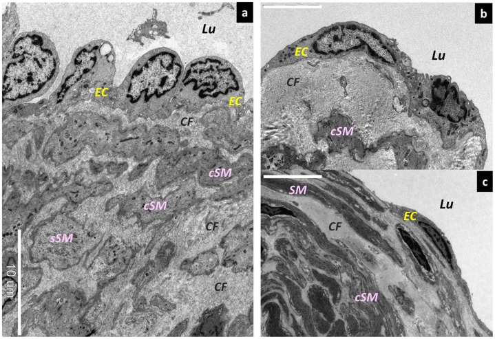
Introduction: To date, an electron microscopy study of the collecting lymphatic vessels has not been conducted to examine the early stages of lymphedema. However, such histological studies could be useful for elucidating the mechanism of lymphedema onset. The aim of this study was to clarify the changes occurring in collecting lymphatic vessels after lymphadenectomy.

Methods: The study was conducted on 114 specimens from 37 patients who developed lymphedema of the lower limbs after receiving surgical treatment for gynecologic cancers and who consulted the University of Tokyo Hospital and affiliated hospitals from April 2009 to March 2011. Lymphatic vessels that were not needed for lymphatico venous anastomosis surgery were trimmed and subsequently examined using electron microscopy and light microscopy.

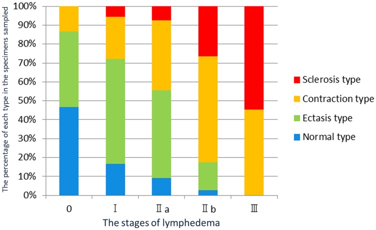
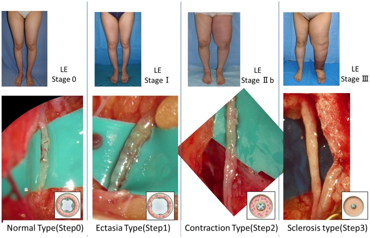
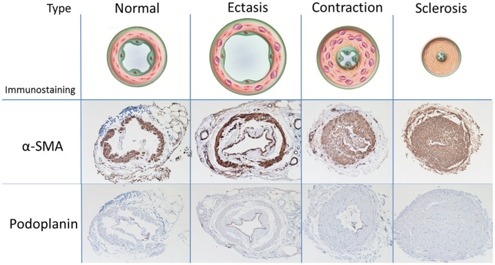
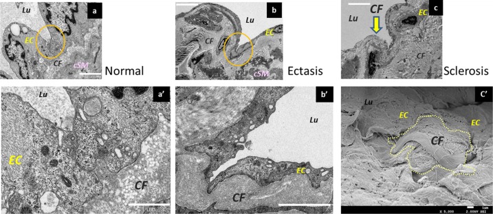
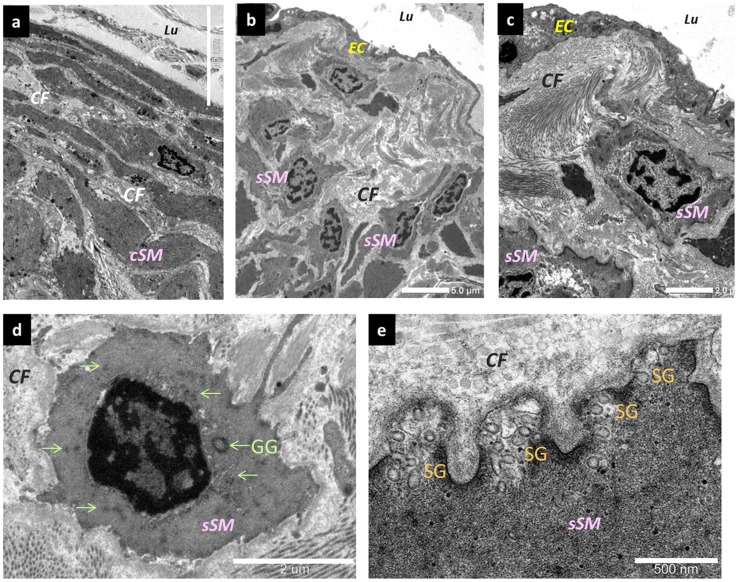
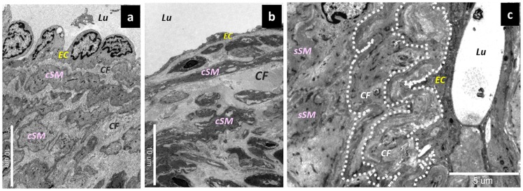
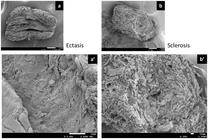
Results: Based on macroscopic findings, the histochemical changes in the collecting lymphatic vessels were defined as follows: normal, ectasis, contraction, and sclerosis type (NECST). In the ectasis type, an increase in endolymphatic pressure was accompanied by a flattening of the lymphatic vessel endothelial cells. In the contraction type, smooth muscle cells were transformed into synthetic cells and promoted the growth of collagen fibers. In the sclerosis type, fibrous elements accounted for the majority of the components, the lymphatic vessels lost their transport and concentrating abilities, and the lumen was either narrowed or completely obstructed.

Conclusions: The increase in pressure inside the collecting lymphatic vessels after lymphadenectomy was accompanied by histological changes that began before the onset of lymphedema.


Conflict of interest statement

Competing Interests: The authors have declared that no competing interests exist.

Figures

Figure 1
Figure 1. Association between the classification of the types of collecting lymphatic vessels and the staging of lymphedema.
The horizontal axis shows the stages of lymphedema. The vertical axis shows the percentage of each type in the specimens sampled at each stage of the disease.
Figure 2
Figure 2. Staging of lymphedema and the macroscopic anatomical findings in the collecting lymphatic vessels associated with the stages.
In the normal type, microvascular networks were found to nourish the largely developed walls of the collecting lymphatic vessels. The microvascular networks were gradually lost with the progression of the disease stages. In addition, the lymphatic vessel lumen was found to be dilated in the ectasis type, which was associated with an increase in endolymphatic pressure. Because increases in smooth muscle cells and collagen fibers are the major causes of the cloudiness and thickening of the lymphatic vessel wall, they were found to be prominent in the contraction type and the sclerosis type. LE: lymphedema.
Figure 3
Figure 3. Classification of the types of collecting lymphatic vessels and the corresponding immunostaining findings.
The first row: Findings from alpha- smooth muscle actin (αSMA) staining. In the normal type, several layers of smooth muscle cells were present. In the ectasis type, the smooth muscle cell layer was moderately thin. In the contraction type, a very thick layer of smooth muscle was present directly beneath the endothelium, and the lumen was narrowed to a certain degree. In the sclerosis type, the lumens were narrowed. The second row: Findings from podoplanin staining. The normal type was positive for podoplanin staining in the endothelium; the ectasis type was strongly positive; the contraction type, slightly positive; the sclerosis type, negative.
Figure 4
Figure 4. Morphological changes in lymphatic vessel endothelial cells.
Scanning electron microscopy (SEM) findings. (a) Lymphatic vessel endothelial cells protruding into the lymphatic lumen in the normal type. Bar = 10 µm. (b) Lymphangiectasia at its early stages. The lymphatic vessel endothelial cells are slightly flattened. Bar = 5 µm. (c) Medium-stage lymphangiectasia. The lymphatic vessel endothelial cells are markedly flattened. Bar = 10 µm. Lu; Lumen, EC; endothelium cells, sSM; synthetic smooth muscle cells, cSM; contractile smooth muscle cells, CF; collagen fibers.
Figure 5
Figure 5. Changes in the desmosomes of lymphatic vessel endothelial cells as shown by electron microscopy.
(a) Desmosomes in the normal type. In a three-dimensional structure, they adhere to the adjacent lymphatic vessel endothelial cells. Bar = 2 µm. (a’) Magnified image. Bar = 2 µm. (b) Desmosomes of lymphatic vessel endothelial cells in the ectasis type. The cell-to-cell adhesions have gradually loosened and disassembled, so that the cells adhere only at points. Bar = 10 µm. (b’) Magnified image. Bar = 2 µm. (c) Lymphatic endothelial cells in the collecting lymphatic vessels in the sclerosis type. The desmosomes are disassembled, and the collagen fibers under the lymphatic vessel endothelium are exposed in the lymphatic vessel lumen (yellow arrow). Bar = 5 µm. (c’) Collagen fibers exposed in the lymphatic vessel lumen (Electron microscopy, SEM). Lymphatic vessel endothelial cells are missing, and fascicles of collagen fibers are exposed in the lymphatic vessel lumen. Bar = 1 µm. EC; endothelium cells, sSM; synthetic smooth muscle cells, cSM; contractile smooth muscle cells, CF; collagen fibers.
Figure 6
Figure 6. Changes in smooth muscle cells.
(a) Normal type. They are present in the medial layer of the collecting lymphatic vessels, as contractile smooth muscle cells. Synthetic smooth muscle cells are only partially present. Bar = 10 µm. (b) Changes affecting the smooth muscle cells in the contraction type. Synthetic smooth muscle cells migrated directly beneath the endothelial cells and proliferated. Bar = 5 µm. (c) Synthetic smooth muscle cells stimulated collagen fiber hyperplasia directly beneath the lymphatic vessel endothelial cells. Bar = 2 µm. (d) Synthetic smooth muscle cells. An increase in glycogen granules inside the cells is evident. (e) The periphery of synthetic smooth muscle cells. Secretory granules are present in large numbers. Bar = 500 nm. EC; endothelium cells, sSM; synthetic smooth muscle cells, cSM; contractile smooth muscle cells, CF; collagen fibers, GG; glycogen granules (green arrows), SG; secretory granules.
Figure 7
Figure 7. Changes affecting collagen fibers in collecting lymphatic vessels.
(a) Collagen fibers in the normal type. Contractile smooth muscle cells and collagen fibers were mixed together in the medial layer. Bar = 10 µm. (b) Collagen fibers in the ectasis type. The dilation of the lymphatic vessel was accompanied by an elongation of collagen fibers, causing the slightly thickened aspect. Bar = 10 µm. (c) Collagen fibers in the contraction type. Once the collagen fibers were elongated, they became multilayered and tortuous (dotted line), in association with the hypertrophy of the walls of the collecting lymphatic vessels. Bar = 5 µm. EC; endothelium cells, seSM; synthetic smooth muscle cells, sySM; synthetic smooth muscle cells, cSM; contractile smooth muscle cells, CF; collagen fibers.
Figure 8
Figure 8. Electron microscopic and SEM findings.
(a) Normal type. Bar = 100 µm. (a’) Ectasis type. The lymphatic vessel lumen was dilated and entirely covered with lymphatic vessel endothelial cells. Bar = 10 µm. (b) Sclerosis type. Bar = 100 µm. (b’) The lymphatic vessel wall was thickened because of the hyperplasia of smooth muscles and collagen fibers. The lymphatic vessel lumen was narrowed and as shaped like a pinhole. Bar = 10 µm.

References

    1. Rockson SG (2001) Lymphedema. Am J Med . 110: 288–295. - PubMed
    1. Jensen MR, Simonsen L, Karlsmark T, Bulow J (2010) Lymphoedema of the lower extremities–background, pathophysiology and diagnostic considerations. Clin Physiol Funct Imaging . 30: 389–398. - PubMed
    1. Unno N, Inuzuka K, Suzuki M, Yamamoto N, Sagara D, et al. (2007) Preliminary experience with a novel fluorescence lymphography using indocyanine green in patients with secondary lymphedema. J Vasc Surg . 45: 1016–1021. - PubMed
    1. Unno N, Nishiyama M, Suzuki M, Yamamoto N, Inuzuka K, et al. (2008) Quantitative lymph imaging for assessment of lymph function using indocyanine green fluorescence lymphography. Eur J Vasc Endovasc Surg . 36: 230–236. - PubMed
    1. Unno N, Nishiyama M, Suzuki M, Tanaka H, Yamamoto N, et al. (2010) A novel method of measuring human lymphatic pumping using indocyanine green fluorescence lymphography. J Vasc Surg . 52: 946–952. - PubMed

Publication types

Substances