. 2012;7(7):e41242.
doi: 10.1371/journal.pone.0041242. Epub 2012 Jul 24.

Potential role for PAD2 in gene regulation in breast cancer cells

Affiliations

Potential role for PAD2 in gene regulation in breast cancer cells

Brian D Cherrington et al. PLoS One. 2012.

Abstract

The peptidylarginine deiminase (PAD) family of enzymes post-translationally convert positively charged arginine residues in substrate proteins to the neutral, non-standard residue citrulline. PAD family members 1, 2, 3, and 6 have previously been localized to the cell cytoplasm and, thus, their potential to regulate gene activity has not been described. We recently demonstrated that PAD2 is expressed in the canine mammary gland epithelium and that levels of histone citrullination in this tissue correlate with PAD2 expression. Given these observations, we decided to test whether PAD2 might localize to the nuclear compartment of the human mammary epithelium and regulate gene activity in these cells. Here we show, for the first time, that PAD2 is specifically expressed in human mammary gland epithelial cells and that a portion of PAD2 associates with chromatin in MCF-7 breast cancer cells. We investigated a potential nuclear function for PAD2 by microarray, qPCR, and chromatin immunoprecipitation analysis. Results show that the expression of a unique subset of genes is disregulated following depletion of PAD2 from MCF-7 cells. Further, ChIP analysis of two of the most highly up- and down-regulated genes (PTN and MAGEA12, respectively) found that PAD2 binds directly to these gene promoters and that the likely mechanism by which PAD2 regulates expression of these genes is via citrullination of arginine residues 2-8-17 on histone H3 tails. Thus, our findings define a novel role for PAD2 in gene expression in human mammary epithelial cells.


Conflict of interest statement

Competing Interests: Dr. Coonrod is a PLoS ONE Editorial Board member. This does not alter the authors‘ adherence to all the PLoS ONE policies on sharing data and materials.

Figures

Figure 1
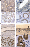
Figure 1. PAD2 is expressed in human endometrial, kidney, breast ductal and luminal epithelial cells.
(A) Endometrial, (B) kidney, (C) mammary duct, and (D) mammary alveolar epithelial cells express PAD2, while staining is absent (or weak) in adjacent tissue. Tissue sections were probed with anti-PAD2 antibody or equal concentration of rabbit IgG as a control and counterstained with hematoxylin. The black arrows indicate epithelial cells with varying PAD2 staining in the nucleus between different tissues.
Figure 2
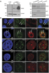
Figure 2. PAD2 is detected in the cytoplasm and nucleus of human breast luminal epithelial cells.
(A) PAD2 is expressed in luminal but not myoepithelial human breast cells. Normal human breast tissue sections were probed with anti-PAD2 (green fluorescent signal), anti-cytokeratin (luminal marker - red fluorescent signal), and anti-p63 (myoepithelial marker - red fluorescent signal) antibodies or equal concentration of rabbit and mouse IgG as a control and nuclei are stained with DAPI. (B) A portion of PAD2 staining in human breast epithelial cells is detected in the nucleus. Normal human breast tissue sections were probed with anti-PAD2 (green fluorescent signal) and nuclei are stained with DAPI. The white arrow illustrates a breast epithelial cell with robust PAD2 staining in the nucleus.
Figure 3
Figure 3. PAD2 associates with chromatin in the nucleus of MCF-7 cells.
(A) PAD2 is endogenously expressed in MCF-7 cells and is detected in multiple cellular compartments. Wild type and PAD2 over-expressing MCF-7 whole cell lysates were subject to SDS-PAGE and probed with an anti-PAD2 antibody. Anti-Rabbit IgG was used as a negative control. (left panel). Endogenous MCF-7 cellular proteins were also separated into cytoplasmic, chromatin, and soluble nuclear pools by fractionation methods and examined by western blot (right panel). Cleanliness of fractionation was determined by stripping membranes and re-probing with antibodies for TBP (nuclear) and SOD4 (cytoplasmic) proteins. (B) Punctate PAD2 expression is detected in the nucleus of MCF-7 cells. MCF-7 cells were subject to IF using anti-PAD2 (green), anti-cytokeratin (luminal-red), anti-H3K9 acetyl (euchromatin-red), anti-HP1 (heterochromatin-red), anti-H3K9 dimethyl (facultative heterochromatin-red), and anti-H3K27 trimethyl (facultative heterochromatin-red) antibodies while DAPI nuclear stain is in blue. White arrows denote punctuate PAD2 staining in DAPI poor nuclear regions.
Figure 4
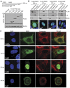
Figure 4. Truncated FLAG-tagged PAD2 proteins reveal regions necessary for nuclear localization.
(A) The schematic at the top indicates the different truncated PAD2 proteins of 140, 278 and 437 amino acids. Truncated FLAG-PAD2 constructs were validated by overexpressing in HEK 293 cells with lysates analyzed by SDS-PAGE. Membranes were probed with an anti-FLAG antibody which detected predicted molecular weight of truncated proteins while β-actin shows loading control. (B) Truncation of PAD2 reveals regions of the protein necessary for subcellular localization by cellular fractionation. Truncated FLAG-PAD2 constructs were overexpressed in HEK 293 cells after which cellular proteins were separated by fractionation methods and examined by western blot. Cleanliness of fractionation was determined by stripping membranes and re-probing with antibodies for Histone H3 (nuclear) and SOD4 (cytoplasmic) proteins. Truncated FLAG-PAD2 constructs were also transiently transfected into HEK 293 cells and subcellular localization examined by IF (bottom panel of B) using an anti-FLAG antibody (green fluorescent signal) to corroborate cellular fractionation studies. (C) Truncation of PAD2 reveals regions of the protein necessary for subcellular localization by IF in mammary epithelial cells. Truncated FLAG-PAD2 constructs were over expressed in MCF-7 cells and examined by IF using an anti-PAD2 (green) and anti-cytokeratin (red) antibodies while DAPI nuclear stain is in blue.
Figure 5
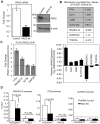
Figure 5. PAD2 plays a role in gene expression in MCF-7 cells.
(A) PAD2 levels are significantly reduced in PAD2 knock down MCF-7 cells compared to shRNA controls. Whole cell lysates from shRNA control and PAD2 knock down MCF-7 cells were analyzed by western blot using an anti-PAD2 antibody and an anti-β-actin antibody for loading control. RNA was purified from PAD2 knock down MCF-7 and shRNA control cells, reverse transcribed, and resulting cDNA used in qPCR reactions containing TaqMan assays to PAD2 and GAPDH as the reference gene control. Data represent the means ± SEM of four independent experiments performed in triplicate. All values are normalized to shRNA control samples and bars represent the means ± SEM. Means were separated by Student’s T-Test and ** represent significant differences (P<0.01). (B) Microarray and qPCR validation studies indicate that PAD2 is involved in gene regulation in MCF-7 cells. The table shows a sampling of genes whose expression profiles were significantly altered between shRNA control and PAD2 knockdown MCF-7 cells. Numbers represent log2 ratio of fold change of expression between control and PAD2 knockdown cell lines with P<0.001 and FDR<0.01. To validate microarray studies, total RNA was harvested from shRNA control and PAD2 knockdown MCF-7 cells and reverse transcribed. Resulting cDNA was used in qPCR reactions with specific primers listed in Table S1. The graph shows fold change of log2 ratio of expression between control and PAD2 knockdown cell lines with P<0.05. (C) PAD2 associates with the PTN and MAGEA12 gene promoters in MCF-7 cells stably overexpressing FLAG-tagged PAD2. ChIP-qPCR studies were done using an anti-FLAG antibody and data is presented as fold change of signal from MCF-7 cells overexpressing FLAG-tagged PAD2 over control cells with β-actin and GAPDH serving as controls. Data represent the means ± SEM of three independent experiments. (D) The PTN and MAGEA12 gene promoters show citrullination of histone H3, but not histone H4 tail arginine residues using shRNA control and PAD2 knockdown MCF-7 cells. ChIP-qPCR studies were done using an anti-H3 cit 2–8–17 and H4 cit 3 antibodies and data is presented as % input with the GAPDH promoter serving as the control. Data represent the means ± SEM of three independent experiments and means separated by one-tailed T-tests.
Figure 6
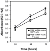
Figure 6. Knockdown of endogenous PAD2 in MCF-7 cells alters cellular proliferation rates versus control cells.
Equal numbers of shRNA control and PAD2 knockdown MCF-7 cells were plated in 96-well plates and allowed to grow for 24, 48, or 72 hours. At time of harvest, dye solution from MTT assay kit was allowed to incubate with cells for one hour at which point solubilization reagent was added and resulting formazan product measured at 570 nm. Data represent the means ± SEM of three independent experiments performed in triplicate. Values represent the means ± SEM.

References

    1. Suzuki A, Yamada R, Chang X, Tokuhiro S, Sawada T, et al. Functional haplotypes of PADI4, encoding citrullinating enzyme peptidylarginine deiminase 4, are associated with rheumatoid arthritis. Nat Genet. 2003;34:395–402. - PubMed
    1. Mastronardi FG, Mak B, Ackerley CA, Roots BI, Moscarello MA. Modifications of myelin basic protein in DM20 transgenic mice are similar to those in myelin basic protein from multiple sclerosis. J Clin Invest. 1996;97:349–358. - PMC - PubMed
    1. Ishigami A, Ohsawa T, Hiratsuka M, Taguchi H, Kobayashi S, et al. Abnormal accumulation of citrullinated proteins catalyzed by peptidylarginine deiminase in hippocampal extracts from patients with Alzheimer’s disease. J Neurosci Res. 2005;80:120–128. - PubMed
    1. Chang X, Han J. Expression of peptidylarginine deiminase type 4 (PAD4) in various tumors. Mol Carcinog. 2006;45:183–196. - PubMed
    1. Luo Y, Knuckley B, Lee YH, Stallcup MR, Thompson PR. A fluoroacetamidine-based inactivator of protein arginine deiminase 4: design, synthesis, and in vitro and in vivo evaluation. J Am Chem Soc. 2006;128:1092–1093. - PMC - PubMed

Publication types