. 2012;7(7):e41633.
doi: 10.1371/journal.pone.0041633. Epub 2012 Jul 23.

Local oxidative stress expansion through endothelial cells--a key role for gap junction intercellular communication

Affiliations

Local oxidative stress expansion through endothelial cells--a key role for gap junction intercellular communication

Ilan Feine et al. PLoS One. 2012.

Abstract

Background: Major circulation pathologies are initiated by oxidative insult expansion from a few injured endothelial cells to distal sites; this possibly involves mechanisms that are important to understanding circulation physiology and designing therapeutic management of myocardial pathologies. We tested the hypothesis that a localized oxidative insult of endothelial cells (ECs) propagates through gap junction inter-cellular communication (GJIC).

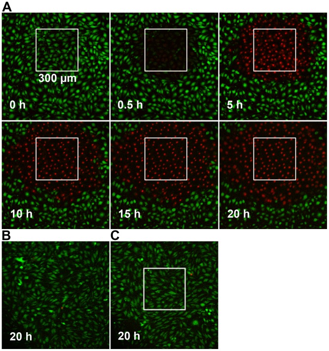
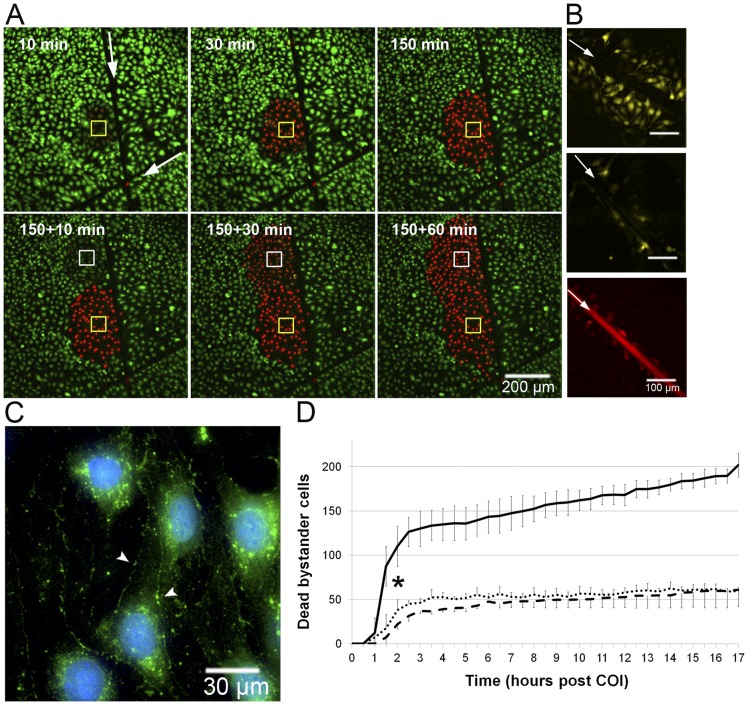
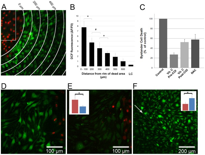
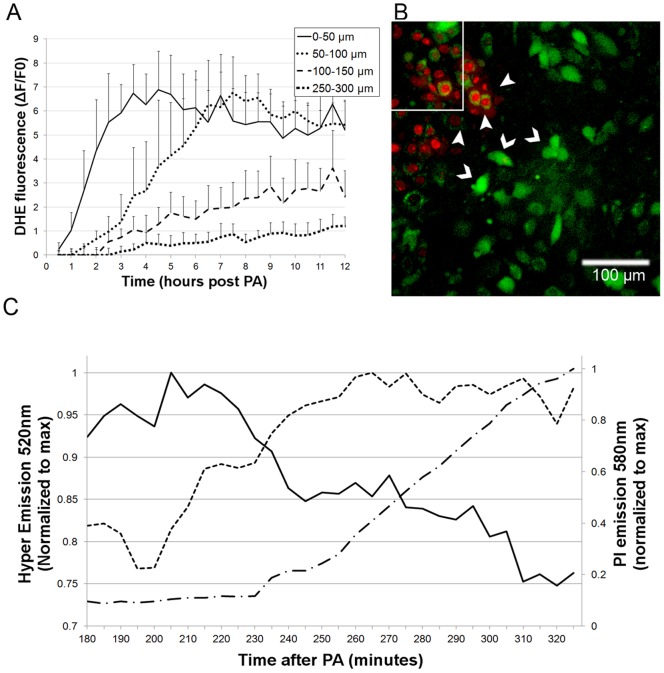
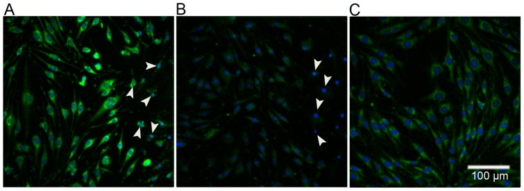
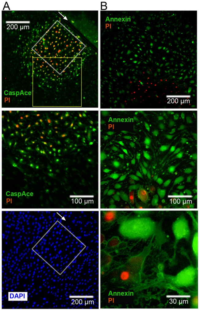
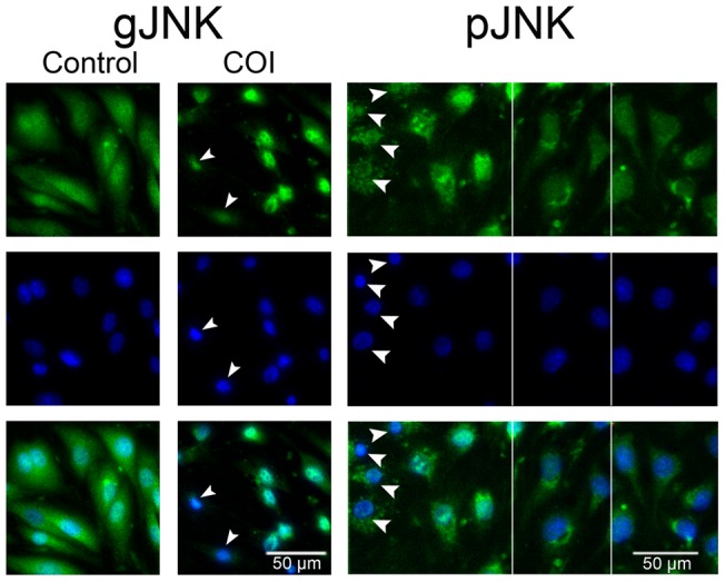
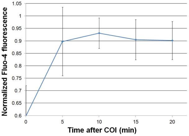
Methodology/principal findings: Cultures comprising the bEnd.3 cell line, that have been established and recognized as suitable for examining communication among ECs, were used to study the propagation of a localized oxidative insult to remote cells. Spatially confined near infrared illumination of parental or genetically modified bEnd.3 cultures, pretreated with the photosensitizer WST11, generated O(2)•(-) and •OH radicals in the illuminated cells. Time-lapse fluorescence microscopy, utilizing various markers, and other methods, were used to monitor the response of non-illuminated bystander and remote cells. Functional GJIC among ECs was shown to be mandatory for oxidative insult propagation, comprising de-novo generation of reactive oxygen and nitrogen species (ROS and RNS, respectively), activation and nuclear translocation of c-Jun N-terminal kinase, followed by massive apoptosis in all bystander cells adjacent to the primarily injured ECs. The oxidative insult propagated through GJIC for many hours, over hundreds of microns from the primary photogeneration site. This wave is shown to be limited by intracellular ROS scavenging, chemical GJIC inhibition or genetic manipulation of connexin 43 (a key component of GJIC).

Conclusion/significance: Localized oxidative insults propagate through GJIC between ECs, while stimulating de-novo generation of ROS and RNS in bystander cells, thereby driving the insult's expansion.


Conflict of interest statement

Competing Interests: A.S. and Y.S. are the inventors of WST11; patent owned by the Weizmann Institute of Science, Rehovot, Israel, licensed to Steba Biotech, France (Scherz A, Brandis A, Mazor O, Salomon Y, Scheer H, (2011) Water-soluble anionic bacteriochlorophyll derivatives and their uses, US patent 7,947,672, reference #33). A.S. and Y.S. serve as consultants to Steba Biotech. This does not alter adherence to all the PLoS ONE policies on sharing data and materials.

Figures

Figure 1
Figure 1. Propagation of cell death following COI.
A – bEnd.3 monolayers were subjected to COI. Cell death was monitored by changes in the fluorescence of membrane viability probes, CaAMg and PI, as followed by TLFM. Dead cells are defined as CaAMg negative (green), PI positive (red). Time zero represents termination of COI. B – Dark control (with WST11, no illumination). C – Light control (Illumination, of same rectangle, no WST11). Both control images were taken at 20h. All other details are as described in the Materials and Methods section.
Figure 2
Figure 2. Death propagation among bystander cells requires cell-cell contact and connexins.
A – Cell contact in bEnd.3 monolayer was impaired by surgical scalpel scratch (white arrows, scratch extends far beyond the field of view), and the culture was subjected to COI (60×60 µm, top left image, yellow rectangle) in one side of the scratch. Cell death (monitored as described in Fig. 1) propagates only in that side. Lower panel – an additional COI site was imposed at t = 150 min in the same side of the scratch (white rectangle). B – bEnd.3 cells were incubated with GZA (inactive analog, upper panel) or the GJ uncoupler CBX (middle panel). After 30min Lucifer yellow (LY, pseudo yellow) was added and SLDT was carried out (n = 12). White arrow marks the linear scratch. Rhodamine-dextran (red, lower panel) served as initial loading site control. C – bEnd.3 monolayers immunostained for Cx43, (pseudo-colored green) seen on cell membranes (arrow heads) and around the nuclei (counter stained with DAPI, pseudo-colored blue). Control experiments showed no non-specific binding of the secondary antibody (n = 3). D – The graph illustrates time dependent registration of PI+ cells beyond the COI rectangle in GZA treated (solid line), CBX treated (dashed line) and bEnd.3-D2 cells (dotted line) following COI. Values are displayed as means ± SD. * P<0.01 between the GZA and CBX/ bEnd.3-D2 experiments from t = >1.5h onwards (n = 4). All other details are described in Materials and Methods section.
Figure 3
Figure 3. De-novo ROS generation in endothelial bystander cells and the effect of ROS scavengers and GJIC inhibition.
bEnd.3 monolayers were incubated with DCFH-DA, rinsed with fresh culture medium and subjected to COI. After illumination the plates were placed in the incubator for 3h and then imaged for PI (pseudo-colored red) and DCF (ROS, pseudo-colored green) fluorescence. A – An overlay of two pseudo-colored captures, the white arches represent distance increments of 100µm from the COI (white rectangle), representative of n = 3. PI+ bystander cells on the top left corner are at 50–100µm from the rim of the COI. Adjacent to them on the first arch (0–100µm) are DCF positive, PI negative bystander cells. B – A plot of DCF fluorescence intensity (mean ± SD of five cells in three separate experiments). LC – light control, cells exposed to laser illumination, without WST11 incubation, imaged for DCF fluorescence 3h later. C – bEnd.3 monolayers were subjected to COI under the following conditions: Control: encompassing standard COI; Vit. C Pre-COI: encompassing incubation with 100µmol/L vitamin C for 1h, rinse and then subjected to COI; Vit. C Post-COI: encompassing the addition of 100µmol/L vitamin C immediately after COI; NAC: incubation with 10mmol/L NAC prior to COI. 3h after COI, cell death was determined by PI fluorescence. Notably, in all cases COI resulted in a complete cell death within the primary illuminated square. Values represent averaged percent of bystander cell death (mean ± SD of at least n = 3 separate experiments in each group) relative to the control. The control values are statistically significant higher than all treated groups (P<0.01). D, E – bEnd.3 monolayers were incubated with GZA (D) or CBX (E), respectively, subjected to COI and probed with DCFH-DA. Dead bystander cells (red) at a distance of 60–80µm from the rim of COI, are accompanied by PI, DCF+ (representative of n = 3 experiments) in (D) but not in (E). The insert in (E) illustrates the mean DCF fluorescence of CBX compared to GZA treated cells (right and left colum respectively, n = 10 cells) at equal distances from the dead bystander cells. *- P<0.01 between the treatments. # - P<0.05. F – bEnd.3 monolayers were scratched by a surgical scalpel (arrow), underwent COI (near the upper right corner), and placed in the incubator for 10h. Intra-cellular ROS propagation (DCF+) is blocked at the scratch. Insert – right and left columns representing mean DCF fluorescence intensities in the photoactivation side and beyond the scratch, respectively). * P<0.01.
Figure 4
Figure 4. Superoxide anion (O2 •−) and H2O2 detection in bystander cells following localized oxidative insult.
A – Time dependent DHE fluorescence increase (superoxide generation) in bEnd.3 cells at four selected distances from the COI boundary, within the monolayers (n = 10 cells, ± SD). B, C – H5V-Hyper monolayers supplemented with PI and 100UI/ml catalase, were subjected to COI and imaged. B – An overlay of Hyper protein fluorescence (H2O2 elevation, intense green, 520nm emission, chevrons) and PI+ (red, at a distant of 30–40µm from the white rectangle COI rim, arrowheads) emissions acquired 3h after the COI. C – Time dependent fluorescence intensity of Hyper-low (solid line), Hyper-high (dashed line) and PI (dashed dotted line) levels, in a single bystander H5V-Hyper cell adjacent to PI positive cells that were normalized to maximum. The Hyper low fluorescence decreased by 20%, while the Hyper-high emission elevated within 90 minutes by ∼25%.
Figure 5
Figure 5. Peroxynitrite generation in bEnd.3 bystander cells following COI.
A – Monolayers were subjected to COI, placed back in the incubator for three hours and then subjected to anti-nitrotyrosine antibodies first, and then to fluorescing secondary antibodies (pseudo-colored green). Arrowheads mark dead bystander cells (PI+); blue depicts DAPI nuclear stain. B – Control, as in (A) but with secondary antibodies only, right side arrows mark dead bystander cells. C – Control, untreated monolayers subjected to primary and secondary antibodies. All images represent at least three similar experiments.
Figure 6
Figure 6. Apoptotic cell death in bystander cells following COI.
A – bEnd.3 monolayers were linearly scratched by a surgical scalpel (arrow), subjected to COI (white rectangle, left of scratch upper panel) and placed back in the incubator for 3h. Then, CaspACE FITC-VAD-FMK, PI and DAPI were added to the cells. Upper and lower panels are snapshots of the same field of view. Middle panel is a higher magnification snapshot of the yellow rectangle in upper panel. Cell death (PI+) is preceded by CaspACE staining at 100–200µm beyond the COI, left of the scratch only. B – bEnd.3 monolayers were placed in the incubator after COI for 3h, and then subjected to Annexin-V-FITC (for phosphatidylserine depolarization) and PI. Phosphatidylserine depolarization ((Annexin-V)+ cells (green)) clearly precedes cell death and permeabilization (PI+). Panels show different magnifications. Images are a representative of n = 3 experiments.
Figure 7
Figure 7. JNK activation and nuclear translocation in bystander cells.
bEnd.3 monolayers, were subjected to COI or left untreated and returned into the incubator for 3 more h, then fixed with 4% PFA and stained for gJNK or pJNK (pseudo-colored green) as indicated. After immune-staining, the cell nuclei were counter stained by DAPI (pseudo-colored blue) and imaged by fluorescence microscope. gJNK: Untreated cells exhibit JNK distribution throughout the cell. In treated monolayers, bystander cells adjacent to PI+ cells (marked by arrowheads, 60–80µm away from rim of the COI) show distinct JNK nuclear localization. pJNK: a collage comprised of three snapshots separated by vertical white lines. Bystander cells adjacent to PI+ cells show pJNK in the nucleus (left side) while distant cells have no pJNK staining (right side). Lowest panels are a merge of the upper ones.
Figure 8
Figure 8. Cytosolic Ca2+ ions elevation in bystander cells.
bEnd.3 monolayers were incubated with Fluo-4, for Ca2+ staining, subjected to COI and then followed by TLFM at 5min intervals. The plot illustrates the maximal normalized Fluo-4 emission intensity in n = 5 bystander cells close to the rim of the COI rectangle.

References

    1. Bogdan C, R llinghoff M, Diefenbach A. Reactive oxygen and reactive nitrogen intermediates in innate and specific immunity. Current opinion in immunology. 2000;12:64–76. - PubMed
    1. Miller G, Schlauch K, Tam R, Cortes D, Torres MA, et al. The plant NADPH oxidase RBOHD mediates rapid systemic signaling in response to diverse stimuli. Science's STKE. 2009;2:ra45. - PubMed
    1. Ruiz-Gines J, Lopez-Ongil S, Gonzalez-Rubio M, Gonzalez-Santiago L, Rodriguez-Puyol M, et al. Reactive oxygen species induce proliferation of bovine aortic endothelial cells. Journal of cardiovascular pharmacology. 2000;35:109. - PubMed
    1. Simon HU, Haj-Yehia A, Levi-Schaffer F. Role of reactive oxygen species (ROS) in apoptosis induction. Apoptosis. 2000;5:415–418. - PubMed
    1. Irani K. Oxidant signaling in vascular cell growth, death, and survival: a review of the roles of reactive oxygen species in smooth muscle and endothelial cell mitogenic and apoptotic signaling. Circulation research. 2000;87:179. - PubMed

Publication types

MeSH terms