. 2012 Nov;30(11):2487-97.
doi: 10.1002/stem.1207.

Stem-like epithelial cells are concentrated in the distal end of the fallopian tube: a site for injury and serous cancer initiation

Affiliations

Stem-like epithelial cells are concentrated in the distal end of the fallopian tube: a site for injury and serous cancer initiation

Daniel Y Paik et al. Stem Cells. 2012 Nov.

Abstract

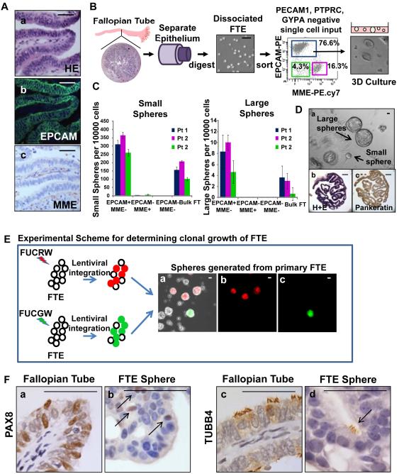
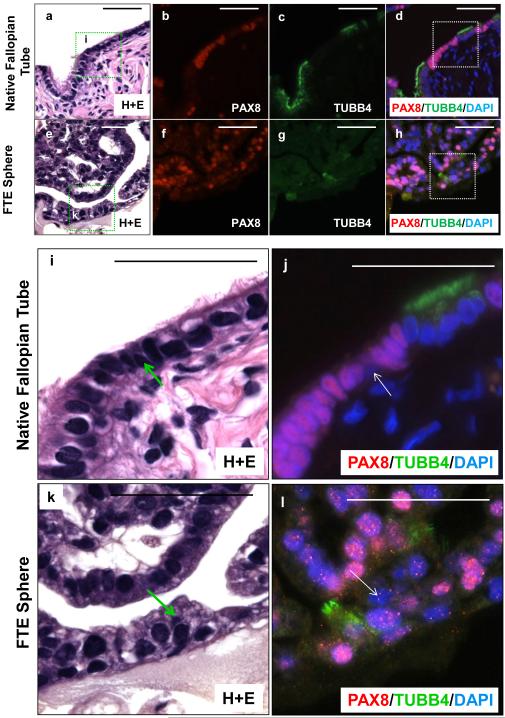
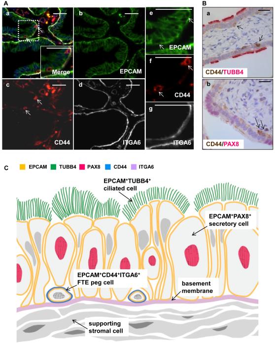
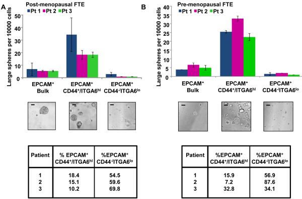
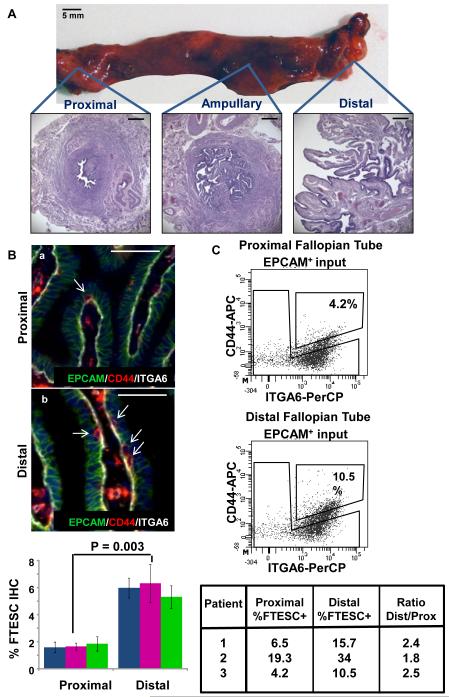
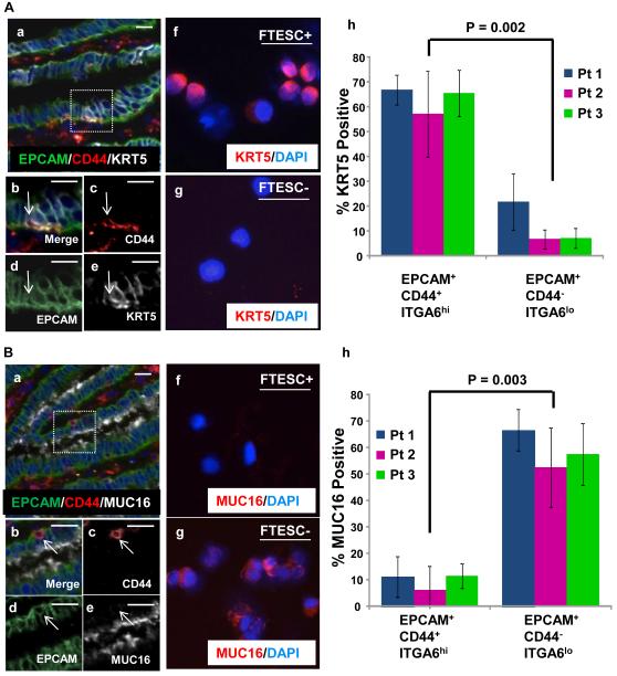
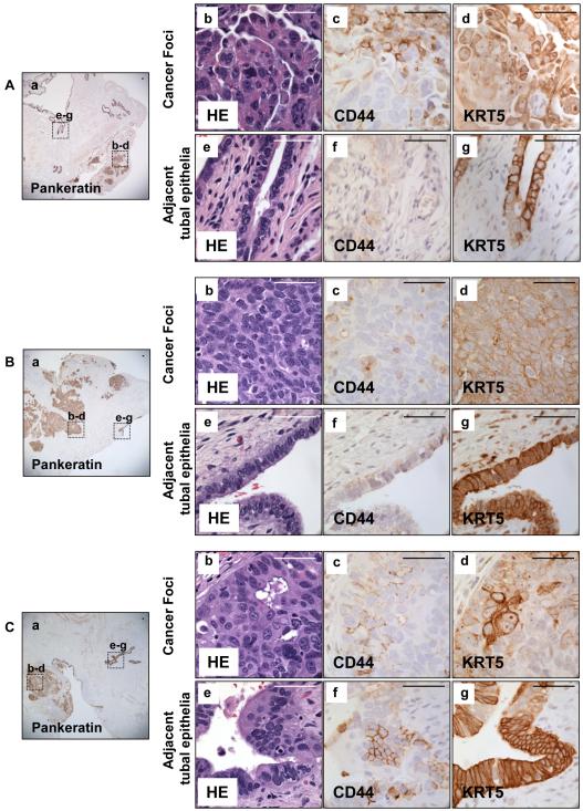
The reproductive role of the fallopian tube is to transport the sperm and egg. The tube is positioned to act as a bridge between the ovary where the egg is released and the uterus where implantation occurs. Throughout reproductive years, the fallopian tube epithelium undergoes repetitive damage and regeneration. Although a reservoir of adult epithelial stem cells must exist to replenish damaged cells, they remain unidentified. Here, we report isolation of a subset of basally located human fallopian tube epithelia (FTE) that lack markers of ciliated (β-tubulin; TUBB4) or secretory (PAX8) differentiated cells. These undifferentiated cells expressed cell surface antigens: epithelial cell adhesion molecule, CD44, and integrin α 6. This FTE subpopulation was fivefold enriched for cells capable of clonal growth and self-renewal suggesting that they contain the FTE stem-like cells (FTESCs). A twofold enrichment of the FTESC was found in the distal compared to the proximal end of the tube. The distal fimbriated end of the fallopian tube is a well-characterized locus for initiation of serous carcinomas. An expansion of the cells expressing markers of FTESC was detected in tubal intraepithelial carcinomas and in fallopian tubes from patients with invasive serous cancer. These findings suggest that FTESC may play a role in the initiation of serous tumors. Characterization of these stem-like cells will provide new insight into how the FTE regenerate, respond to injury, and may initiate cancer.


Figures

Figure 1
Figure 1. Isolation and in vitro clonal growth of adult human fallopian tube epithelia
(A) All FTE expressed the EPCAM cell surface marker (a,b), while supporting stroma expressed MME (a,c). (B) Expression of stromal and epithelial markers were examined by flow cytometry in mechanically isolated cells from fresh fallopian tube specimens. Based on EPCAM and MME expression, three populations were identified and plated in a matrigel culture assay. Subsets of cells generated spheroid structures. (C) Only the EPCAM+MMELin epithelial fraction was capable of generating large and small spheres. (D) FTE generated both large (>250 µm) and small (<250 µm) spheres (a), that were single layered and contain papillary structures reminiscent of the fallopian tube (b). Sphere cells were epithelial based on expression of pankeratin (c). (E) FTE cells were infected with either FUCRW (marked with red fluorescent protein [RFP]) or FUCGW (marked with green fluorescent protein [GFP]) lentivirus and mixed prior to plating. Regenerated spheres were either unlabeled (uninfected), RFP positive or GFP positive. Chimeric spheres were not detected suggesting that one cell gives rise to each sphere. Scale bars equal 100 µm. (F) Similar to native fallopian tube, subsets of FTE sphere cells were secretory based on PAX8 expression (a vs. b) or ciliated based on TUBB4 expression (c vs. d). All scale bars equal 50 µm.
Figure 2
Figure 2. Multi-lineage differentiation of dissociated human FTE cells evidenced by formation of all three fallopian tube cell types
Sections of both native fallopian tube (a-d, i-j) and FTE spheres (e-h, k-l) co-stained for secretory marker PAX8 and ciliated marker TUBB4 demonstrated that most cells were either secretory or ciliated. Rare basally located cells found in both native fallopian tube (magnified image i-j) and FTE spheres (magnified image k-l) were negative for both PAX8 and TUBB4. All scale bars equal 50 µm.
Figure 3
Figure 3. A subpopulation of CD44 positive cells adjacent to the basement membrane do not express markers of ciliated or secretory cells
(A) Immunohistochemistry revealed EPCAM positive epithelial cells (a,b,e) were bound by a basement membrane expressing ITGA6 (a,d,g). Rare, small basally located cells immediately adjacent to the basement membrane expressed both EPCAM and CD44 (a, c, f). (B) Basally located CD44 positive cells expressed neither the ciliated marker TUBB4 (a) or the secretory marker PAX8 (b). (C) Schematic of a third undifferentiated cell interspersed among the ciliated and secretory cells, marked with EPCAM, CD44, and ITGA6. This cell marked with EPCAM, CD44, and ITGA6 is the peg cell of the fallopian tube. All scale bars equal 25 µm.
Figure 4
Figure 4. The EPCAM+CD44+ITGA6hiLin population are the stem-like cells of the FTE
(A) In postmenopausal specimens, 15% (± 4.1%) of FTE were EPCAM+ CD44+ITGA6hiLin. These cells contained the predominance of large sphere forming potential, and were enriched 4.8 fold (± 0.1x) for growth compared to bulk FTE and 16.5 fold (± 4.3x) compared to the depleted fraction. (B) Similarly, in pre-menopausal patients undergoing post-partum tubal ligation, the EPCAM+CD44+ITGA6hiLin signature was identified in 19% (± 13.0%) of FTE. This cellular fraction was enriched 5.7 fold (± 1.0x) for in vitro growth compared to bulk FTE and a 16.8 fold (± 0.4x) compared to the depleted fractions. No significant difference was seen between the percentages of cells expressing the EPCAM+CD44+ITGA6hiLin antigenic profile under these differing hormonal conditions (P = 0.65 by t test). All scale bars equal 100 µm.
Figure 5
Figure 5. FTESC are distributed across the fallopian tube, but are concentrated in the distal end
(A) A gross example of a human fallopian tube and its cross sectional histology. Scale bars equal 5 mm and 500 µm. (B) Proportions of EPCAM+CD44+ITGA6hi cells in matched distal and proximal FT specimens were evaluated by IHC. A 3-fold increase in the FTESC positive population was observed in the distal compared to proximal fallopian tube. Scale bars equal 50 µm. (C) A two-fold greater percentage of FTESC was observed in the distal compared to proximal FTE analyzed by flow cytometry.
Figure 6
Figure 6. Predominance of FTESC express KRT5 but not MUC16
(A) In stained fallopian tube sections, many CD44 positive epithelial cells were found to co-express KRT5 (a-e). Cytospin analysis of FACS isolated cells confirmed that the majority of FTESC positive cells also expressed KRT5 (f,h), while most FTESC negative cells did not express KRT5 (g,h). (B) Basally located EPCAM positive CD44 positive epithelial cells in human fallopian tube sections were negative for MUC16 expression (a-e). Cytospin analysis performed on flow cytometry isolated cells demonstrated that the majority of isolated FTESC were MUC16 negative (f,h). Conversely, FTESC negative populations were predominantly positive for MUC16 expression (g,h). Scale bars equal 25 µm.
Figure 7
Figure 7. Expansion of CD44 and KRT5 positive cells in tubal epithelium and adjacent tumor nodules was detected in three patients with serous carcinoma
Histologic sections were obtained from fallopian tubes of three patients with serous carcinoma. Pankeratin marked all epithelia (Aa, Ba, Ca). Many CD44 and KRT5 positive cells were detected in serous tumor samples (A b-d, B b-d, C b-d). Expansion of both KRT5 and CD44 positive cells was observed in normal appearing tubal epithelia adjacent to these tumor foci (A e-g, B e-g, C e-g). Scale bars equal 50 µm.

Similar articles

Cited by

References

    1. Critoph FN, Dennis KJ. Ciliary activity in the human oviduct. Obstet Gynecol Surv. 1977;32:602–603. - PubMed
    1. Mahmood T, Saridogan E, Smutna S, et al. The effect of ovarian steroids on epithelial ciliary beat frequency in the human Fallopian tube. Hum Reprod. 1998;13:2991–2994. - PubMed
    1. Lyons RA, Djahanbakhch O, Mahmood T, et al. Fallopian tube ciliary beat frequency in relation to the stage of menstrual cycle and anatomical site. Hum Reprod. 2002;17:584–588. - PubMed
    1. Leese HJ, Tay JI, Reischl J, et al. Formation of Fallopian tubal fluid: role of a neglected epithelium. Reproduction. 2001;121:339–346. - PubMed
    1. Crow J, Amso NN, Lewin J, et al. Morphology and ultrastructure of fallopian tube epithelium at different stages of the menstrual cycle and menopause. Hum Reprod. 1994;9:2224–2233. - PubMed

Publication types

MeSH terms