. 2012 Sep 4;109(36):14520-5.
doi: 10.1073/pnas.1201153109. Epub 2012 Aug 21.

Conserved tyrosine kinase promotes the import of silencing RNA into Caenorhabditis elegans cells

Affiliations

Conserved tyrosine kinase promotes the import of silencing RNA into Caenorhabditis elegans cells

Antony M Jose et al. Proc Natl Acad Sci U S A. .

Abstract

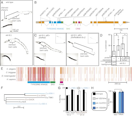
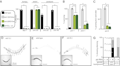
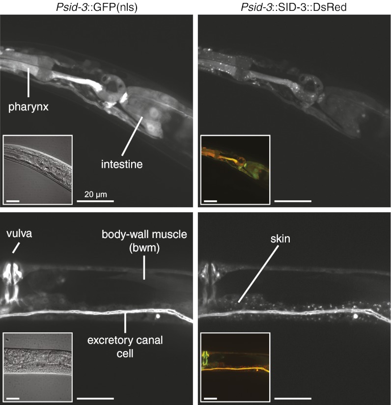
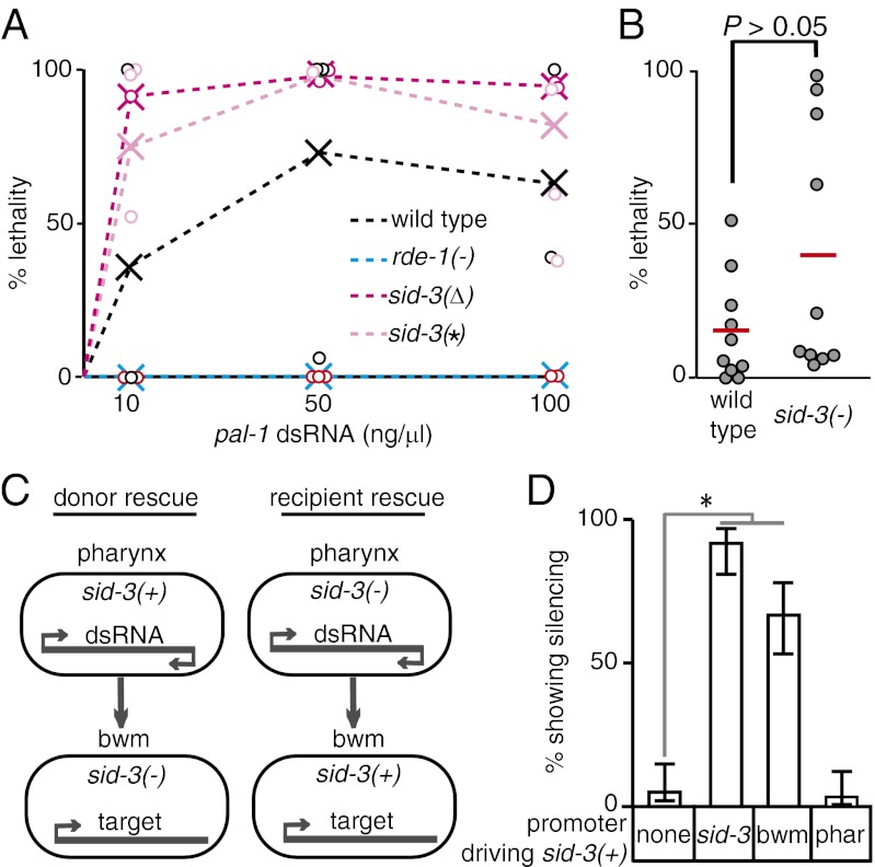
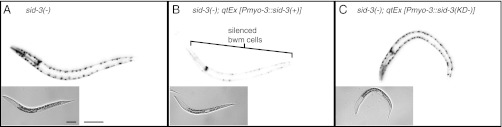
RNA silencing in Caenorhabditis elegans is transmitted between cells by the transport of double-stranded RNA (dsRNA). The efficiency of such transmission, however, depends on both the cell type and the environment. Here, we identify systemic RNAi defective-3 (SID-3) as a conserved tyrosine kinase required for the efficient import of dsRNA. Without SID-3, cells perform RNA silencing well but import dsRNA poorly. Upon overexpression of SID-3, cells import dsRNA more efficiently than do wild-type cells and such efficient import of dsRNA requires an intact SID-3 kinase domain. The mammalian homolog of SID-3, activated cdc-42-associated kinase (ACK), acts in many signaling pathways that respond to environmental changes and is known to directly associate with endocytic vesicles, which have been implicated in dsRNA transport. Therefore, our results suggest that the SID-3/ACK tyrosine kinase acts as a regulator of RNA import into animal cells.


Conflict of interest statement

The authors declare no conflict of interest.

Figures

Fig. 1.
Fig. 1.
sid-3 encodes a tyrosine kinase that regulates mobile RNA silencing. (A) sid-3(−) animals are defective in systemic RNAi. Representative wild-type (Upper) and sid-3(−) (Lower) animals where GFP expression in the pharynx and bwm cells is silenced (brackets) because of both pharynx-expressed and ingested gfp-dsRNA. Insets are brightfield images. (B) Gene structure of sid-3 showing mutations in isolated sid-3(−) alleles and the deduced changes in the SID-3 protein. ∆, deletion; *, stop codon. sid-3(tm342) was obtained from the C. elegans stock center. The tyrosine kinase domain (blue), SH3 domain (green), and CRIB domain (pink) are shown. (C) The silencing defect in sid-3(−) animals is rescued by sid-3(+) expression. Unlike expression of the fluorescent protein DsRed alone (Left, qtEx[DsRed]), coexpression of sid-3(+) (Right, qtEx[DsRed&sid-3(+)]) results in robust silencing in the bwm cells of sid-3(−) animals. Left Insets are brightfield images and Right Insets are red channel images. (D) Penetrance of silencing depicted in A and C. For each strain, the proportions of animals that lack detectable silencing are shown. Error bars indicate 95% confidence intervals. n = 40; *P < 0.05; §P = 0.054. (E) Schematic of multiple sequence alignment of SID-3 with its orthologs from Caenorhabditis briggsae, Drosophila melanogaster, and Homo sapiens. Known domains as in B and amino acid residues identical in three (orange) or in four (red) species are shown. (F) Phylogenetic relationship between the kinase domains of SID-3 and that of its paralog and homologs. Scale indicates amino acid substitution rate. (G) Similarity across the whole protein between SID-3, its paralog, and homologs in other species. SID-3 (black) shows greater identity than ARK-1 (white) to the human and fly ACK homolgs. (H) ark-1 mutants do not have a significant defect in silencing the skin gene dpy-7 by feeding RNAi. See Fig 2 for details of the assay.
Fig. 2.
Fig. 2.
sid-3 mutants are defective for feeding RNAi in all tested tissues. (A–C) Feeding RNAi of endogenous genes. Fourth larval stage (L4) animals of wild-type, sid-1(−), and two sid-3(−) strains with mutations that result in early stop codons [sid-3(W310*) and the tm342 deletion sid-3(∆→*)] were fed bacteria expressing dsRNA that target skin (dpy-7), muscle (unc-22 and unc-45) (A), embryonic (par-1 and pos-1) (B), or intestinal (act-5) genes (C). Percentage of affected (for skin and muscle genes) or surviving (for embryonic and intestinal genes) progeny are shown. Error bars indicate 95% confidence intervals. n ≥ 100; *P < 0.05; §sid-3(W310*) vs. wild-type (P = 0.02) and sid-3(∆→*) vs. wild-type (P = 0.053). (D–G) Feeding RNAi of gfp expression in transgenic animals. L4 animals that express GFP in all somatic nuclei (sur-5::gfp) in sid-1(−) (D), wild-type (E), or sid-3(tm342) (F) backgrounds were fed bacteria expressing gfp-dsRNA. Short lines indicate unsilenced gut nuclei (black). Proportions of progeny that show increasing extents of silencing (hatch < gray < black) are indicated along with representative schematics (G). n = 25–50 worms; Insets are brightfield images. (Scale bars, 100 μm.)
Fig. 3.
Fig. 3.
SID-3 is a widely expressed cytoplasmic protein. Fluorescence images of animals that coexpress nuclear-enriched GFP (Left) and a rescuing SID-3::DsRed fusion protein (Right) under the control of the sid-3 promoter and 3′ UTR. Left Insets are differential interference contrast images and Right Insets are merged red and green channel images. Fluorescence from SID-3::DsRed fusion was detected diffusely throughout the cytoplasm and in cytoplasmic foci. Similar diffuse and focal expression was also observed using a SID-3::GFP fusion protein (Fig. S6). Note that extrachromasomal arrays, which express the fluorescent proteins above, are lost mitotically, resulting in mosaic expression. For the more stable extrachromosomal arrays, the mosaic expression patterns largely match the known cell lineage. (Scale bars, 20 μm.)
Fig. 4.
Fig. 4.
Efficient import of dsRNA requires sid-3. (A) Response to different concentrations of dsRNA injected into the germline. Adult animals of wild-type, rde-1(−), and two sid-3(−) genotypes [sid-3(W310*)sid-3(*) and the tm342 deletion–sid-3(∆)] were injected with a similar volume of the indicated pal-1 dsRNA concentrations. pal-1-RNAi is embryonic lethal. The proportion of dead embryos laid by each injected animal (circle) and the average pooled proportion of dead embryos for each concentration and genotype (X) is plotted. (B) Response to limiting amounts of dsRNA (10 ng/μL) injected into the germ line of wild-type and sid-3(W310*) animals. Red bars and circles indicate average and individual proportions of dead embryos laid, respectively. P value is based on Mann–Whitney U test. (C) Schematic of experiment to test the role of SID-3 in exporting and importing tissues. (D) SID-3 is not required in the exporting tissue but is required in the importing tissue for silencing because of mobile RNA. sid-3(−) animals that express GFP in the pharynx and in bwm cells but express gfp-dsRNA only in the pharynx were transformed with constructs that express sid-3(+) under the control of its own (sid-3), bwm-specific (bwm), or pharynx-specific (phar) promoter and the percentage of transgenic animals that show gfp silencing in bwm cells was determined. Error bars indicate 95% confidence intervals. n = 100 L4 animals; *P < 0.05.
Fig. 5.
Fig. 5.
The kinase domain of SID-3 is required for the efficient import of dsRNA into cells. sid-3(−) animals that express GFP in the pharynx and in bwm cells but that express gfp-dsRNA only in the pharynx (A) were transformed with constructs that express either wild-type SID-3 [sid-3(+)] (B) or a kinase-dead version of SID-3 [sid-3(KD−)] (C) in bwm cells. Representative fluorescence images of silencing in response to gfp feeding RNAi in third larval-staged animals of each of the above three genotypes are shown. Only animals that express sid-3(+) in bwm showed silencing of bwm cells (brackets). Insets are brightfield images. (Scale bars, 50 μm.)

References

    1. Melnyk CW, Molnar A, Baulcombe DC. Intercellular and systemic movement of RNA silencing signals. EMBO J. 2011;30:3553–3563. - PMC - PubMed
    1. Winston WM, Molodowitch C, Hunter CP. Systemic RNAi in C. elegans requires the putative transmembrane protein SID-1. Science. 2002;295:2456–2459. - PubMed
    1. Wolfrum C, et al. Mechanisms and optimization of in vivo delivery of lipophilic siRNAs. Nat Biotechnol. 2007;25:1149–1157. - PubMed
    1. Duxbury MS, Ashley SW, Whang EE. RNA interference: A mammalian SID-1 homologue enhances siRNA uptake and gene silencing efficacy in human cells. Biochem Biophys Res Commun. 2005;331:459–463. - PubMed
    1. Valadi H, et al. Exosome-mediated transfer of mRNAs and microRNAs is a novel mechanism of genetic exchange between cells. Nat Cell Biol. 2007;9:654–659. - PubMed

Publication types

MeSH terms