Axon guidance in the developing ocular motor system and Duane retraction syndrome depends on Semaphorin signaling via alpha2-chimaerin
- PMID: 22912401
- PMCID: PMC3437829
- DOI: 10.1073/pnas.1116481109
Axon guidance in the developing ocular motor system and Duane retraction syndrome depends on Semaphorin signaling via alpha2-chimaerin
Abstract
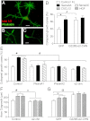
Eye movements depend on correct patterns of connectivity between cranial motor axons and the extraocular muscles. Despite the clinical importance of the ocular motor system, little is known of the molecular mechanisms underlying its development. We have recently shown that mutations in the Chimaerin-1 gene encoding the signaling protein α2-chimaerin (α2-chn) perturb axon guidance in the ocular motor system and lead to the human eye movement disorder, Duane retraction syndrome (DRS). The axon guidance cues that lie upstream of α2-chn are unknown; here we identify candidates to be the Semaphorins (Sema) 3A and 3C, acting via the PlexinA receptors. Sema3A/C are expressed in and around the developing extraocular muscles and cause growth cone collapse of oculomotor neurons in vitro. Furthermore, RNAi knockdown of α2-chn or PlexinAs in oculomotor neurons abrogates Sema3A/C-dependent growth cone collapse. In vivo knockdown of endogenous PlexinAs or α2-chn function results in stereotypical oculomotor axon guidance defects, which are reminiscent of DRS, whereas expression of α2-chn gain-of-function constructs can rescue PlexinA loss of function. These data suggest that α2-chn mediates Sema3-PlexinA repellent signaling. We further show that α2-chn is required for oculomotor neurons to respond to CXCL12 and hepatocyte growth factor (HGF), which are growth promoting and chemoattractant during oculomotor axon guidance. α2-chn is therefore a potential integrator of different types of guidance information to orchestrate ocular motor pathfinding. DRS phenotypes can result from incorrect regulation of this signaling pathway.
Conflict of interest statement
The authors declare no conflict of interest.
Figures


Similar articles
-
The Rac-GAP alpha2-Chimaerin Signals via CRMP2 and Stathmins in the Development of the Ocular Motor System.J Neurosci. 2021 Aug 4;41(31):6652-6672. doi: 10.1523/JNEUROSCI.0983-19.2021. Epub 2021 Jun 24. J Neurosci. 2021. PMID: 34168008 Free PMC article.
-
α2-Chimaerin regulates a key axon guidance transition during development of the oculomotor projection.J Neurosci. 2013 Oct 16;33(42):16540-51. doi: 10.1523/JNEUROSCI.1869-13.2013. J Neurosci. 2013. PMID: 24133258 Free PMC article.
-
Human CHN1 mutations hyperactivate alpha2-chimaerin and cause Duane's retraction syndrome.Science. 2008 Aug 8;321(5890):839-43. doi: 10.1126/science.1156121. Epub 2008 Jul 24. Science. 2008. PMID: 18653847 Free PMC article.
-
Ocular congenital cranial dysinnervation disorders (CCDDs): insights into axon growth and guidance.Hum Mol Genet. 2017 Aug 1;26(R1):R37-R44. doi: 10.1093/hmg/ddx168. Hum Mol Genet. 2017. PMID: 28459979 Free PMC article. Review.
-
Molecular basis of semaphorin-mediated axon guidance.J Neurobiol. 2000 Aug;44(2):219-29. doi: 10.1002/1097-4695(200008)44:2<219::aid-neu11>3.0.co;2-w. J Neurobiol. 2000. PMID: 10934324 Review.
Cited by
-
Motor neurons are dispensable for the assembly of a sensorimotor circuit for gaze stabilization.bioRxiv [Preprint]. 2024 Jan 27:2024.01.25.577261. doi: 10.1101/2024.01.25.577261. bioRxiv. 2024. Update in: Elife. 2024 Nov 20;13:RP96893. doi: 10.7554/eLife.96893. PMID: 38328255 Free PMC article. Updated. Preprint.
-
Oculomotor nerve guidance and terminal branching requires interactions with differentiating extraocular muscles.Dev Biol. 2021 Aug;476:272-281. doi: 10.1016/j.ydbio.2021.04.006. Epub 2021 Apr 24. Dev Biol. 2021. PMID: 33905720 Free PMC article.
-
α2-chimaerin is required for Eph receptor-class-specific spinal motor axon guidance and coordinate activation of antagonistic muscles.J Neurosci. 2015 Feb 11;35(6):2344-57. doi: 10.1523/JNEUROSCI.4151-14.2015. J Neurosci. 2015. PMID: 25673830 Free PMC article.
-
Childhood Onset Strabismus: A Neurotrophic Factor Hypothesis.J Binocul Vis Ocul Motil. 2021 Apr-Jun;71(2):35-40. doi: 10.1080/2576117X.2021.1893585. Epub 2021 Apr 19. J Binocul Vis Ocul Motil. 2021. PMID: 33872122 Free PMC article.
-
Loss of CXCR4/CXCL12 Signaling Causes Oculomotor Nerve Misrouting and Development of Motor Trigeminal to Oculomotor Synkinesis.Invest Ophthalmol Vis Sci. 2018 Oct 1;59(12):5201-5209. doi: 10.1167/iovs.18-25190. Invest Ophthalmol Vis Sci. 2018. PMID: 30372748 Free PMC article.
References
-
- Guthrie S. Patterning and axon guidance of cranial motor neurons. Nat Rev Neurosci. 2007;8:859–871. - PubMed
Publication types
MeSH terms
Substances
Grants and funding
LinkOut - more resources
Full Text Sources
Molecular Biology Databases

