

Review
doi: 10.1242/dev.063495.
Partitioning the heart: mechanisms of cardiac septation and valve development
Affiliations
- PMID: 22912411
- PMCID: PMC3424040
- DOI: 10.1242/dev.063495
Item in Clipboard
Review
Partitioning the heart: mechanisms of cardiac septation and valve development
Development.
2012 Sep.
Abstract
Heart malformations are common congenital defects in humans. Many congenital heart defects involve anomalies in cardiac septation or valve development, and understanding the developmental mechanisms that underlie the formation of cardiac septal and valvular tissues thus has important implications for the diagnosis, prevention and treatment of congenital heart disease. The development of heart septa and valves involves multiple types of progenitor cells that arise either within or outside the heart. Here, we review the morphogenetic events and genetic networks that regulate spatiotemporal interactions between the cells that give rise to septal and valvular tissues and hence partition the heart.
Figures
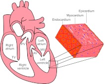
The structure of a mammalian heart. A mature mammalian heart contains four chambers (right atrium, left atrium, right ventricle, left ventricle) and four valves (pulmonary valve, PV; tricuspid valve, TV; atrial valve, AV; mitral valve, MV). The wall of each chamber consists of three tissue layers: endocardium, myocardium and epicardium.
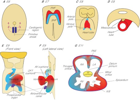
The formation of a mouse heart. (A) Ventral view of a mouse embryo at E6. The heart originates from mesodermal cells in the primitive streak. During gastrulation, mesodermal cardiac progenitor cells migrate to the splanchnic mesoderm to form the cardiac crescent. (B) Ventral view at E7. One subset of cardiac progenitors forms a horseshoe-shaped cardiac crescent (the first heart field, FHF; red). Another subset of cardiac progenitor cells forms the secondary heart field (SHF; blue), which is located posteriorly and medially to the FHF. (C) Ventral view at E8. Cells in the FHF merge in the midline to form the heart tube, which then elongates on both arterial and venous poles via the addition of progenitor cells from the SHF. (D) Transverse section at E8. The developing heart tube is suspended from the body wall by the dorsal mesocardium, which later dissolves except at the poles of heart tube, allowing the tube to loop rightward. (E,F) Ventral (E) and left lateral (F) views at E9. The looped heart tube contains four anatomical segments: atrium, atrioventricular canal, ventricle and outflow tract (OFT). Within the AVC and OFT, AV cushions (yellow) and OFT cushions (orange) develop. The proepicardial organ (purple) houses epicardial progenitors that later migrate to the heart and give rise to the epicardium. (G) Transverse section at E11. At this stage, the heart is partially partitioned by the primitive atrial septum (PAS), interventricular septum (IVS) and atrioventricular cushions (AV cushions) into a prototypic four-chamber heart. The AVC is divided into tricuspid and mitral orifices, forming ventricular inlets that connect the respective atrium to the ventricle. The opening between the PAS and AVC is the ostium primum. RA, right atrium; LA, left atrium; RV, right ventricle; LV, left ventricle.
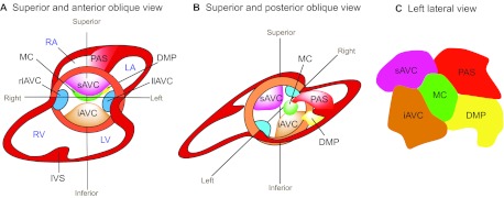
Endocardial cushion development. (A,B) Superior and anterior oblique view (A) and superior and posterior oblique view (B) of the heart. The superior and inferior atrioventricular cushions (sAVC and iAVC) are the two major cushions that develop in the central portion of the AVC. Two minor cushions, left and right lateral AV cushions (llAVC and rlAVC), form laterally at the AVC. The mesenchymal cap (MC) is a tissue that caps the leading edge of primary atrial septum (PAS) that grows from the atrial roof towards the AV canal. The dorsal mesenchymal protrusion (DMP) protrudes from the dorsal mesocardium into the atrial chamber. RA, right atrium; LA, left atrium; RV, right ventricle; LV, left ventricle; IVS, interventricular septum. (C) The composition of the atrial septum (left lateral view).
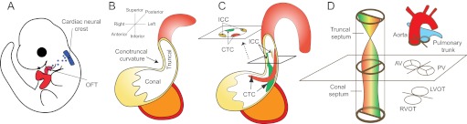
Septation of the cardiac outflow tract. (A) Left lateral view of an E10 mouse embryo. The neural crest at rhombomere 6-8 gives rise to cells (blue) that migrate to and colonize the distal cardiac outflow tract (OFT). (B) The cardiac OFT contains conal (proximal) and truncal (distal) cushions. The boundary between the conal and truncal cushions is marked by an outer curvature of the OFT (the conotruncal curvature). (C) The conotruncal cushions (CTCs) and intercalated cushions (ICCs) develop within the OFT. These cushions occupy four quadrants of the OFT (shown in cross-section). The conotruncal cushions fuse to septate the OFT, as shown in D. (D) Fusion of the conotruncal cushions forms a spiral septum, the truncal part of which divides the OFT into aorta and pulmonary trunk, whereas the conal part septates the OFT into left and right ventricular outlets (LVOT, RVOT). The aortic valves (AV) and pulmonic valves (PV) develop at the conotruncal junction.
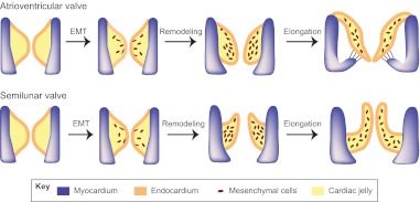
Epithelial-to-mesenchymal transformation and valve elongation. Endocardial cells in the AV cushions and conal cushions undergo epithelial-to-mesenchymal transformation (EMT) and generate mesenchymal cells that populate the cushions. The mesenchymal cushions then remodel and elongate themselves to from primitive valves that mature into thin valve leaflets (shown here for the atrioventricular valves and the semilunar valves).
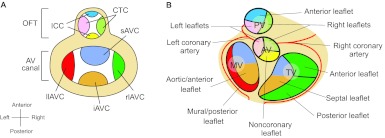
Endocardial cushions and heart valve leaflets. (A) Schematic of endocardial cushions in the atrioventricular (AV) canal and the outflow tract (OFT). The figure is a superior view of the heart with atria removed. The cushions are color coded to correspond to their derived valve leaflets illustrated in B. CTC. conotruncal cushions; ICC. intercalated cushions; sAVC. superior AV cushion; iAVC. inferior AV cushion; rlAVC. right lateral AV cushion; llAVC. left lateral AV cushion. (B) Schematic (superior view) of atrioventricular and semilunar valve leaflets that develop from the corresponding cushions color coded in A. PV, pulmonary valve; AV, aortic valve; TV, tricuspid valve; MV, mitral valve.
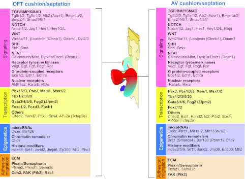
Genes and pathways essential for cardiac septation and valve development. Cushion and valve development, and hence septation, in the outflow tract (OFT) and the atrioventricular (AV) canal require similar molecular pathways. Factors required include those involved in signaling, transcription, epigenetics and cell adhesion/migration.
References
-
- Abedin M., Tintut Y., Demer L. L. (2004). Mesenchymal stem cells and the artery wall. Circ. Res. 95, 671–676 - PubMed
-
- Abu-Issa R., Kirby M. L. (2007). Heart field: from mesoderm to heart tube. Annu. Rev. Cell Dev. Biol. 23, 45–68 - PubMed
-
- Akhurst R. J., Lehnert S. A., Faissner A., Duffie E. (1990). TGF beta in murine morphogenetic processes: the early embryo and cardiogenesis. Development 108, 645–656 - PubMed
Publication types
MeSH terms
LinkOut - more resources
Full Text Sources
