. 2012 Sep;139(18):3326-31.
doi: 10.1242/dev.079418.

Slit2 and Robo3 modulate the migration of GnRH-secreting neurons

Affiliations

Slit2 and Robo3 modulate the migration of GnRH-secreting neurons

Anna Cariboni et al. Development. 2012 Sep.

Abstract

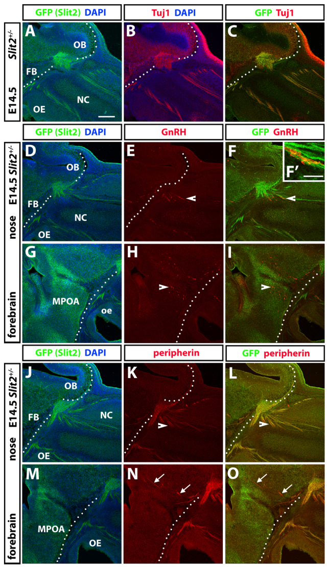
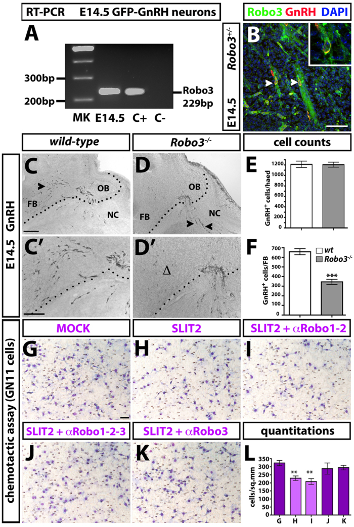
Gonadotropin-releasing hormone (GnRH) neurons are born in the nasal placode and migrate along olfactory and vomeronasal axons to reach the forebrain and settle in the hypothalamus, where they control reproduction. The molecular cues that guide their migration have not been fully identified, but are thought to control either cell movement directly or the patterning of their axonal substrates. Using genetically altered mouse models we show that the migration of GnRH neurons is directly modulated by Slit2 and Robo3, members of the axon guidance Slit ligand and Robo receptor families. Mice lacking Slit2 or Robo3 have a reduced number of GnRH neurons in the forebrain, but a normal complement of their supporting axons, pointing to a direct role for these molecules in GnRH neuron migration.


Figures

Fig. 1.
Fig. 1.
Slit2 expression during GnRH neuron migration. Expression of Slit2 in the NC and FB of embryonic mouse heads. Contiguous sagittal sections from E14.5 Slit2+/– animals were colabelled with GFP and TuJ1/GnRH/peripherin antibodies to visualise co-expression (arrows and arrowheads) of Slit2 and Tuj1 in nasal axons and FB neurons (A-C), migrating GnRH neurons (D-I), and OLF/VN axons (J-O), respectively. The boundary between the NC and FB is indicated by a dotted line. NC, nasal compartment; OE, olfactory epithelium; OB, olfactory bulb; FB, forebrain; MPOA, medial preoptic area. Scale bars: 150 μm, except 50 μm in F′.
Fig. 2.
Fig. 2.
Reduced number of GnRH neurons in the forebrain of Slit2–/– mice. (A) Schematic of the mouse head area displayed in B,C. (B,C) Sagittal sections of E12.5 mouse heads were immunolabelled for GnRH to reveal neurons migrating in the NC and FB. The FB boundary is delineated by a dotted line. Arrows (B,C) indicate areas displayed at higher magnification in insets. Arrow in C also points to abnormal accumulation of cells in the NC of Slit2–/– mice as compared with wild type (Δ in B). (D) Counts of GnRH neurons in the FB of E12.5 wild-type and Slit2–/– mice revealed a significant reduction in the mutant. (E-G′) Schematic (E) and sagittal sections (F,G) of E14.5 mouse heads immunolabelled for GnRH to reveal neurons migrating in the NC and FB. Arrows indicate areas displayed at high magnification in F′,G′. (H-J) Schematic (H) and representative images of sagittal sections (I,J) showing migrating GnRH neurons in the basal FB of E14.5 wild-type and Slit2–/– mice. There is a reduction in cells in the FB of the mutant (Δ in J). Arrowheads indicate examples of migrating GnRH neurons. (K) Counts of GnRH neurons in the FB of E14.5 wild-type and Slit2–/– mice revealed a statistically significant reduction in the mutant. (L-N,P-R) Schematic drawings (L,P) and coronal sections (M,N,Q,R) of E18.5 mouse brains immunolabelled for GnRH to reveal neurons projecting to the MPOA (M,N) and extending their axons to the ME (Q,R). Arrowheads indicate examples of migrating GnRH neurons (M,N) or GnRH+ fibres projecting to the ME (Q,R). The paucity of such projections in the Slit2–/– mice is indicated by Δ. (O) Counts of GnRH neurons in the brain of E18.5 wild-type and Slit2–/– mice revealed a statistically significant reduction in the mutant. (S) Optical density measurement of GnRH neuron fibres projecting to the ME of E18.5 wild-type and Slit2–/– mice revealed a statistically significant reduction in the mutant as compared with wild-type littermates. Mean ± s.e.m. *P<0.05, **P<0.01. ME, median eminence; LV, lateral ventricle; 3v, third ventricle. Scale bars: 100 μm in B,G; 75 μm in I,J,M,N,Q,R.
Fig. 3.
Fig. 3.
Slit and Robo in the GnRH neuronal system. (A-F) Expression of Slit2 and Slit1 in GnRH neurons. Sagittal sections from E12.5 Slit2+/– (A-C) and Slit1+/– (D-F) mouse heads were immunostained for GnRH (A,D) and GFP (B,E); colocalisation between GnRH (red) and Slit2/Slit1 (green) is indicated with arrowheads in the corresponding higher magnification images (C,F). The boundary between NC and FB is indicated by a dotted line. (G) RT-PCR on FACS-sorted E14.5 GFP-GnRH neurons revealed expression of Slit2 (210 bp), Robo1 (338 bp) and Robo2 (823 bp). Gapdh, positive control (188 bp). (H) Coronal section taken from E14.5 mouse head shows that Slit1 is expressed in GnRH neurons emerging from the VNO (yellow; arrowheads). (I,J) Coronal sections through the FB of E16.5 wild-type and Robo1–/– Robo2–/– littermates were immunolabelled to visualise GnRH neurons in the MPOA. (K) Counts of labelled cells did not show statistically significant differences between the wild-type and Robo1–/– Robo2–/– littermates. Mean ± s.e.m. VNO, vomeronasal organ. Scale bars: 150 μm in A,B,D,E; 50 μm in C,F,H; 100 μm in I,J.
Fig. 4.
Fig. 4.
Robo3 in the GnRH neuronal system. (A,B) Expression of Robo3 in GnRH neurons. (A) RT-PCR on FACS-sorted E14.5 GFP-GnRH neurons revealed the expression of Robo3 (229 bp). C+, positive control E14.5 mouse head; C–, negative control water. (B) Sagittal sections from E14 Robo3+/– animals were immunostained for GnRH and GFP. Colocalisation of GnRH (red) and Robo3 (green) is indicated (arrowheads and inset). (C-F) Robo3–/– mice have a reduced number of GnRH neurons in the FB. (C-D′) Sagittal sections of E14.5 mouse heads were immunolabelled for GnRH to reveal migrating neurons (arrowheads) in NC and FB. Higher magnification images are shown in C′,D′. There is a reduction of GnRH neurons in the FB of Robo3–/– mice (Δ in D′). (E,F) Counts of GnRH+ neurons in the head (E) and FB (F) of E14.5 wild-type and Robo3–/– mice revealed no differences in total number (E), but a statistically significant reduction in the FB of Robo3 mutants compared with wild-type littermates (F). (G-K) Chemomigratory response of GN11 cells towards Slit2. Representative images of migrated GN11 cells in a Boyden chamber assay with mock conditioned media of COS-7 cells (G) and Slit2-expressing COS-7 cells in the absence (H) or presence of Robo1/2 (I), Robo1-3 (J) or Robo3 (K) antibodies. (L) Counts of migrated GN11 cells/mm2 towards the indicated stimuli. Mean ± s.e.m. **P<0.01, ***P<0.001. Scale bars: 50 μm in B; 100 μm in C,D; 75 μm in C′,D′; 50 μm in G-K.

Similar articles

Cited by

References

    1. Bauer K., Dowejko A., Bosserhoff A. K., Reichert T. E., Bauer R. (2011). Slit-2 facilitates interaction of P-cadherin with Robo-3 and inhibits cell migration in an oral squamous cell carcinoma cell line. Carcinogenesis 32, 935–943 - PMC - PubMed
    1. Cariboni A., Rakic S., Liapi A., Maggi R., Goffinet A., Parnavelas J. G. (2005). Reelin provides an inhibitory signal in the migration of gonadotropin-releasing hormone neurons. Development 132, 4709–4718 - PubMed
    1. Cariboni A., Maggi R., Parnavelas J. G. (2007a). From nose to fertility: the long migratory journey of gonadotropin-releasing hormone neurons. Trends Neurosci. 30, 638–644 - PubMed
    1. Cariboni A., Hickok J., Rakic S., Andrews W., Maggi R., Tischkau S., Parnavelas J. G. (2007b). Neuropilins and their ligands are important in the migration of gonadotropin-releasing hormone neurons. J. Neurosci. 27, 2387–2395 - PMC - PubMed
    1. Cariboni A., Davidson K., Rakic S., Maggi R., Parnavelas J. G., Ruhrberg C. (2011). Defective gonadotropin-releasing hormone neuron migration in mice lacking SEMA3A signalling through NRP1 and NRP2: implications for the aetiology of hypogonadotropic hypogonadism. Hum. Mol. Genet. 20, 336–344 - PubMed

Publication types

MeSH terms