. 2012 Jul;12(4):180-5.
doi: 10.1016/s0972-6292(16)30524-1. Epub 2012 Jul 28.

What next after failed septal ventricular tachycardia ablation?

Affiliations

What next after failed septal ventricular tachycardia ablation?

Laurent Roten et al. Indian Pacing Electrophysiol J. 2012 Jul.

Abstract

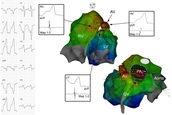
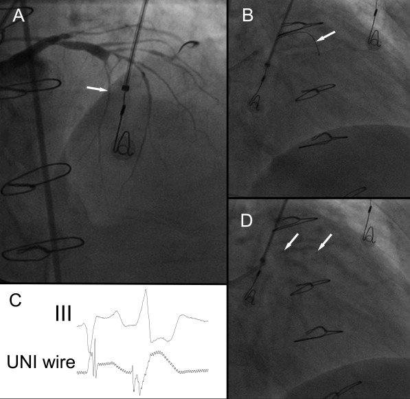
Ablation of ventricular tachycardia (VT) by conventional radiofrequency ablation can be impossible if the ventricular wall at the targeted ablation site is very thick, as for example the ventricular septum. We present a case of a patient with incessant, non-sustained slow VT originating from the septal part of the lower outflow tracts. Radiofrequency catheter ablation from both ventricles as well as from the anterior cardiac vein were not successful. Both high power radiofrequency ablation and bipolar radiofrequency ablation neither were successfull. Finally, ethanol ablation of the first septal perforator successfully terminated arrhythmia. We discuss the possibilities to overcome failed conventional radiofrequency VT ablation of a septal focus.

Keywords: bipolar radiofrequency ablation; ethanol ablation; ventricular tachycardia.


Figures

Figure 1
Figure 1
On the left side, the 12-lead ECG of the ventricular tachycardia is shown. On the right side, two views of an activation map of the VT are presented. Activation during VT in both ventricles as well as in the great cardiac vein (GV) and anterior cardiac vein (AV) is demonstrated. Recordings of distal ablation catheter (Map 1-2) and lead aVF at the earliest activation sites in the left ventricle (LV; 10 ms before QRS onset), in the right ventricle (RV, -20 ms) and in the AV (-20 ms) are shown. Brown dots represent ablation points, pink dots ablation points partially effective at higher power ablation. PA=pulmonary artery.
Figure 2
Figure 2
Antero-posterior fluoroscopic view of bipolar catheter ablation from the left and right ventricular outflow tracts. A first ablation catheter is positioned in the left ventricular outflow tract via a retrograde, aortic approach (catheter A) and a second ablation catheter in the right ventricular outflow tract via a long sheath (catheter B). Both catheters are placed at sites of earliest activation in the respective outflow tracts. A third mapping catheter is situated high in the right ventricular outflow tract (catheter C). Bipolar ablation was performed between the distal tips of both ablation catheters (A and B) with one catheter acting as cathode, and the other as anode.
Figure 3
Figure 3
Ethanol ablation of the first septal perforator. A: Angiogram of the left coronary arteries (RAO 45º view). The left anterior descending coronary artery is diseased and stenotic, a first septal perforator is opacified (white arrow). B: A guidewire (white arrow) is introduced into the proximal part of the first septal perforator (RAO cranial view). C: Unipolar recording from the guidewire and electrocardiogram of lead III. The guidewire during this recording was in the position as shown in picture B. Earliest activation at this site preceded QRS onset by minus 50 ms. D: A balloon is introduced into the very proximal part of the first septal perforator (white arrows denote margins of balloon), and 1.5 ml of ethanol infused into the first septal perforator, successfully eliminating VT (RAO cranial view).

References

    1. Sacher F, et al. Transcoronary ethanol ventricular tachycardia ablation in the modern electrophysiology era. Heart Rhythm. 2008;5:62. - PubMed
    1. Kay GN, et al. Intracoronary ethanol ablation for the treatment of recurrent sustained ventricular tachycardia. J Am Coll Cardiol. 1992;19:159. - PubMed
    1. Segal OR, et al. Intra-coronary guidewire mapping-a novel technique to guide ablation of human ventricular tachycardia. J Interv Card Electrophysiol. 2007;18:143. - PubMed
    1. Sivagangabalan G, et al. Bipolar ablation of the interventricular septum is more efficient at creating a transmural line than sequential unipolar ablation. Pacing Clin Electrophysiol. 2010;33:16. - PubMed
    1. Rigopoulos AG, et al. A decade of percutaneous septal ablation in hypertrophic cardiomyopathy. Circ J. 2010;75:28. - PubMed

LinkOut - more resources