. 2012;8(8):e1002852.
doi: 10.1371/journal.ppat.1002852. Epub 2012 Aug 9.

HSV-1 genome subnuclear positioning and associations with host-cell PML-NBs and centromeres regulate LAT locus transcription during latency in neurons

Affiliations

HSV-1 genome subnuclear positioning and associations with host-cell PML-NBs and centromeres regulate LAT locus transcription during latency in neurons

Frédéric Catez et al. PLoS Pathog. 2012.

Abstract

Major human pathologies are caused by nuclear replicative viruses establishing life-long latent infection in their host. During latency the genomes of these viruses are intimately interacting with the cell nucleus environment. A hallmark of herpes simplex virus type 1 (HSV-1) latency establishment is the shutdown of lytic genes expression and the concomitant induction of the latency associated (LAT) transcripts. Although the setting up and the maintenance of the latent genetic program is most likely dependent on a subtle interplay between viral and nuclear factors, this remains uninvestigated. Combining the use of in situ fluorescent-based approaches and high-resolution microscopic analysis, we show that HSV-1 genomes adopt specific nuclear patterns in sensory neurons of latently infected mice (28 days post-inoculation, d.p.i.). Latent HSV-1 genomes display two major patterns, called "Single" and "Multiple", which associate with centromeres, and with promyelocytic leukemia nuclear bodies (PML-NBs) as viral DNA-containing PML-NBs (DCP-NBs). 3D-image reconstruction of DCP-NBs shows that PML forms a shell around viral genomes and associated Daxx and ATRX, two PML partners within PML-NBs. During latency establishment (6 d.p.i.), infected mouse TGs display, at the level of the whole TG and in individual cells, a substantial increase of PML amount consistent with the interferon-mediated antiviral role of PML. "Single" and "Multiple" patterns are reminiscent of low and high-viral genome copy-containing neurons. We show that LAT expression is significantly favored within the "Multiple" pattern, which underlines a heterogeneity of LAT expression dependent on the viral genome copy number, pattern acquisition, and association with nuclear domains. Infection of PML-knockout mice demonstrates that PML/PML-NBs are involved in virus nuclear pattern acquisition, and negatively regulate the expression of the LAT. This study demonstrates that nuclear domains including PML-NBs and centromeres are functionally involved in the control of HSV-1 latency, and represent a key level of host/virus interaction.


Conflict of interest statement

The authors have declared that no competing interests exist.

Figures

Figure 1
Figure 1. In situ detection of the HSV-1 genome in mouse TG sections.
(A) Models of latent infection. (B) Schematic representation of the HSV-1 genome and the LAT locus. The RNA- and DNA-FISH probes used in this study are indicated in grey. (C) Control DNA-FISH experiments were performed on mock-infected mouse tissue by using an HSV-1-specific probe and on HSV-1-infected mouse tissue by using an empty cosmid vector (Cos64) probe. Stained tissue sections were imaged by wide-field microscopy. Dashed lines indicate the position of the nucleus. Scale bar = 5 µm. (D) Illustration of the main latent HSV-1 genome pattern. DNA-FISH was performed on TG sections obtained from infected mice 28 d.p.i. as in (A), using a mix of HSV-1 cosmids 14, 28, and 56 as indicated in (B). Stained sections were imaged by wide-field microscopy. Dashed lines indicate position of the nucleus. Scale bar = 5 µm. (E) Quantification of HSV-1 genome patterns during latency. Two groups of mice were infected according to the two models presented in (A). For SC16/lip-infected mice, n = 6 (4671 infected neurons); for 17syn+/eye mice, n = 4 (1000 infected neurons). Bars show the standard error of the mean. (F) Distribution of latently infected neurons along the TG. The data obtained in (E) from five SC16/lip-infected mice were plotted as total number of HSV-1 genome-positive neurons per section. (G) Distribution of the single/single+ and multiple/super-multiple patterns along the TG. Data from one mouse shown in (F) is shown as example.
Figure 2
Figure 2. LAT expression correlates with HSV-1 genome pattern.
(A) TG sections obtained from SC16/lip-infected mice at 28 d.p.i. were processed for RNA-FISH using a 2-kb LAT RNA-FISH probe and an HSV-1 DNA-FISH probe as in Figure 1D. The dotted lines outline the nucleus. Each labeling is shown as separated channels on the right panel. Wide-field imaging. Scale bar = 5 µm. (B) TG sections obtained from three mice at 28 d.p.i. were processed as in (A). The 2-kb LAT RNA-FISH signal and DNA-FISH pattern were quantified and plotted as the fraction of LAT+ neurons among neurons with the various patterns. (C) Same data set as in (B), plotted as the fraction of each pattern in LAT+ neurons. Bars show the standard error of the mean.
Figure 3
Figure 3. Latent HSV-1 genomes co-localize with centromeres in neuron nuclei.
(A) Co-detection of HSV-1 genomes with telomeres and centromeres. TG sections from mock-infected and latently infected (SC16/lip) mice were stained by dual-color DNA-FISH using HSV-1-, telomere- and centromere-specific (Minor satellite, MiSat) probes. Tissues were counterstained with Hoechst 33258, which revealed aggregated pericentromeres in mouse cells (see text for details). The boxed areas show single-channel images. Arrowheads show co-localization of the HSV-1 signal with the minor satellite signal. Scale bar = 5 µm. (B) TG sections from two mice were stained with HSV-1 genome- and centromere-specific probes as in (A), and neurons in which the HSV-1 genome signal was associated with centromeres and pericentromeres were counted. Bars show the standard error of the mean. (C–D) TG sections from latently infected (SC16/lip) mice were stained by immuno-FISH with an HSV-1 genome-specific probe and anti-CENP-A antibody. Pericentromeres were counterstained with Hoechst. The images show that HSV-1 genome spots precisely co-localize with the CENP-A signal at the surface of pericentromeres. Scale bar = 5 µm.
Figure 4
Figure 4. PML-NBs form around HSV-1 genomes in neurons.
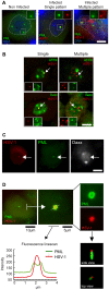
(A) Co-localization of HSV-1 genomes with PML protein. TG sections from mock-infected and latently infected (SC16/lip) mice (28 d.p.i.) were stained by immuno-FISH to co-detect the HSV-1 genome and PML protein. The boxed areas show single-channel images. Scale bar = 5 µm. (B) The HSV-1 genome associates with major PML-NB components. TG sections from latently infected mice were stained by immuno-DNA-FISH using anti-ATRX or anti-Daxx antibody and HSV-1 probe. HSV-1 genomes associated with ATRX or Daxx are indicated by arrows and enlarged in the insets. (C) TG sections from the same mouse as in (B) were stained by dual immuno-FISH using anti-PML and anti-Daxx antibodies and HSV-1 probe. The arrow shows an HSV-1-containing PML-NB stained with anti-Daxx antibody. (D) Confocal microscopic analysis of an HSV-1-containing PML-NB, showing (by fluorescence line-scan measurement and 3D reconstruction of a confocal Z-stack) that the HSV-1 DNA-FISH signal is localized within the PML-NB.
Figure 5
Figure 5. PML/PML-NBs control the distribution of incoming and latent HSV-1 genomes.
(A–B) PML signals increase in neuronal and non-neuronal cells during the acute phase. (A) TG sections from mock-infected and infected mice sacrificed during the acute phase (6 d.p.i.) or latency (28 d.p.i.) were stained by immuno-FISH using HSV-1 probe and anti-PML antibody. In the SC16/lip model, inoculation of the virus on the upper left side induced asymmetrical acute and latent infection (see Materials and methods). All images were collected using identical gain and exposure settings. For acute infection, images of the left and right TG were collected from the same section. (B) Western blot analysis of PML protein in left and right TG harvested from mock-infected and infected mice during acute infection (6 d.p.i.). NIH3T3 cells were used as a positive control. A pan-HSV-1 serum was used to confirm ongoing acute infection in the left TG, through detection of lytic HSV-1 protein (arrows). Lytic infection is known to occur in early acute phase in some neurons and accessory cells, and is cleared as latency is being established , and see figure S3). Actin was used as a loading control. (C) HSV-1-containing PML-NBs were detected during acute phase. TG sections from infected (SC16/lip) mice sacrificed at 6 d.p.i. were stained by immuno-FISH using HSV-1 probe and anti-PML (left), anti-ATRX (middle), and anti-Daxx (right) antibodies. The fluorescence plot profiles (dashed lines) are consistent with a ring-shaped PML-NB containing the HSV-1 genome and ATRX and Daxx proteins in its center. a.u. = arbitrary units. Scale bar = 5 µm. (D) The HSV-1 genome pattern is altered in PML-knockout mice. Wild-type, heterozygous, and PML-knockout mice were infected by corneal inoculation with the 17syn+ HSV-1 strain and were sacrificed at 28 d.p.i. HSV-1 genome patterns were quantified in serial sections spanning the whole ganglion. n = 4 mice per genotype (∼1500 infected neurons per genotype). Bars show the standard error of the mean. (E) Same data set as in (D). Fractions of neurons displaying very strong HSV-1 DNA-FISH signals (“super-multiple” pattern, Figure S1) within the multiple-pattern neurons in (D) are shown.
Figure 6
Figure 6. Absence of LAT transcription from PML-NB- and centromere-associated HSV-1 genomes.
(A) Detection of nascent 8.3-kb primary LAT RNA. TG sections obtained from SC16/lip mice at 28 d.p.i. were stained by RNA/DNA-FISH using an HSV-1 genome DNA probe and LAT-2 (2-kb LAT) and LAT-5 (8.3-kb LAT) RNA probes (Figure 1B). (B) HSV-1 genomes producing nascent 8.3-kb LAT are not associated with PML-NBs. TG sections obtained from infected (SC16/lip) mice at 28 d.p.i. were subjected to dual immuno-FISH labeling with HSV-1 genome DNA probe, LAT-5 RNA probe, and anti-PML antibody. Examples of single, single+, and multiple patterns are shown. Dashed line images are single-channel images (right). Scale bar = 5 µm. (C) HSV-1 genomes producing nascent 8.3-kb LAT are not associated with centromeres. TG sections obtained from infected (SC16/lip) mice at 28 d.p.i. were subjected to dual RNA/DNA-FISH labeling with HSV-1 genome DNA probe, LAT-5 RNA probe, and minor satellite DNA probe. Scale bar = 5 µm. (D–E) PML wild-type, heterozygous, and knockout mice were infected (17syn+/eye) and sacrificed at 28 d.p.i. TG sections were stained by RNA/DNA-FISH using LAT-2 RNA probe and HSV-1 DNA probe. Graphs show (D) total numbers of neurons expressing 2-kb LAT RNA and (E) numbers of multiple/super multiple HSV-1 pattern neurons expressing 2-kb LAT RNA in PML+/+, PML+/− and PML−/− mice. n = 7 mice (∼2200 neurons) per genotype. Bars show the standard error of the mean.

References

    1. Roizman B, Knipe D, Whitley R (2007) Herpes Simplex Viruses. In: Fields Virology, 5th edition. Knipe D, Howley P, editors. Philadelphia: Lippincott Williams and Wilkins. 2501–2602.
    1. Knipe DM, Cliffe A (2008) Chromatin control of herpes simplex virus lytic and latent infection. Nat Rev Microbiol 6: 211–221. - PubMed
    1. Bloom DC, Giordani NV, Kwiatkowski DL (2010) Epigenetic regulation of latent HSV-1 gene expression. Biochim Biophys Acta 1799: 246–256. - PMC - PubMed
    1. Maillet S, Naas T, Crepin S, Roque-Afonso AM, Lafay F, et al. (2006) Herpes simplex virus type 1 latently infected neurons differentially express latency-associated and ICP0 transcripts. J Virol 80: 9310–9321. - PMC - PubMed
    1. Kramer MF, Coen DM (1995) Quantification of transcripts from the ICP4 and thymidine kinase genes in mouse ganglia latently infected with herpes simplex virus. J Virol 69: 1389–1399. - PMC - PubMed

Publication types

MeSH terms