. 2012;8(8):e1002874.
doi: 10.1371/journal.pgen.1002874. Epub 2012 Aug 9.

Drosophila Hox and sex-determination genes control segment elimination through EGFR and extramacrochetae activity

Affiliations

Drosophila Hox and sex-determination genes control segment elimination through EGFR and extramacrochetae activity

David Foronda et al. PLoS Genet. 2012.

Abstract

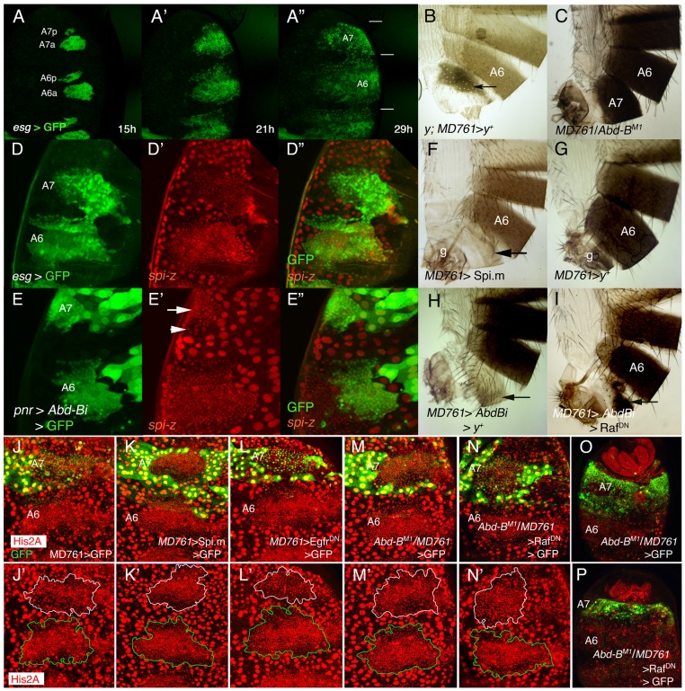
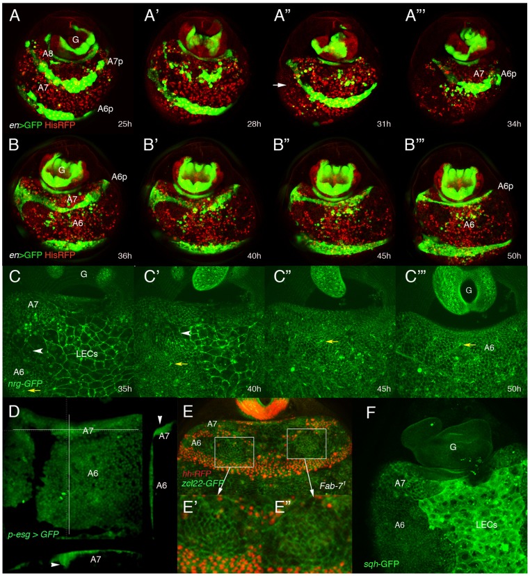
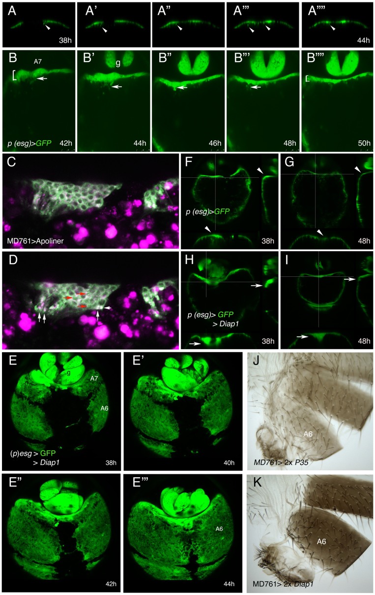
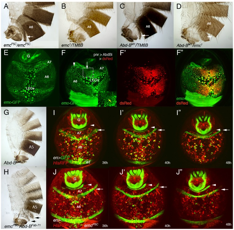
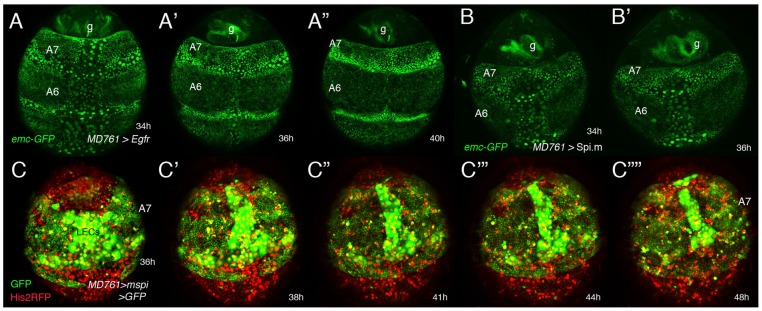
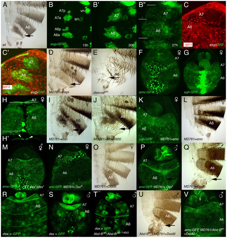
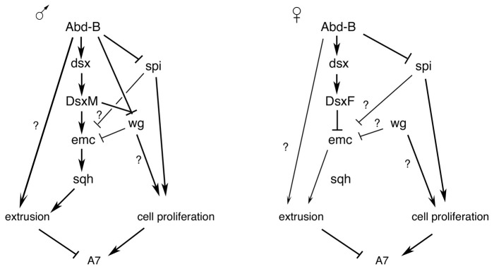
The formation or suppression of particular structures is a major change occurring in development and evolution. One example of such change is the absence of the seventh abdominal segment (A7) in Drosophila males. We show here that there is a down-regulation of EGFR activity and fewer histoblasts in the male A7 in early pupae. If this activity is elevated, cell number increases and a small segment develops in the adult. At later pupal stages, the remaining precursors of the A7 are extruded under the epithelium. This extrusion requires the up-regulation of the HLH protein Extramacrochetae and correlates with high levels of spaghetti-squash, the gene encoding the regulatory light chain of the non-muscle myosin II. The Hox gene Abdominal-B controls both the down-regulation of spitz, a ligand of the EGFR pathway, and the up-regulation of extramacrochetae, and also regulates the transcription of the sex-determining gene doublesex. The male Doublesex protein, in turn, controls extramacrochetae and spaghetti-squash expression. In females, the EGFR pathway is also down-regulated in the A7 but extramacrochetae and spaghetti-squash are not up-regulated and extrusion of precursor cells is almost absent. Our results show the complex orchestration of cellular and genetic events that lead to this important sexually dimorphic character change.


Conflict of interest statement

The authors have declared that no competing interests exist.

Figures

Figure 1
Figure 1. Cell divisions and EGFR activity in the male A7.
(A–A″) Snapshots from Video S1 (esg-Gal4 UAS-nls-myc-GFP pupa), from ∼15 to 29 h APF showing the expansion of the dorsal histoblast nests in the male A6 and A7 segments. In this and subsequent figures (except in adult cuticle preparations) the posterior part of the pupa is at the top. Note that the size of the A7 is smaller than that of the A6 (horizontal bars in A″). Approximate hours of development APF are also indicated. (B) y; MD761-Gal4 UAS-y+/+ female. Note the dark pigmentation in the A7 (arrow). (C) MD761-Gal4/Abd-BM1 male. See that the A7 (absent in the wildtype) is almost completely transformed into the A6. (D–D″) Posterior part of a spi-lacZ esg-Gal4 UASGFP pupa of about 26 h APF showing that spi levels (in red) are reduced in the A7 histoblasts (the A7/A6 ratio of signal intensity is 0,82±0,12; n = 10). Histoblasts are marked by esg in green. (E–E″) If we express a UAS-Abd-BRNAi (UAS-Abd-Bi) construct under the control of the pannier (pnr)-Gal4 driver (domain of expression in green), the levels of spi-lacZ are elevated in this domain (arrow). The arrowhead marks the lower spi-lacZ levels in A7 histoblasts where Abd-B expression is still high (note also the bigger cells). (F) UAS-Spi.m-GFP; MD761-Gal4/+ adult male (cross made at 17°C), with a small A7 segment (compare with a male expressing a UAS-y+ construct (G), which, like the wildtype, has no A7); g, genitalia. (H) UAS-Abd-BRNAi/+; MD761-Gal4 UAS-y+/+adult male, showing the transformation of the A7 into the A4. (I) In UAS-Abd-BRNAi/+; MD761-Gal4/UAS-RafDN the size of the A7 segment is reduced as compared to that shown in H. (J–L′). Posterior abdomens of ∼22–24 h APF male pupae of the following genotypes: His2A-RFP/+; MD761-Gal4 UAS-GFP/+ (J, J′), UAS-Spi.m-GFP; His2A-RFP/+; MD761-Gal4 UAS-GFP/+ (K, K′), and His2A-RFP/UAS-EgfrDN; MD761-Gal4 UAS-GFP/+ (L, L′), showing a slight reduction (L, L′) and an increase (K, K′) in histoblast number in the A7 with respect to the His2A-RFP/+; MD761-Gal4 UAS-GFP/+ pupae (J, J′): at about this time, in the wildtype, the histoblast number A7/A6 ratio is 0’47±0’07 (n = 4), it is 0’95±0’19 (n = 4) when we express mspi in the A7, and 0’31±0’04 (n = 4) when the RafDN product is present in the same segment. (M, M′) In MD761-Gal4 UAS-GFP/Abd-BM1 male pupa the number of histoblasts in the A7a dorsal nests approaches that of the A6a, and this number is reduced in MD761-Gal4 UAS-GFP/UAS-RafDN Abd-BM1 pupae (N, N′). At later stages, the A7 of the MD761-Gal4 UAS-GFP/Abd-BM1 pupae (O; marked in green) is bigger than the wildtype and it is strongly reduced when a RafDN protein is concomitantly expressed in this genetic background (P). Nuclei are marked in red and the A7 delimited by GFP expression (in green). In the lower panels (J′, K′, L′, M′, N′) the A7 dorsal nests are delineated in white and the A6 ones in green.
Figure 2
Figure 2. Dorsal histoblasts of the male A7 extrude through the epithelium.
(A–B″′) Stills from Videos S2 and S3 showing the progressive elimination, first of A8 LECs and then of A7 LECs and histoblasts, in His2A-RFP/en-Gal4 UAS-GFP male pupae from about 25 h till about 50 h APF. En is expressed in posterior compartments, (marked in green by GFP), and nuclei are marked in red. In this and other movies (and snapshots from them) marked with His2A-RFP, histoblasts are difficult to see under the moving cells (macrophages or hemocytes), which present a strong red signal. A histoblast nest is indicated by an arrow in A″. A8 cells disappear first and A7 cells follow, so that A6p histoblasts end up contacting the genitalia (G), which rotates during this period. Numbers indicate approximate hours APF. (C–C″′) Snapshots from Video S5 (approx. 35–50 h APF) showing the apparent disappearance of A7 histoblasts in an nrg-GFP male pupa. The yellow arrows indicate the position of a bristle precursor and the white arrowheads shows the LECs separating the A6p and A7a histoblast nests. Note how both marks move posteriorly as the A7 histoblasts are eliminated. Numbers indicate approximate hours APF. (D) Snapshot from a movie showing a (p)esg-Gal4 UAS-GFP male pupa of about 40 h APF; cross sections, to the right and below (the plane of section indicated by white lines) show the accumulation of histoblasts as bulges under the epidermis (arrowheads). (E) Still taken from video S6, showing an Abd-BFab7-1 homozygous male pupa in which Hh-RFP marks posterior compartments (in red) and the membrane marker zcl22-GFP is in green. E′, E″ are details of the squares in E, showing the constriction of cells in this optical section in two regions of the histoblast nests before histoblast invagination. (F) sqh-GFP expression is higher in the A7 histoblast nests of a ∼36 h APF male pupa than in the A6.
Figure 3
Figure 3. Inhibition of cell death does not prevent delamination of histoblasts.
(A–A″″) Snapshots from video S9, from about 38 to 44 h APF, showing how the histoblasts from the male A7 left and right anterior dorsal nests of a (p)esg-Gal4 UAS-GFP male pupa delaminate as the nests meet at the central midline. The arrows indicate the delamination of some histoblasts. (B–B″″) Stills from video S10, made in a (p)esg-Gal4 UAS-GFP male pupa of about 42–50 h APF, showing delamination of A7 histoblasts (arrows). See how the “width” of the A7 segment (brackets at 42 h and 50 h) is reduced as delamination proceeds; g, genitalia. Numbers indicate approximate hours APF. (C, D) Two optical sections of a movie sequence in which the Apoliner construct, which reveals cell death , is expressed under the control of the MD761-Gal4 driver in A7 histoblasts. The panel C is from about 40 h APF and panel D from about 42 h APF. The red arrows indicate three cells where the GFP reporter has nuclear localization (indicating apoptosis) and the white arrows point to what could be apoptotic bodies. (E–E′″) Snapshots from video S11 showing the invagination of the male A7 in (p)esg-Gal4 UAS-GFP UAS-Diap1 (esg-Gal4 act>y+>Gal4/UAS-Diap1; UAS-flp/UAS-Diap1) pupae. The invagination takes place as in the wildtype although it may be delayed. Numbers indicate approximate hours APF. (F–I) Optical sections of (p)esg-Gal4 UAS-GFP (F, G) and (p)esg-Gal4 UAS-Diap1 UAS-GFP (H, I) male pupae. Cross-sections (white lines) to the right and below in each figure show that in (p)esg-Gal4 UAS-GFP pupae there are some histoblasts under the epithelium at about 38 h APF (F), but they are not longer there by 48 h APF (G) (arrowheads). By contrast, in (p)esg-Gal4 UAS-Diap1 UAS-GFP male pupae, the number of A7 histoblasts that remain under the epithelium is higher at about 38 h APF (H) and have not been completely eliminated by 48 h APF (I) (arrows). (J, K) The inhibition of cell death in UAS-P35/+; UAS-P35/MD761-Gal4 (J) and UAS-Diap1/+; UAS-Diap1/MD761-Gal4 (K) males does not prevent A7 elimination.
Figure 4
Figure 4. emc regulates extrusion of the male A7 histoblasts.
(A, B) emcP5C homozygous males show a small A7 segment (A), absent in emc heterozygous conditions (B). (C) Males heterozygous for an Abd-B mutation present a small A7 , , and the trans-heterozygous combination Abd-BM1/emc1 enhances this phenotype (D). Arrows indicate the A7 segment. (E) Distribution of emc-GFP in the A7 and A6 segments of a ∼38 h APF male pupa. There is emc expression in LECs and histoblasts, with higher levels in A7 than in A6 histoblasts. We have measured the difference in signal intensity between nuclei of the two segments and found that the A7/A6 signal ratio is 1, 32±0,15 (n = 4). See also higher levels at the periphery of histoblast nests. (F–F″) UAS-DsRed/+; emc-GFP pnr-Gal4/UAS-Abd-BRNAi male pupa of about 36 h APF showing that in the central region of pnr expression (red in F′, F″), where Abd-B levels are reduced, emc-GFP levels are also reduced (arrow in F). Levels remain high where Abd-B has not been eliminated (arrowhead in F) but also in midline cells (also with high expression in anterior segments; yellow arrow in inset). (G) Abd-BFab7-1 homozygous adult male. The A6 disappears as it is transformed into the A7 . (H) Abd-BFab7-1 emcP5C homozygous male: there are small A6 and A7 segments (arrows), showing the emcP5C mutation is epistatic over the Abd-BFab7-1 mutation. (I–I″) Snapshots from Video S12 in a ∼36–48 h APF en-Gal4 UAS-GFP/His2A-RFP male, showing the progressive disappearance of the A7 segment. Posterior compartments show en expression (in green), whereas nuclei are labeled in red. The arrow marks the A6p band of expression and the arrowhead the A8p cells. (J–J″) Snapshots from Video S13, taken at similar stages and with the same markers but in an emc mutant background (en-Gal4 UAS-GFP/His2A-RFP; emcP5/emcP5male). Note that, contrary to the previous panels (I–I″), the A6p en band does not move posteriorly, indicating that the A7 segment is not being extruded. Numbers indicate approximate hours APF.
Figure 5
Figure 5. Genetic interactions between emc and the EGRF pathway.
(A–B′) Stills from two videos (S14 and another one, not shown) done in pupae of the following genotypes: UAS-Egfr/+; emc-GFP MD761-Gal4/+ (A–A″) and emc-GFP MD761-Gal4/UAS-Spi.m-HRP (B, B′). The size of the A7 is increased in A–A″ and B, B′ with respect to the wildtype and emc-GFP expression seems slightly reduced in the A7 (compare with Figure 4E). (C–C″″) Snapshots from video S15 (∼35–48 h APF) showing a male pupa of the genotype UAS-Spi.m-GFP; MD761-Gal4 UAS-GFP/+. There is an excess of histoblasts in this segment (marked in green). The extrusion of LECs takes longer and that of histoblasts seems to be largely prevented. Numbers indicate approximate hours APF.
Figure 6
Figure 6. Relationship between sex determination and Hox information in the development of an A7.
(A) Wildtype female adult, showing the small A7 segment (arrow). (B–B″) Stills taken from a movie in which the histoblasts of a ∼15–27 h APF (p)esg-Gal4 UAS-GFP female pupa are marked in green. Note that at the end of this period the A7 is slightly smaller than the A6 (segments separated by white lines). vn and sn indicate ventral nest and spiracular nests, respectively. Numbers indicate approximate hours of development APF. (C, C′). Posterior abdomen of an esg-Gal4 UAS-GFP/spi-lacZ female pupa from ∼25 h APF showing that the expression of spi-lacZ in the A7 (C, C′ in red) is reduced compared with that of the A6. Histoblasts are marked by esg expression in green (C′). (D) In UAS-Spi.m-GFP/+; MD761-Gal4/+ females the A7 is slightly bigger than in the wildtype (arrow; compare with A). (E) Posterior abdomen of a dsx-Gal4/UAS-RafDN female, in which the A7 segment is reduced. The larvae were grown at 25°C and transferred to 29°C at the third larval stage. (F, G) The expression of emc-GFP (F) and sqh-GFP (G) in the A7 of female pupae of about 36–38 h APF is not higher than in the A6 (compare with the male expression in Figs. 4E and 2F, respectively). (H) Posterior abdomen of a female pupa marked with (p)esg-Gal4 UAS-GFP at about 36 h APF. The optical section below (the white line indicates the plane of section) shows a slight accumulation of histoblasts in the central region of the segment under the epithelium (arrows in H′). This central dorsal region is partially absent in the adult female (see A). (I) Posterior abdomen of a UAS-emcRNAi/+; MD761-Gal4/+ female showing a small size increase in the dorsal region of the A7 (arrow: compare with Figure 6A). (J) Females expressing the MbsN300 protein in their A7 also show an enlarged dorsal domain (arrow). (K) In UAS-emc/+; MD761-Gal4/+ female pupae the expression of sqh-GFP is increased in the A7 (compare with G) and this segment disappears in the adult female (L). (M) X BSY; dsx1/emc-GFP dsx1 intersex pupa of ∼36 h APF, in which emc expression in the A7 is not up-regulated as in males. (N–Q) The expression of DsxM in the female A7 increases emc-GFP signal (N) and prevents the formation of the segment (O), whereas the expression of DsxF in the male A7 down-regulates emc-GFP expression (P) and develops an A7 (Q). (R, S) In female (R) or male (S) dsx-Gal4 UAS-GFP late pupae, the levels of dsx are higher in the A7 than in the A6. In the males, measurements show that the A7/A6 ratio in GFP signal intensity is 3,29±0,99 (n = 5). (T) In UAS-GFP/+; dsx-Gal4 Abd-BM1/Abd-Biab-7MX2 male pupae, in which the A7 and A6 are transformed into A5, the levels of dsx in the A7 and A6 are similar. The round cells expressing dsx-Gal4 in R-T are probably fat cells . (U) If the DsxM protein is expressed in an Abd-B mutant background (UAS-DsxM/+; MD761-Gal4/Abd-BM1), it almost completely rescues the transformation induced by the loss of Abd-B (compare with Figure 1C). 5 males of this genotype show this phenotype whereas 4 other males present a small A7 segment, sometimes in only one side. Cross made at 17°C. (V) In a male pupa of the UAS-DsxM/+; emc-GFP MD761-Gal4/Abd-BM1 genotype the levels of emc-GFP expression in the A7 are high.
Figure 7
Figure 7. Schemes of genetic regulation in male and female A7.
Summary of the results obtained together with those of Ref. 15. The different regulatory inputs take place in some cases at different times in development. See text for details.

References

    1. Williams TM, Carroll SB (2009) Genetic and molecular insights into the development and evolution of sexual dimorphism. Nat Rev Genet 10: 797–804. - PubMed
    1. Pearson JC, Lemons D, McGinnis W (2005) Modulating Hox gene functions during animal body patterning. Nat Rev Genet 6: 893–904. - PubMed
    1. Sánchez-Herrero E, Vernós I, Marco R, Morata G (1985) Genetic organization of Drosophila bithorax complex. Nature 313: 108–113. - PubMed
    1. Tiong S, Bone LM, Whittle RS (1985) Recessive lethal mutations within the bithorax-complex in Drosophila . Mol Gen Genet 200: 335–342. - PubMed
    1. Casanova J, Sánchez-Herrero E, Morata G (1985) Identification and characterization of a parasegment specific regulatory element of the Abdominal-B gene of Drosophila. Cell 47: 627–36. - PubMed

Publication types

MeSH terms