doi: 10.3410/B4-16.
Epub 2012 Aug 1.
Spatial regulation of translation through RNA localization
- PMID: 22912650
- PMCID: PMC3412389
- DOI: 10.3410/B4-16
Item in Clipboard
Spatial regulation of translation through RNA localization
F1000 Biol Rep.
2012.
Abstract
RNA localization is a mechanism to post-transcriptionally regulate gene expression. Eukaryotic organisms ranging from fungi to mammals localize mRNAs to spatially restrict synthesis of specific proteins to distinct regions of the cytoplasm. In this review, we provide a general summary of RNA localization pathways in Saccharomyces cerevisiae, Xenopus, Drosophila and mammalian neurons.
Figures
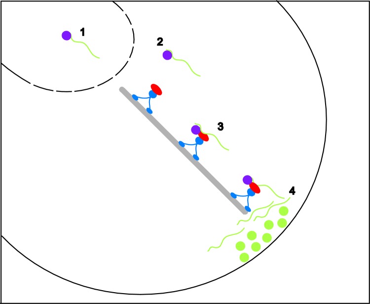
Step 1 – RNA localization substrates (green line) can be identified in
the nucleus by RNA-binding proteins (purple ball). Step 2 – the
RNA-protein complex is exported from the nucleus to the cytoplasm through
the nuclear pore complex. Step 3 – once in the cytoplasm, the RNA can
associate with additional proteins (red) that interface the RNA with motor
proteins (blue) that transport the RNA along cytoskeletal filaments (gray)
to the site of localization. Step 4 – upon reaching the correct
destination, translational repression is relieved and the corresponding
protein (green ball) is synthesized at the site of action.
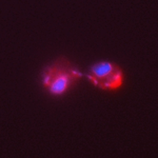
The distribution of ASH1 mRNA is shown in red, and the
positions of the nuclei are shown in blue.

A schematic representation of the Drosophila egg chamber
(top panel) oskar mRNA is transcribed in the nurse cells,
transported into the oocyte and localized at the posterior pole (red). The
bottom panel shows oskar mRNA (red) localization as
detected by in situ hybridization. DAPI stained DNA is
shown in blue.
References
LinkOut - more resources
Full Text Sources
Molecular Biology Databases