. 2012;7(8):e43174.
doi: 10.1371/journal.pone.0043174. Epub 2012 Aug 17.

Broad expression analysis of human ANTXR1/TEM8 transcripts reveals differential expression and novel splizce variants

Affiliations

Broad expression analysis of human ANTXR1/TEM8 transcripts reveals differential expression and novel splizce variants

Micaela Vargas et al. PLoS One. 2012.

Erratum in

  • PLoS One. 2012; 7(10): 10.1371/annotation/cebf633f-19e7-496b-b370-c0f1b1aea888

Abstract

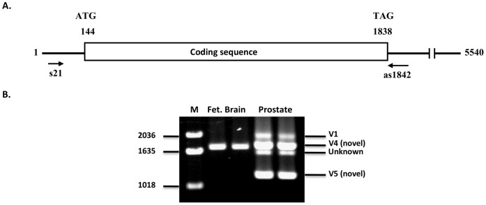
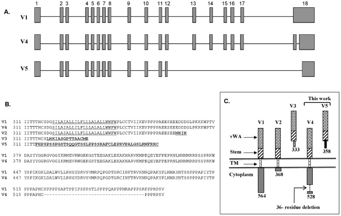
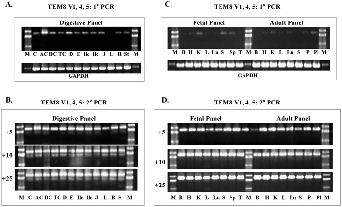
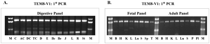
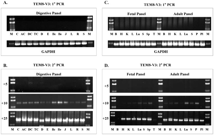
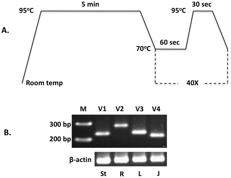
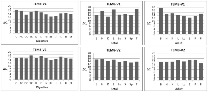
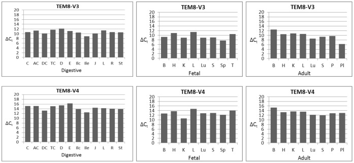
Tumor endothelial marker 8 (TEM8; ANTXR1) is one of two anthrax toxin receptors; the other is capillary morphogenesis gene 2 protein (CMG2; ANTXR2). TEM8 shows enhanced expression in certain tumor endothelia, and is thought to be a player in tumor vasculature formation. However, a comprehensive expression profile of individual TEM8 variants in normal or cancerous tissues is lacking. In this work we carried out an extensive analysis of all splice variants of human TEM8 in 12 digestive tissues, and 8 each fetal and adult tissues, 6 of them cognate pairs. Using variant-specific primers, we first ascertained the status of full-length transcripts by nested PCR. We then carried out quantitative analysis of each transcript by real-time PCR. Three splice variants of TEM8 were reported before, two single-pass integral membrane forms (V1 and V2) and one secreted (V3). Our analysis has revealed two new variants, one encoding a membrane-bound form of the receptor and the other secreted, which we have designated V4 and V5, respectively. All tissues had V1, V2, V3, and V4, but only prostate had V5. Real-time PCR revealed that all variants are present at different levels in various tissues. V3 appeared the most abundant of all. To ascertain its functionality for anthrax toxin, we expressed the newly identified form V4 in a receptor-negative host cell, and included V1 and V2 for comparison. Cytotoxicity, toxin binding, and internalization assays showed V4 to be as efficient a receptor as V1 and V2.


Conflict of interest statement

Competing Interests: The authors have declared that no competing interests exist.

Figures

Figure 1
Figure 1. Amplification of ANTXR1/TEM8 from prostate and fetal brain cDNA with V1-specific primers. A
) Schematic representation of V1 transcript features. The positions of nested PCR primers s21 and as1842 are shown. Numbering is based on the original sequence reported by St. Croix et al. (40). B) Analysis of amplicons following nested PCR. The expected V1 amplicon was 1848 bp. But the primers amplified a unique 1740 bp new splice variant, which we have designated V4. Prostate had V4 as well as a new variant, V5. Repeated attempts to sequence the unknown fragment failed. The PCR was carried out in duplicate for each template. M, molecular weight markers.
Figure 2
Figure 2. Comparison of ANTXR1/TEM8 variantsV1, V4 and V5 differential splicing.
A) Differential splicing of new variants V4 and V5 relative to V1. The gene comprises 18 exons with respect to V1 mRNA. V4 results from split splicing within exon 18, excluding a 108-bp segment, but it has the same stop codon as V1. V5 differential splicing occurs by partial skipping of exon 12 and 18 and complete skipping of exons 13–17. This results in a frame shift and consequently V5 acquires a new downstream stop codon. B) Sequence comparison of TEM8 receptor variants. The transmembrane helix (TM)) is underlined. The sequence differences are in underlined bold. V4 differs from V1 only in that it has a 36-residue in-frame deletion. V5, like V3, has no TM, and is therefore likely secreted. C) A schematic representation of membrane-bound and secreted variants of ANTXR1/TEM8. Same patterns signify identity of sequences and different ones divergence. The putative secreted variants V3 and V5 do not have any alternative membrane-spanning helices in their unique carboxyl-terminal segments.
Figure 3
Figure 3. Analysis of TEM8 V1, V4, and V5 expression in human tissue panels.
A & B, Digestive tissue cDNA panel. C & D, Fetal and Adult tissue cDNA panels. The primer pairs used were as for the prostate and fetal brain analysis (Figure 1). There was little or no amplification following 1°PCR, but 2° PCR resulted in definite bands. The number of 2° PCR cycles is shown on the left of each set. The results show that only V4 amplified; V1 and V5 transcripts were absent. Digestive tissue panel: C, cecum; AC, ascending colon; DC, descending colon; TC, transverse colon; D, duodenum; E, esophagus; Ilc, ileocecum; Ile, ileum; J, jejunum; L, liver; R, rectum; St, stomach. Fetal and adult tissue panel: B, brain; H, heart; K, kidney; L, liver; Lu, lung; P, pancreas; Pl, placenta; S, skeletal muscle; Sp, spleen; T, thymus.
Figure 4
Figure 4. Amplification of V1 with unique V1-specific antisense primers.
The sense primer was the same as for primary PCR of other variants, but the antisense primers were specific for the 108-bp segment absent in V4, as well as other variants. Because the amplification was quite robust upon primary PCR, nested PCR was not required. A, Digestive tissue panel: C, cecum; AC, ascending colon; DC, descending colon; TC, transverse colon; D, duodenum; E, esophagus; Ilc, ileocecum; Ile, ileum; J, jejunum; L, liver; R, rectum; St, stomach. B, Fetal and adult tissue panels: B, brain; H, heart; K, kidney; L, liver; Lu, lung; P, pancreas; Pl, placenta; S, skeletal muscle; Sp, spleen; T, thymus.
Figure 5
Figure 5. Analysis of TEM8 V2 transcript by stepwise nested PCR.
A & B, Digestive tissue cDNA panel. C & D, Fetal and Adult tissue cDNA panels. The primer pairs used for 1° and 2° PCR were s3+as1273 and s21+as1251, respectively. The number of 2° PCR cycles is shown on the left of each gel picture. Digestive tissue panel: C, cecum; AC, ascending colon; DC, descending colon; TC, transverse colon; D, duodenum; E, esophagus; Ilc, ileocecum; Ile, ileum; J, jejunum; L, liver; R, rectum; St, stomach. Fetal and adult tissue panels: B, brain; H, heart; K, kidney; L, liver; Lu, lung; P, pancreas; Pl, placenta; S, skeletal muscle; Sp, spleen; T, thymus.
Figure 6
Figure 6. Nested PCR analysis of TEM8 V3 in human tissue panels. A & B,
digestive tissue cDNA panel. C & D, Fetal and Adult tissue cDNA panel. The primer pairs used for 1° and 2° PCR were s3+as1374 and s21+as1352, respectively. The number of 2° PCR cycles is shown on the left of each gel picture. V3 shows broad expression; all tissues have it, although the amounts vary. Digestive tissue panel: C, cecum; AC, ascending colon; DC, descending colon; TC, transverse colon; D, duodenum; E, esophagus; Ilc, ileocecum; Ile, ileum; J, jejunum; L, liver; R, rectum; St, stomach. Fetal and adult tissue panels: B, brain; H, heart; K, kidney; L, liver; Lu, lung; P, pancreas; Pl, placenta; S, skeletal muscle; Sp, spleen; T, thymus.
Figure 7
Figure 7. Quantitative analysis of TEM8 splice variants. A,
Real-time PCR protocol. B, Agarose gel analysis of amplicons. β-actin was used as the normalization standard. The expected amplicons were: V1, 231 bp; V2, 281 bp; V3, 234 bp; V4, 218 bp. Several experiments were done to set the appropriate conditions, and to ensure that the real-time PCR worked satisfactorily and that the expected fragments were amplified. Different tissue cDNAs were used for this purpose. St, stomach; R, rectum; L, liver; J, jejunum. We got no reliable results with V5, and therefore excluded it from real-time PCR analysis.
Figure 8
Figure 8. Quantitative analysis of TEM8 splice variants V1 and V2.
The analysis was carried out according to the real-time PCR protocol in Figure 7, and as described in Materials and Methods. ΔCt values are the averages of two independent real-time PCRs, calculated by subtracting the Ct values for each variant from the Ct values for β-actin. Digestive tissue panel: C, cecum; AC, ascending colon; DC, descending colon; TC, transverse colon; D, duodenum; E, esophagus; Ilc, ileocecum; Ile, ileum; J, jejunum; L, liver; R, rectum; St, stomach. Fetal and adult tissue panels: B, brain; H, heart; K, kidney; L, liver; Lu, lung; P, pancreas; Pl, placenta; S, skeletal muscle; Sp, spleen; T, thymus.
Figure 9
Figure 9. Real-time PCR analysis of TEM8 splice variants V3 and V4. Digestive tissue panel:
C, cecum; AC, ascending colon; DC, descending colon; TC, transverse colon; D, duodenum; E, esophagus; Ilc, ileocecum; Ile, ileum; J, jejunum; L, liver; R, rectum; St, stomach. Fetal and adult tissue panels: B, brain; H, heart; K, kidney; L, liver; Lu, lung; P, pancreas; Pl, placenta; S, skeletal muscle; Sp, spleen; T, thymus.
Figure 10
Figure 10. Assessment of TEM8 functionality as an anthrax toxin receptor. A,
Sensitivity of cells expressing V1, V2, or V4. The plasmid-encoded receptors were expressed in the receptor-negative host PR230 as described in Materials and Methods, followed by cytotoxicity assays with PA+FP59. B, Toxin concentrations required for 50% cell death (IC50, ng/mL) for PR230 expressing V1, V2, or V4. The means of 2–3 independent experiments are shown in parentheses. The greater sensitivity of the parental cells, WTB111, is probably due to expression of both receptors, especially CMG2/ANTXR2, which has about 10 times greater affinity for anthrax toxin protective antigen than does TEM8 , , .
Figure 11
Figure 11. V1, V2, and V4 capacities for anthrax toxin protective antigen (PA, 83 kDa) binding, processing, and internalization. A,
PA binding to normal cells (WTB111). At 4°C, PA83 binds the receptors and is cleaved by furin to yield PA63. At 37°C, PA83 not only binds and is cleaved by furin, but enters cells and forms SDS-resistant oligomers. B, PA binding and processing at 4°C to PR230 expressing V1, V2, or V4. C, PA binding, processing, internalization, and SDS-resistant oligomer formation at 37°C. Two blots of a typical experiment are shown.

Similar articles

Cited by

References

    1. St Croix B, Rago C, Velculescu V, Traverso G, Romans KE, et al. (2000) Genes expressed in human tumor endothelium. Science 289: 1197–1202. - PubMed
    1. Bradley KA, Mogridge J, Mourez M, Collier RJ, Young JA (2001) Identification of the cellular receptor for anthrax toxin. Nature 414: 225–229. - PubMed
    1. Liu S, Leppla SH (2003) Cell surface tumor endothelium marker 8 cytoplasmic tail-independent anthrax toxin binding, proteolytic processing, oligomer formation, and internalizations. J Biol Chem 278: 5227–5234. - PubMed
    1. Scobie HM, Rainey GJ, Bradley KA, Young JA (2003) Human capillary morphogenesis protein 2 functions as an anthrax toxin receptor. Proc Natl Acad Sci USA, 100 (9); 5170–5174. - PMC - PubMed
    1. Scobie HM, Young JA (2005) Interactions between anthrax toxin receptors and protective antigen. Curr Opin Microbiol 8(1): 106–112. - PubMed

Publication types

MeSH terms

LinkOut - more resources