. 2012;7(8):e43464.
doi: 10.1371/journal.pone.0043464. Epub 2012 Aug 17.

Hmgb3 is regulated by microRNA-206 during muscle regeneration

Affiliations

Hmgb3 is regulated by microRNA-206 during muscle regeneration

Simona Maciotta et al. PLoS One. 2012.

Abstract

Background: MicroRNAs (miRNAs) have been recently involved in most of human diseases as targets for potential strategies to rescue the pathological phenotype. Since the skeletal muscle is a spread-wide highly differentiated and organized tissue, rescue of severely compromised muscle still remains distant from nowadays. For this reason, we aimed to identify a subset of miRNAs major involved in muscle remodelling and regeneration by analysing the miRNA-profile of single fibres isolated from dystrophic muscle, which was here considered as a model of chronic damage.

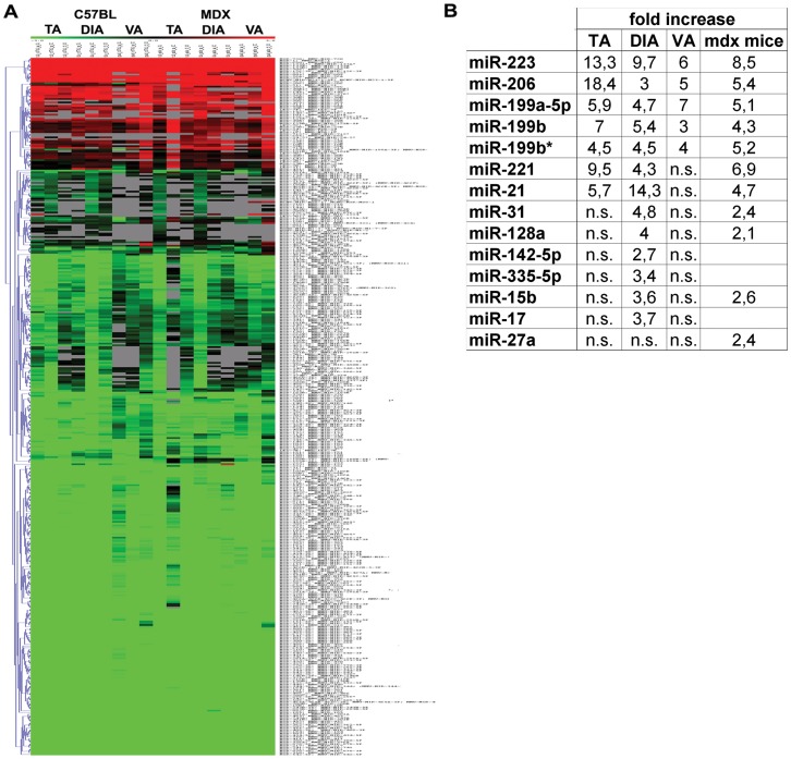
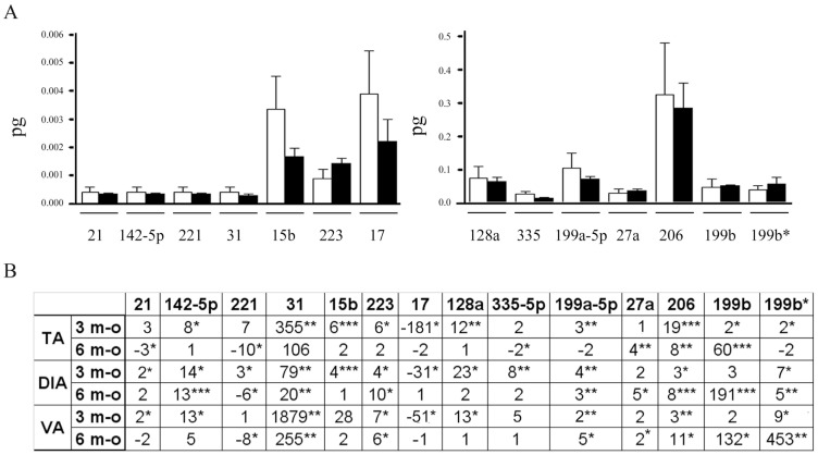
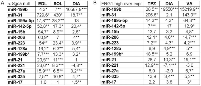
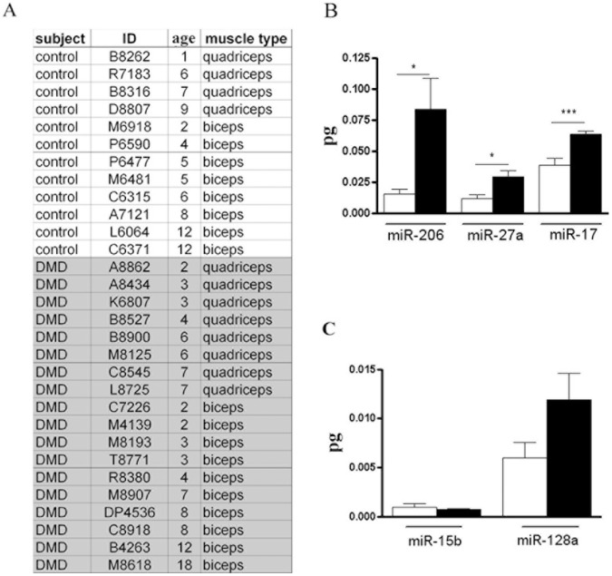
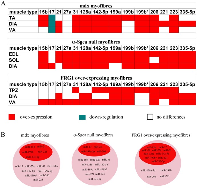
Methodology/principal findings: The miRNA-signature associated to regenerating (newly formed) and remodelling (resting) fibres was investigated in animal models of muscular dystrophies and acute damage, in order to distinguish which miRNAs are primary related to muscle regeneration. In this study we identify fourteen miRNAs associated to dystrophic fibres responsible for muscle regeneration and remodelling, and confirm over-expression of the previously identified regeneration-associated myomiR-206. In particular, a functional binding site for myomiR-206 was identified and validated in the 3'untranslated region (3'UTR) of an X-linked member of a family of sequence independent chromatin-binding proteins (Hmgb3) that is preferentially expressed in hematopoietic stem cells. During regeneration of single muscle fibres, Hmgb3 messenger RNA (mRNA) and protein expression was gradually reduced, concurrent with the up-regulation of miR-206.

Conclusion/significance: Our results elucidate a negative feedback circuit in which myomiR-206 represses Hmgb3 expression to modulate the regeneration of single muscle fibres after acute and chronic muscle damage. These findings suggest that myomiR-206 may be a potential therapeutic target in muscle diseases.


Conflict of interest statement

Competing Interests: The authors have declared that no competing interests exist.

Figures

Figure 1
Figure 1. MiRNA profile of single fibres isolated from the mdx mouse.
(A) Clustered heat map showing the expression ratios of miRNAs in TA, DIA and VA of age- and sex-matched c57bl (n = 3) and mdx (n = 3) mice. Expression data were normalized on Universal Reference. A total of 14 miRNAs were found over-expressed in dystrophic samples with distinction among myofibres isolated from different muscle type. (B) Fold change values of the 14 up-regulated miRNAs were reported in the table (ns = no significant).
Figure 2
Figure 2. Dystrophin absence is not responsible for the over-expression of several muscle-enriched miRNAs in the mdx mouse.
(A) MiRNAs that were observed over-expressed in the adult mdx mouse (3½ months-old) by the array analysis were quantified by Q-PCR in single fibres isolated from the hind limb of newborn mdx (n = 10) and c5bl (n = 10) mice. The absolute quantity (pg) of each miRNA was represented in the histogram as white columns for single fibres of c57bl mice and black bars for dystrophic muscle fibres. Single fibres of newborn mdx mice were characterized by control levels of muscle-enriched miRNAs, demonstrating no correlation with the genetic defect of DMD. (B) MiRNAs that were observed over-expressed in the adult mdx mouse (3½ months-old) by the array analysis were quantified by Q-PCR in single fibres isolated from the TA, DIA and VA of 6 months-old mdx mice and normalized on control samples. In the table were represented the fold change values of tested miRNAs in myofibres of 3½ and 6 months-old mdx mice in comparison to c57bl myofibers. Tested miRNAs showed a heterogeneous behaves depending on the muscle type and/or disease progression. (Two-wail parametric t-test; p value <0, 05 **p value <0, 01 ***p value <0,001).
Figure 3
Figure 3. Different MDs are characterized by the same muscle-enriched miRNA-dysregulation.
MiRNAs that were observed over-expressed in the adult 3½ months-old mdx mouse by the array analysis were quantified by absolute Q-PCR in single fibres isolated from EDL, SOL and DIA of α-Sgca null mice (A) and in single fibres isolated from TPZ, DIA and VA of FRG1 high over-expressing mice (B). MiRNAs average fold changes in muscle fibres of α-Sgca null mice (A) and of FRG1 over-expressing mice were shown in the tables in a linear scale. Tested miRNAs were similarly dysregulated in the animal model of different MDs. (Two-tail parametric t-test; *p value <0, 05; **p value <0, 01; ***p value <0,001).
Figure 4
Figure 4. Identification of new murine dystrophic-fibres associated-miRNAs in muscle biopsies of DMD subjects.
(A) The age and type of muscle of healthy subjects (12) and DMD patients (18) were listed. Moreover the type of muscle from which the biopsies were isolated were also reported. (B–C) MiR-15b, miR-128a, miR-206, miR-17 and miR-27a were quantified by absolute Q-PCR in muscle biopsies of control and dystrophic subjects listed in A. The absolute values (pg) of tested miRNAs were represented in the histogram for healthy (white bars) and dystrophic single fibres (black bars). (B) Quantitative analysis showed that only miR-128a, miR-206 and miR-17 were up-regulated in human dystrophic single fibres independently of the muscle type. (Two-tail parametric t-test; *p value <0, 05; **p value <0, 01; ***p value <0,001).
Figure 5
Figure 5. Hmgb3 expression-pattern in single fibres.
(A) Immunoblotting experiments confirmed the presence of Hmgb3 protein in single muscle fibers isolated from the TA and VA of c57bl (n = 3) and mdx (n = 3) mice. (B) Densitometric analysis on WB bands evidenced a decreased expression of Hmgb3 in the TA and VA (p = 0.0133) of mdx mice. (C) Quantification of Hmgb3 mRNA by qRT-PCR in single fibres isolated from the TA and VA of mdx mice (n = 10) and of c57bl (n = 10) mice confirmed a down-regulation of Hmgb3 in dystrophic muscle. (Two-tail parametric t-test; *p value <0, 05; **p value <0, 01; ***p value <0,001).
Figure 6
Figure 6. MyomiR-206 modulates Hmgb3 expression during myogenesis.
(A) Hek cells were co-transfected with mimic-miR-206 at a concentration of 25 nM for 48 h and with pGLO-control or pGLOHmgb3 or pGLOHmgb3Mut. MiR-206 down-regulated the expression of luciferase of 32% (p = 0, 0151) in Hek cells co-transfected with pGLOHmgb3. Instead no down-regulation happened when the binding site of miR-206 was mutated. (B) 3T3 cells were transfected with mimic miR-206 at a concentration of 25 nM for 48 h, evidencing a strong down-regulation of endogenous Hmgb3 mRNAs. (C) Quantification of Hmgb3 mRNA by qRT-PCR in single fibres isolated from CTX-injured TA at day 2, 5, 7 and 10 from the injection evidenced a strong down-regulation of Hmgb3 during muscle regeneration. (D–E) Hmgb3 mRNA (D) and miR-206 (E) were respectively quantified by relative qRT-PCR and by absolute Q-PCR in proliferating C1C12 myoblasts cell line (MB) versus differentiated C1C12 myotubes (MT). The quantification analysis highlighted an opposite expression trend for Hmgb3 and miR-206, further validating inhibition of Hmgb3 by miR-206 (AU = arbitrary units). (Two-tail parametric t-test; *p value <0, 05; **p value <0,01; ***p value <0,001).
Figure 7
Figure 7. Expression profile of miRNAs associated to dystrophic single muscle fibres.
(A) The expression profiles of miRNAs associated to dystrophic fibres of mdx, α-Sgca null and FRG1 over-expressing mice are reported as over-expressed (red block), down-regulated (green block) or similarly expressed (white block) to normal C57/BL fibres. (B) MiRNAs whose dysregulation in dystrophic fibres of mdx, α-Sgca null and FRG1 over-expressing mice depended on the muscle-type are listed in the red circle. Otherwise, miRNAs that were mis-modulated in dystrophic fibres of mdx, α-Sgca null and FRG1 over-expressing mice independently to the muscle-type are listed in the pink circle.

References

    1. Bartel DP (2004) MicroRNAs: genomics, biogenesis, mechanism, and function. Cell 116: 281–297. - PubMed
    1. Singh SK, Pal Bhadra M, Girschick HJ, Bhadra U (2008) MicroRNAs–micro in size but macro in function. Febs J 275: 4929–4944. - PubMed
    1. Crist CG, Buckingham M (2009) Megarole for microRNA in muscle disease Cell Metab. 12: 425–426. - PubMed
    1. Eisenberg I, Alexander MS, Kunkel LM (2009) MiRNAS in normal and diseased skeletal muscle J Cell Mol Med. 13: 2–11. - PMC - PubMed
    1. Thum T, Gross C, Fiedler J, Fischer T, Kissler S, et al. (2008) MicroRNA-21 contributes to myocardial disease by stimulating MAP kinase signalling in fibroblasts Nature. 456: 980–984. - PubMed

Publication types

MeSH terms

Associated data