. 2012;7(8):e43768.
doi: 10.1371/journal.pone.0043768. Epub 2012 Aug 17.

Mesenchymal stem cells attenuate peritoneal injury through secretion of TSG-6

Affiliations

Mesenchymal stem cells attenuate peritoneal injury through secretion of TSG-6

Nan Wang et al. PLoS One. 2012.

Abstract

Background: Mesothelial cell injury plays an important role in peritoneal fibrosis. Present clinical therapies aimed at alleviating peritoneal fibrosis have been largely inadequate. Mesenchymal stem cells (MSCs) are efficient for repairing injuries and reducing fibrosis. This study was designed to investigate the effects of MSCs on injured mesothelial cells and peritoneal fibrosis.

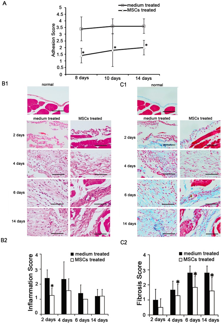
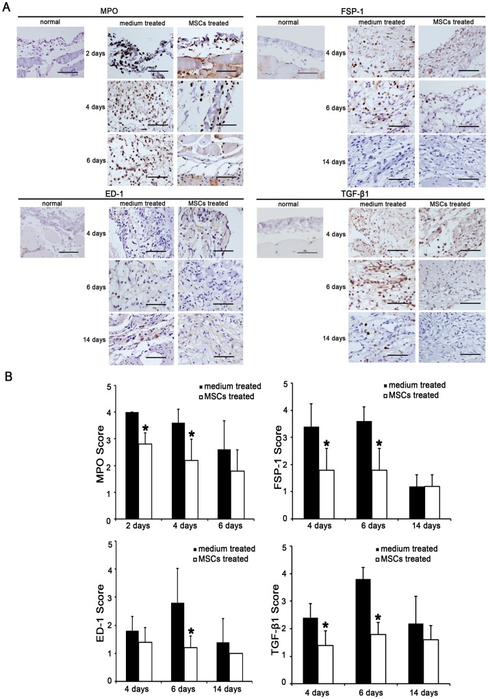
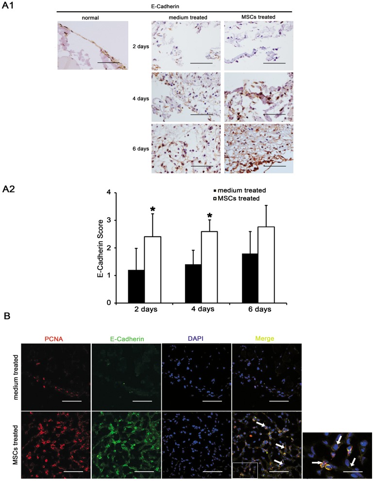
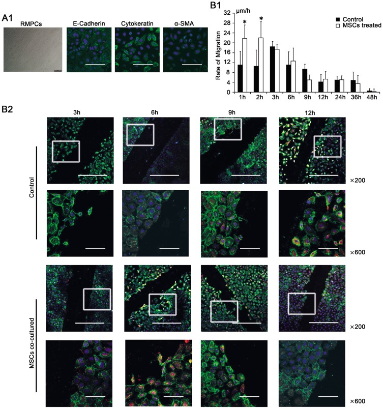
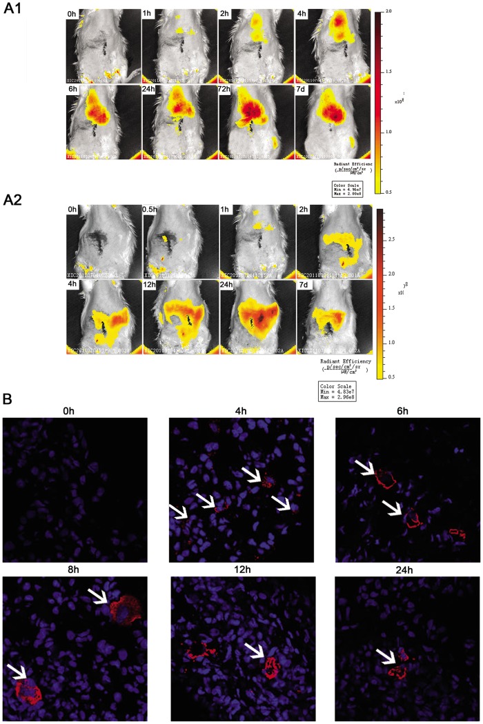
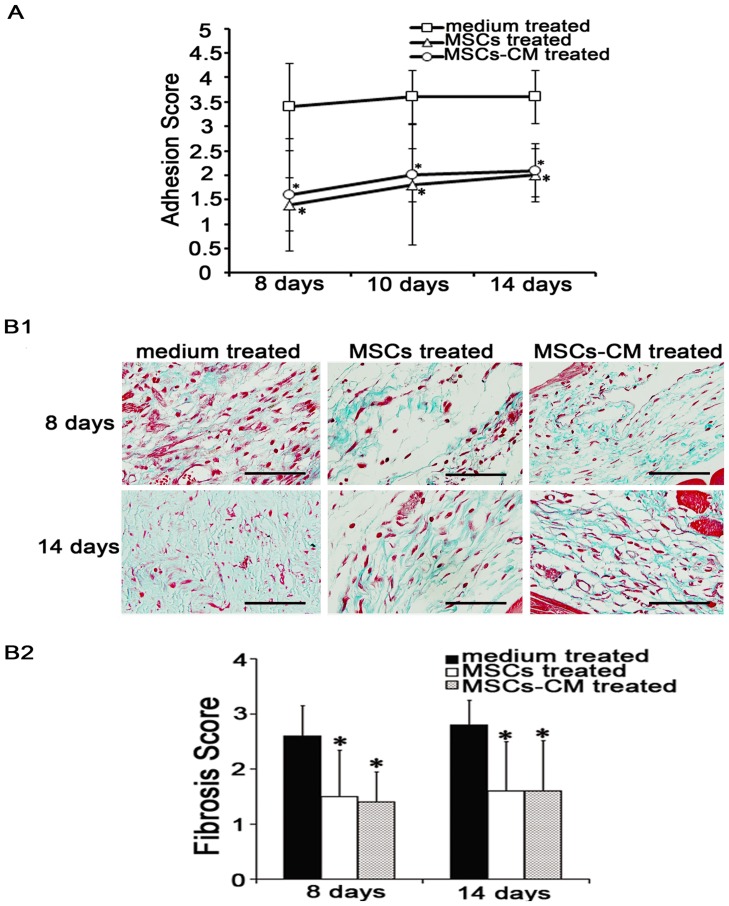
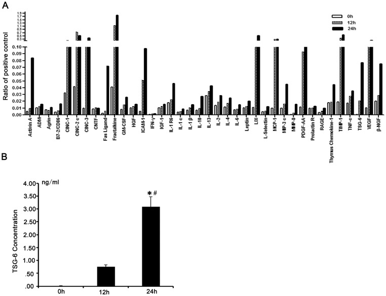
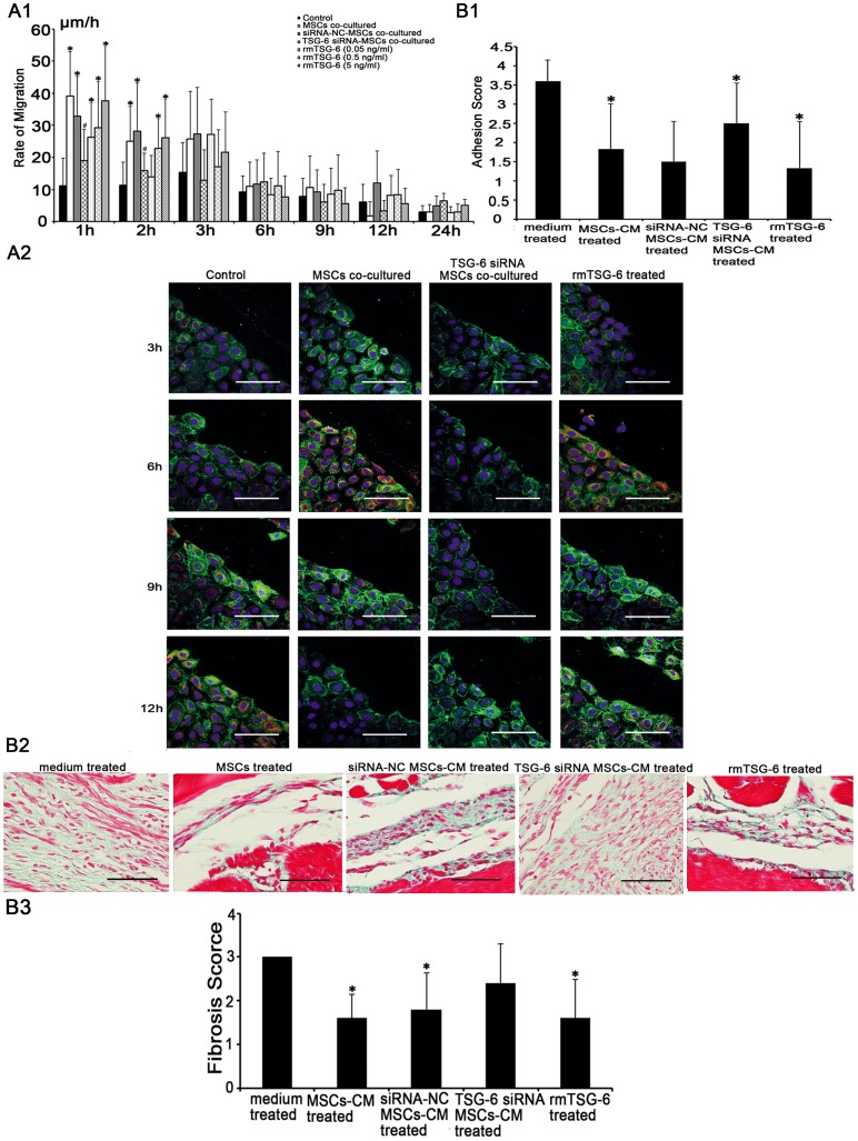
Methodology/principal findings: Rat bone marrow-derived MSCs (5 × 10(6)) were injected into Sprague-Dawley (SD) rats via tail vein 24 h after peritoneal scraping. Distinct reductions in adhesion formation; infiltration of neutrophils, macrophage cells; number of fibroblasts; and level of transforming growth factor (TGF)-β1 were found in MSCs-treated rats. The proliferation and repair of peritoneal mesothelial cells in MSCs-treated rats were stimulated. Mechanically injured mesothelial cells co-cultured with MSCs in transwells showed distinct increases in migration and proliferation. In vivo imaging showed that MSCs injected intravenously mainly accumulated in the lungs which persisted for at least seven days. No apparent MSCs were observed in the injured peritoneum even when MSCs were injected intraperitoneally. The injection of serum-starved MSCs-conditioned medium (CM) intravenously reduced adhesions similar to MSCs. Antibody based protein array of MSCs-CM showed that the releasing of TNFα-stimulating gene (TSG)-6 increased most dramatically. Promotion of mesothelial cell repair and reduction of peritoneal adhesion were produced by the administration of recombinant mouse (rm) TSG-6, and were weakened by TSG-6-RNA interfering.

Conclusions/significance: Collectively, these results indicate that MSCs may attenuate peritoneal injury by repairing mesothelial cells, reducing inflammation and fibrosis. Rather than the engraftment, the secretion of TSG-6 by MSCs makes a major contribution to the therapeutic benefits of MSCs.


Conflict of interest statement

Competing Interests: The authors have declared that no competing interests exist.

Figures

Figure 1
Figure 1. Effects of mesenchymal stem cells (MSCs) on the severity and the histological changes of acute peritoneal adhesions.
(A). The MSCs treated group had lower adhesion scores. The size and severity of peritoneal adhesion were evaluated macroscopically by an independent observer on a scale of 0–4 (0, 0%; 1, <25%; 2, 25–49%; 3, 50–74%; and 4, 75–100% adhesions). * compared with medium treated group, p <0.05, n  = 6, respectively. (B). HE staining revealed the changes of inflammation in acute peritoneal adhesions. (B1). Peritoneal inflammations after scraping were reduced by injecting MSCs. Magnification  = ×400. (B2). The scores of peritoneal inflammation days 2 after scraping were reduced by injecting MSCs. * compared with medium treated group, p <0.05, n  = 6, respectively. (C). Masson's trichrome staining revealed the changes of fibrosis in acute peritoneal adhesions. (C1). Peritoneal fibroses after scraping were reduced by injecting MSCs. Magnification  = ×400. (C2). The scores of peritoneal fibrosis days 4, 6, 14 after scraping were reduced by injecting MSCs. * compared with medium treated group, p <0.05, n  = 6, respectively. The sections were evaluated from five randomly selected fields under a magnification of ×100 by an independent pathologist. The extents of inflammation (HE staining) and fibrosis (masson's trichrome staining) were scored as 0 (negative), 1 (weak), 2 (medium), or 3 (intensive), respectively.
Figure 2
Figure 2. Effects of mesenchymal stem cells (MSCs) on the inflammation and fibrosis of acute peritoneal adhesions.
(A). Immunohistochemical evaluation revealed that the numbers of fibroblasts (FSP-1), neutrophils (MPO), and macrophage cells (ED-1), and the level of transforming growth factor (TGF)-β1 during the active phase in acute peritoneal adhesions were decreased by injecting MSCs. Magnification  = ×400. (B). The immunohistochemical scores of MPO days 2, 4, ED-1 days 6, FSP-1 and TGF-β1 days 4, 6 after scraping were reduced by injecting MSCs. * compared with medium treated group, p <0.05, n  = 6, respectively. Sections were evaluated from five randomly selected fields under a magnification of ×100 by an independent pathologist. The extent of staining was scored as 0 (0%), 1 (1–20%), 2 (21–50%), 3 (51–80%), and 4 (81–100%), indicating the percentage of positive staining in the adhesion tissue.
Figure 3
Figure 3. Effects of mesenchymal stem cells (MSCs) on the repair of peritoneal mesothelial cells.
(A). Immunohistochemical evaluation of peritoneal mesothelial cells (E-Cadherin) in acute peritoneal adhesions. (A1). The number of mesothelial cells after peritoneal scraping was increased by injecting MSCs. Magnification  = ×400. (A2). The immunohistochemical scores of E-Cadherin days 2, 4 after scraping were increased by injecting MSCs. * compared with medium treated group, p <0.05, n  = 6, respectively. Sections were evaluated from five randomly selected fields under a magnification of ×100 by an independent pathologist. The extent of staining was scored as 0 (0%), 1 (1–20%), 2 (21–50%), 3 (51–80%), and 4 (81–100%), indicating the percentage of positive staining in the adhesion tissue. (B). Immunofluorescence evaluation of injured peritoneum days 4 after scraping using antibodies to E-Cadherin (green) and PCNA (red). The nucleus was counterstained with DAPI (blue). Dual-stained cells (indicated as arrows) were predominantly expressed in the MSCs treated group. Magnification  = ×600 and ×1800, when necessary.
Figure 4
Figure 4. Effects of mesenchymal stem cells (MSCs) on the repair of rat peritoneal mesothelial cells (RPMCs) following mechanical injury.
(A). Characteristics of primary RPMCs cultures. (A1). Photomicrograph showing the cobblestone-like appearance of the epithelioid phenotypes. Magnification  = ×100. (A2). An immunofluorescence evaluation revealed that RPMCs expressed E-Cadherin (green) and Cytokeratin (green), but only weakly expressed α-SMA (green). Nuclei were stained with DAPI (blue). Magnification  = ×600. (B). The effects of MSCs on the migratory and proliferative capacities of mechanically injured RPMCs. (B1). Microscopy of RPMCs migrating to the edge of the wound to show the migratory velocity (μm/h). During the first 2 h, injured RPMCs co-cultured with MSCs showed a greater migratory velocity. * compared with control group, p <0.05, n  = 3, respectively. (B2). Immunofluorescence analysis with PCNA (red) and E-Cadherin (green) revealed that the proliferation peak of injured RPMCs co-cultured with MSCs was accelerated to 6 h after scraping, whereas the proliferation peak was 12 h after scraping in control group. Nuclei were stained with DAPI (blue). Magnification  = ×200 and ×600.
Figure 5
Figure 5. Tracking the distribution of mesenchymal stem cells (MSCs) injected into rats.
(A1). In vivo imaging revealed that MSCs injected via tail vein first accumulated in the lungs and gradually in the liver and the spleen. (A2). In vivo imaging revealed that MSCs injected intraperitoneally first accumulated in the liver and gradually in the spleen. No signal was found in the injured peritoneum. (B). An immunofluorescence evaluation of green fluorescent protein (GFP) (red) detected MSCs in the lungs. Nuclei were stained with DAPI (blue). Magnification  = ×1000.
Figure 6
Figure 6. Effects of mesenchymal stem cells (MSCs)-conditioned medium (CM) on acute peritoneal adhesions.
(A). The MSCs-CM treated group had lower adhesion scores days 14 after scraping. The size and severity of peritoneal adhesions were evaluated macroscopically by an independent observer on a scale of 0–4 (0, 0%; 1, <25%; 2, 25–49%; 3, 50–74%; and 4, 75–100% adhesions).* compared with medium treated group, p <0.05, n  = 6, respectively. (B). Masson's trichrome staining revealed that the changes of fibrosis in acute peritoneal adhesions. (B1). The fibrosis in the scraped peritoneum days 8, 14 after scraping was decreased by injecting MSCs-CM. Magnification  =  ×400. (B2). The scores of peritoneal fibrosis were reduced by injecting MSCs-CM, similar to MSC. * compared with medium treated group, p <0.05, n  = 6, respectively. The sections were evaluated from five randomly selected fields under a magnification of ×100 by an independent pathologist. The extent of fibrosis was scored as 0 (negative), 1 (weak), 2 (medium), or 3 (intensive).
Figure 7
Figure 7. Release of TNFα-stimulating gene (TSG)-6 by mesenchymal stem cells (MSCs).
(A). Cytokine profile of 30-folded serum-starved MSCs-conditioned medium (CM) within 24 h was analyzed by rat cytokine antibody array and label-based rat antibody array. Compared with 0 h MSCs-CM, wound healing cytokine TSG-6 was released abundantly by 12 h (52.7-fold) and 24 h (194-fold) serum-starved MSCs. The results were normalized to the positive controls. Similar results were obtained from three independent MSCs-CM samples. (B). TSG-6 in 30-folded serum-starved MSCs-CM within 24 h were measured by Enzyme-linked immunosorbent assays (ELISA). The concentration of TSG-6 in 12 h MSCs-CM was 0.75±0.09 ng/ml (49-fold increase, compared with 0 h MSCs-CM), 24 h MSCs-CM was 3.08±0.22 ng/ml (199.8-fold increase, compared with 0 h MSCs-CM). Three independent samples were placed in three repetitive holes. * compared with 0h MSCs-CM, p <0.05; # compared with 12 h MSCs-CM, p <0.05.
Figure 8
Figure 8. TNF-stimulating gene (TSG)-6 secreted by mesenchymal stem cells (MSCs) ameliorated the peritoneal injury.
(A). The effects of TSG-6 on the repair of mechanically injured rat peritoneal mesothelial cells (RPMCs). (A1). Microscopy of RPMCs migrating to the edge of the wound to show the migratory velocity (μm/h). Injured RPMCs co-cultured with TSG-6-siRNA MSCs showed significant reductions in the migratory capacity. However, migrations were significantly increased dose-dependently in RPMCs cultured in medium containing recombinant mouse (rm) TSG-6. * compared with control group, p <0.05; # compared with MSCs co-cultured group, p <0.05; n  = 3, respectively. (A2). Immunofluorescence analysis with PCNA (red) and E-Cadherin (green) revealed the proliferation of injured RPMCs. RPMCs co-cultured with TSG-6-siRNA MSCs showed no apparent increase in the proliferative capacity. whereas the proliferation peak of RPMCs cultured with rmTSG-6 was accelerated to 6 h after scraping. Nuclei were stained with DAPI (blue). Magnification  = ×600. (B). The effects of TSG-6 on acute peritoneal adhesions. (B1). TSG-6-siRNA MSCs-CM treated group had no significant reduction in adhesion scores days 14 after scraping. However, adhesion scores were reduced in rmTSG-6 treated group. The size and severity of peritoneal adhesions were evaluated macroscopically by an independent observer on a scale of 0–4 (0, 0%; 1, <25%; 2, 25–49%; 3, 50–74%; and 4, 75–100% adhesions). * compared with medium treated group, p <0.05, n  = 6, respectively. (B2). Histological changes were evaluated using masson's trichrome staining. TSG-6-siRNA MSCs-CM treated group revealed no apparent reduction in fibrosis in the scraped peritoneum days 14 after scraping. However, fibrosis were reduced in rmTSG-6 treated group. Magnification  = ×400. (B3). The scores of peritoneal fibrosis days 14 after scraping were reduced by injecting rmTSG-6, but not by injecting TSG-6-siRNA MSCs-CM. * compared with medium treated group, p <0.05, n  = 6, respectively. The sections were evaluated from five randomly selected fields under a magnification of ×100 by an independent pathologist. The extent of fibrosis was scored as 0 (negative), 1 (weak), 2 (medium), or 3 (intensive).

Similar articles

Cited by

References

    1. Herrick SE, Mutsaers SE (2004) Mesothelial progenitor cells and their potential in tissue engineering. Int J Biochem Cell Biol 36: 621–642. - PubMed
    1. Aroeira LS, Aguilera A, Sanchez-Tomero JA, Bajo MA, del Peso G, et al. (2007) Epithelial to mesenchymal transition and peritoneal membrane failure in peritoneal dialysis patients: pathologic significance and potential therapeutic interventions. J Am Soc Nephrol 18: 2004–2013. - PubMed
    1. Saxena R (2008) Pathogenesis and treatment of peritoneal membrane failure. Pediatr Nephrol 23: 695–703. - PubMed
    1. Williams JD, Craig KJ, Topley N, Von Ruhland C, Fallon M, et al. (2002) Morphologic changes in the peritoneal membrane of patients with renal disease. J Am Soc Nephrol 13: 470–479. - PubMed
    1. Williams JD, Craig KJ, von Ruhland C, Topley N, Williams GT (2003) The natural course of peritoneal membrane biology during peritoneal dialysis. Kidney Int Suppl: S43–49. - PubMed

Publication types

MeSH terms

Substances

LinkOut - more resources