. 2012 Aug 16;4(8):356-61.
doi: 10.4253/wjge.v4.i8.356.

Diagnosis of extent of early gastric cancer using flexible spectral imaging color enhancement

Affiliations

Diagnosis of extent of early gastric cancer using flexible spectral imaging color enhancement

Hiroyuki Osawa et al. World J Gastrointest Endosc. .

Abstract

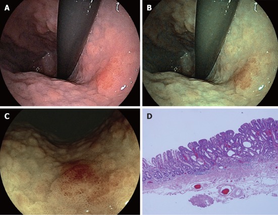
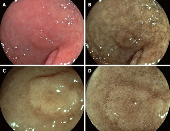
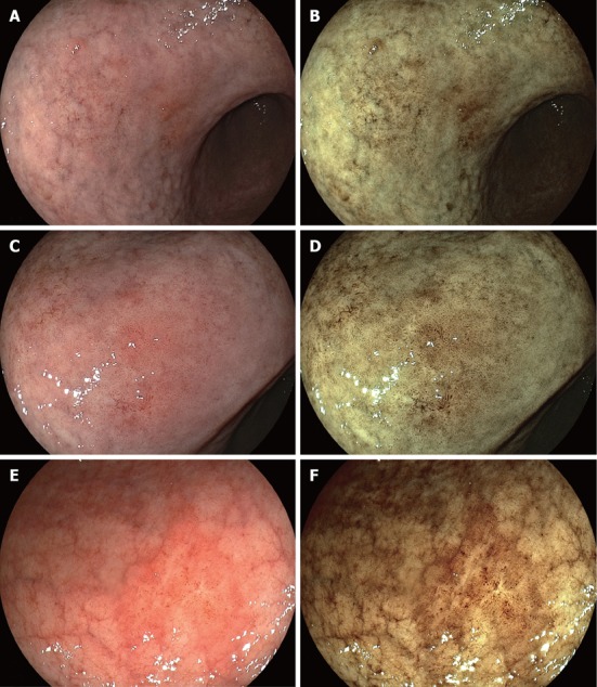
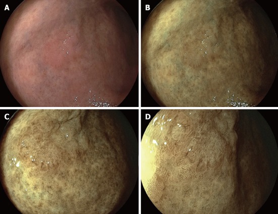
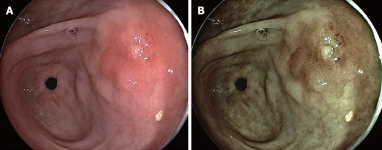
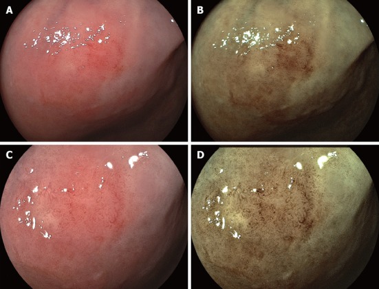
The demarcation line between the cancerous lesion and the surrounding area could be easily recognized with flexible spectral imaging color enhancement (FICE) system compared with conventional white light images. The characteristic finding of depressed-type early gastric cancer (EGC) in most cases was revealed as reddish lesions distinct from the surrounding yellowish non-cancerous area without magnification. Conventional endoscopic images provide little information regarding depressed lesions located in the tangential line, but FICE produces higher color contrast of such cancers. Histological findings in depressed area with reddish color changes show a high density of glandular structure and an apparently irregular microvessel in intervening parts between crypts, resulting in the higher color contrast of FICE image between cancer and surrounding area. Some depressed cancers are shown as whitish lesion by conventional endoscopy. FICE also can produce higher color contrast between whitish cancerous lesions and surrounding atrophic mucosa. For nearly flat cancer, FICE can produce an irregular structural pattern of cancer distinct from that of the surrounding mucosa, leading to a clear demarcation. Most elevated-type EGCs are detected easily as yellowish lesions with clearly contrasting demarcation. In some cases, a partially reddish change is accompanied on the tumor surface similar to depressed type cancer. In addition, the FICE system is quite useful for the detection of minute gastric cancer, even without magnification. These new contrasting images with the FICE system may have the potential to increase the rate of detection of gastric cancers and screen for them more effectively as well as to determine the extent of EGC.

Keywords: Early gastric cancer; Endoscopic submucosal dissection; Flexible spectral imaging color enhancement; Magnified image; Nonmagnified image.


Figures

Figure 1
Figure 1
Image findings and specimen, A: Conventional image with small caliber endoscope (EG530-NW) reveals a slightly reddish mucosal change in the lesser curvature of the upper body; B: Flexible spectral imaging color enhancement (FICE) image with small caliber endoscope enhances a reddish cancerous lesion and can determine with precision a clear line of demarcation between cancer and the yellowish surrounding mucosa; C: FICE image with low magnification (EG590-ZW) also detects much clearer demarcation line; D: Specimen after endoscopic submucosal dissection shows a high density of glandular structure and an apparently irregular microvessel in intervening parts between crypts, which may cause a reddish mucosal change in depressed area.
Figure 2
Figure 2
Some depressed cancers are shown as whitish lesion by conventional endoscopy. A: Conventional image (EG590-ZW) reveals a slightly whitish mucosal change in the anterior wall of antrum; B: Flexible spectral imaging color enhancement (FICE) image enhances a whitish cancerous lesion and can determine a line of demarcation between cancer and surrounding mucosa; C: FICE image in a close-up view (EG590-WR) detects with precision a clearer demarcation line; D: FICE image with half magnification (EG590-ZW) reveals a finer microstructural pattern on mucosal surface of cancer and higher contrasting mucosa between cancer and the surrounding area, leading a clearer demarcation line.
Figure 3
Figure 3
Conventional image and Flexible spectral imaging color enhancement image findings. A: Conventional image (EG590-ZW) reveals a slightly enriched vascular structure of gastric mucosa in the lesser curvature of the lower body; B: Flexible spectral imaging color enhancement (FICE) image enhances such a structure and shows reddish lesions in its anal side near angle; C: Conventional image near angle in a close-up view shows slightly reddish changes on the flat mucosa; D: FICE image near angle in a close-up view shows an irregular structural pattern distinct from the surrounding area, leading to a clearer demarcation line; E: Conventional image (EG590-WR) reveals a slightly enriched vascular structure in nearly flat mucosa in the antrum in a close-up view; F: FICE image enhances such a structure accompanied by a clear margin of cancer distinct from the surrounding mucosa. It is noted that these images can be obtained without magnification.
Figure 4
Figure 4
Most elevated-type early gastric cancers are detected easily as yellowish lesions with clearly contrasting demarcation. A: Conventional image (EG590-ZW) in a distant view reveals a slightly elevated lesion similar to the mucosal color of surrounding area in the anterior wall of lower body; B: Flexible spectral imaging color enhancement (FICE) image shows an uneven surface on the elevated tumor; C: A close-up FICE image without magnification exhibits an irregular microstructural pattern on uneven tumor surface distinct from the surrounding mucosa; D: FICE image with low magnification distinguishes an apparently irregular microstructural pattern of cancer from a normal microstructural pattern of the surrounding mucosa.
Figure 5
Figure 5
In some cases, a partially reddish change is accompanied on the tumor surface similar to depressed type cancer. A: Conventional image (EG590-ZW) in a distant view reveals an elevated lesion with slightly reddish portion in the posterior wall of antrum; B: Flexible spectral imaging color enhancement image enhances a reddish portion on tumor surface with more contrasting demarcation line. In addition, tumor margin of flat area in the right side of this figure can be more clearly visualized than conventional image.
Figure 6
Figure 6
The irregular microvascular pattern of lesions is clearly visualized with half magnification. A: Conventional image (EG590-ZW) in a close-up view reveals a nearly flat lesion with a slightly reddish portion in the anterior wall of middle body; B: Flexible spectral imaging color enhancement (FICE) image enhances a reddish lesion leading to a clear demarcation line; C: Conventional image with half magnification shows an enriched microvascular structure on the tumor surface; D: FICE image with half magnification allows a clear visualization of irregular microvascular and microstructural pattern suggesting gastric cancer with the possible histological type of well-differentiated adenocarcinoma.

Similar articles

Cited by

References

    1. Miyake Y, Sekiya T, Kubo S, Hara T. A new spectrophotometer for measuring the spectral reflectance of gastric mucous membrane. J Photogr Sci. 1989;37:134–138.
    1. Knyrim K, Seidlitz H, Vakil N, Classen M. Perspectives in “electronic endoscopy”. Past, present and future of fibers and CCDs in medical endoscopes. Endoscopy. 1990;22 Suppl 1:2–8. - PubMed
    1. Tsumura N, Tanaka T, Haneishi H, Miyake Y. Optimal design of mosaic color filters for the improvement of image quality in electronic endoscopes. Opt Commun. 1998;145:27–32.
    1. Haneishi H, Shiobara T, Miyake Y. Color correction for colorimetric color reproduction in an electronic endoscope. Opt Commun. 2003;114:57–63.
    1. Osawa H, Yoshizawa M, Yamamoto H, Kita H, Satoh K, Ohnishi H, Nakano H, Wada M, Arashiro M, Tsukui M, et al. Optimal band imaging system can facilitate detection of changes in depressed-type early gastric cancer. Gastrointest Endosc. 2008;67:226–234. - PubMed