Comparative Study
. 2012 Aug;38(4):325-36.
doi: 10.1563/AAID-JOI-D-11-00108.

Si and Ca individually and combinatorially target enhanced MC3T3-E1 subclone 4 early osteogenic marker expression

Affiliations
Comparative Study

Si and Ca individually and combinatorially target enhanced MC3T3-E1 subclone 4 early osteogenic marker expression

Venu G Varanasi et al. J Oral Implantol. 2012 Aug.

Abstract

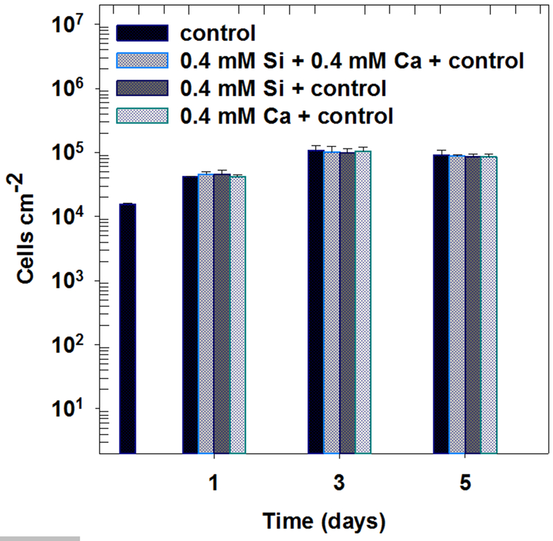
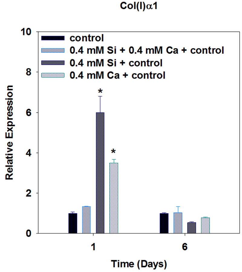
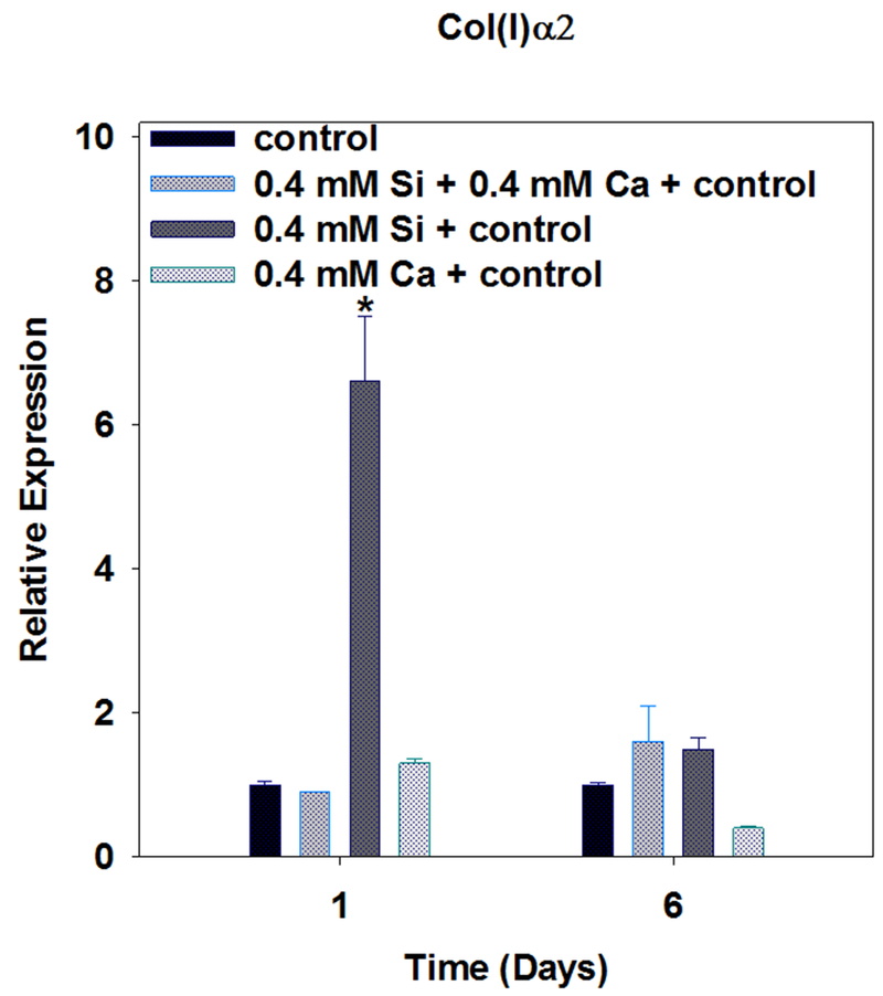
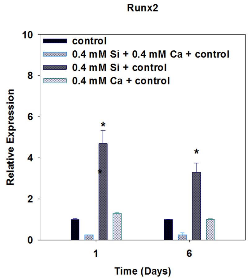
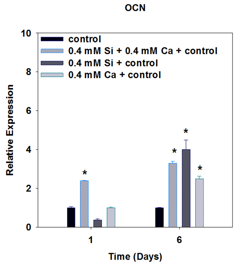
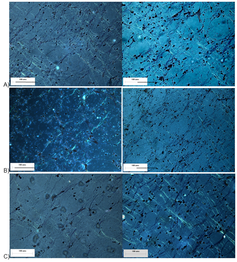
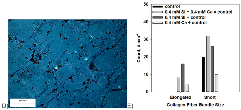
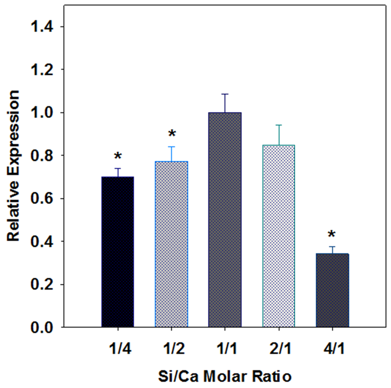
This study tests the hypothesis that silicon and calcium ions combinatorially target gene expression during osteoblast differentiation. MC3T3-E1 subclone 4 osteoblast progenitors (transformed mouse calvarial osteoblasts) were exposed to Si(4+) (from Na(2)SiO(3)) and Ca(2+) (from CaCl(2):H(2)O) ion treatments both individually (0.4 mM each + control treatment) and combinatorially (0.4 mM Si(4+) + 0.4 mM Ca(2+) + control treatment) and compared to control treated (α-minimum essential medium, 10% fetal bovine serum, and 1% penicillin-streptomycin) cells. Cell proliferation studies showed no significant increase in cell density between treatments over 5 days of culture. Cellular differentiation studies involved addition of ascorbic acid (50 mg/L) for all treatments. Relative gene expression was determined for collagen type 1 (Col(I)α1/Col(I)α2), core-binding factor a (cbfa1/Runx2), and osteocalcin (OCN), which indicated osteoblast progenitor differentiation into a mineralizing phenotype. Increased Si(4+) or Ca(2+) ion treatments enhanced Col(I)α1, Col(I)α2, Runx2, and OCN expression, while increased Si(4+) + Ca(2+) ion treatments enhanced OCN expression. Moreover, it was found that a Si(4+)/Ca(2+) ratio of unity was optimal for maximal expression of OCN. Collagen fiber bundles were dense, elongated, and thick within extracellular matrices (ECM) exposed to Si(4+) and Si(4+) + Ca(2+) treatments, while collagen fiber bundles were sparse, short, and thin within Ca(2+) and control treated ECM. These results indicated that individual ions enhance multiple osteogenic gene expression, while combined ion treatments enhance individual gene expression. In addition, these results indicated that Si(4+) enhanced osteoblast gene expression and ECM formation at higher levels than Ca(2+). These results support the larger concept that ions (possibly released from bioactive glasses) could control bone formation by targeting osteoblast marker expression.


Figures

Fig. 1.
Fig. 1.
Effect of Si, Ca, and Si+Ca ion treatments on MC3T3-E1 subclone 4 proliferation (ANOVA, p > 0.05).
Fig. 2
Fig. 2
Effect of Si and Ca ions on MC3T3-E1 subclone 4 expression of Col(I)α1 after 1 and 6 days of culture. (* indicates statistical significance relative to control, ANOVA, p < 0.05).
Fig. 3
Fig. 3
Effect of Si and Ca ions on MC3T3-E1 subclone 4 expression of Col(I)α2 after 1 and 6 days of culture. (* indicates statistical significance relative to control, ANOVA, p < 0.05)..
Fig. 4
Fig. 4
Effect of Si and Ca ions on MC3T3-E1 subclone 4 expression of Runx2 after 1 and 6 days of culture. (* indicates statistical significance relative to control, ANOVA, p < 0.05).
Fig. 5
Fig. 5
Effect of Si and Ca ions on MC3T3-E1 subclone 4 expression of OCN after 1 and 6 days of culture. (* indicates statistical significance relative to control, ANOVA, p < 0.05).
Fig. 6.
Fig. 6.
Effect of individual ion (0.4 mM Si + control + AA (A), 0.4 mM Ca + control + AA (B)), and combined ion (0.4 mM Si + 0.4 mM Ca + control + AA (C)), and control (+ AA (D)) treatments on MC3T3-E1 subclone 4 collagen fiber bundle formation after 6 days of culture. (Elongated fiber bundles indicated with orange stars) Collagen fiber bundle counts (E) confirmed more elongated fiber bundles in the treatments incorporating Si as compared to the treatments that did not incorporate Si.
Fig. 6.
Fig. 6.
Effect of individual ion (0.4 mM Si + control + AA (A), 0.4 mM Ca + control + AA (B)), and combined ion (0.4 mM Si + 0.4 mM Ca + control + AA (C)), and control (+ AA (D)) treatments on MC3T3-E1 subclone 4 collagen fiber bundle formation after 6 days of culture. (Elongated fiber bundles indicated with orange stars) Collagen fiber bundle counts (E) confirmed more elongated fiber bundles in the treatments incorporating Si as compared to the treatments that did not incorporate Si.
Fig. 7
Fig. 7
Effect of Si and Ca ions on MC3T3-E1 subclone 4 expression of OCN after 1 and 6 days of culture. (* indicates statistical significance relative to control, ANOVA, p < 0.05).

Similar articles

Cited by

References

    1. Varanasi VG, Saiz E, Loomer PM, Ancheta B, Uritani N, Ho SP, Tomsia AP, Marshall SJ and Marshall GW, Acta Biomateralia 5 (9), 3536–3547 (2009). - PMC - PubMed
    1. Foppiano S, Marshall SJ, Marshall GW, Saiz E and Tomsia AP, Acta Biomater 3 (5), 765–771 (2007). - PMC - PubMed
    1. Xynos ID, Edgar AJ, Buttery LDK, Hench LL and Polak JM, J Biomed Mater Res 55 (2), 151–157 (2001). - PubMed
    1. Bielby RC, Pryce RS, Hench LL and Polak JM, Tissue Eng 11 (3–4), 479–488 (2005). - PubMed
    1. Hench LL and Polak JM, Science 295 (5557), 1014 (2002). - PubMed

Publication types

MeSH terms