Review
. 2012 Aug;22(4):226-45.
doi: 10.1089/nat.2012.0350.

Uniqueness, advantages, challenges, solutions, and perspectives in therapeutics applying RNA nanotechnology

Affiliations
Review

Uniqueness, advantages, challenges, solutions, and perspectives in therapeutics applying RNA nanotechnology

Peixuan Guo et al. Nucleic Acid Ther. 2012 Aug.

Abstract

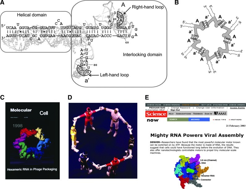
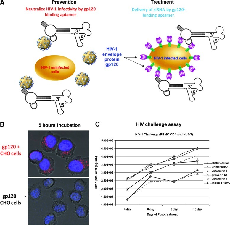
The field of RNA nanotechnology is rapidly emerging. RNA can be manipulated with the simplicity characteristic of DNA to produce nanoparticles with a diversity of quaternary structures by self-assembly. Additionally RNA is tremendously versatile in its function and some RNA molecules display catalytic activities much like proteins. Thus, RNA has the advantage of both worlds. However, the instability of RNA has made many scientists flinch away from RNA nanotechnology. Other concerns that have deterred the progress of RNA therapeutics include the induction of interferons, stimulation of cytokines, and activation of other immune systems, as well as short pharmacokinetic profiles in vivo. This review will provide some solutions and perspectives on the chemical and thermodynamic stability, in vivo half-life and biodistribution, yield and production cost, in vivo toxicity and side effect, specific delivery and targeting, as well as endosomal trapping and escape.


Figures

FIG. 1.
FIG. 1.
Structure of DNA-packaging RNA (pRNA) on the phi29 DNA-packaging motor and the hand-in-hand interaction used to build pRNA nanoparticles. (A) Sequence and secondary structure of phi29 pRNA. Superposition of the 2-dimensional and 3-dimensional structure of the phi29 pRNA Aa′. Uppercase letters represent the right-hand loop and lower-case letters the left-hand loop of pRNA. A pair of upper and lower case (e.g., Aa′) for same letters indicate a pair of complementary loops, whereas a pair of upper- and lowercase from different letters indicate non-complementary loops (see Fig. 2). The 4 bases in the right- and left-hand loops, which are responsible for inter-RNA interactions are boxed. For example, pRNA Aa′ refers to a pRNA with complementary right-hand loop A and left hand loop a′, which can form homo-hexamers (see also Fig. 2 for homo-dimers and trimers). (B) Schematic of pRNA hexamer. (C) Packaging of phi29 DNA through the motor geared by 6 pRNA (Guo et al., ; Zhang et al., 1998). (D) Construction of hexameric pRNA nanoparticles using the hand-in-hand interaction approach (Chen et al., 1999). (E) Elucidation of phi29 pRNA hexamer on the motor. Figures adapted and reproduced with permission: A, Liu et al., , © 2010 ACS; B, Shu et al. , © 2011 Elsevier Inc.; C, © 1998 Cell Press; D, Chen et al., , © RNA Society; E, © 2003 AAAS.
FIG. 2.
FIG. 2.
Construction of therapeutic pRNA nanoparticles via hand-in-hand interaction (see also Fig. 1). Left to right column: Schematic, models, and atomic force microscopy (AFM) image showing the formation of different therapeutic nanoparticles containing small interfering RNA (siRNA), ribozymes, aptamers, and other moieties using bacteriophage phi29 pRNA that possess left- and right-hand interlocking loops; uppercase and lowercase letters represent right-hand and left-hand respectively. Same letter pair (e.g., Aa′) indicates complementarity interlocking loops; different letter (e.g. Ab′) indicates non-coomplementary loops (Hoeprich et al., ; Guo et al., ; Khaled et al., ; Guo et al., ; Shu et al., ; Abdelmawla et al., ; Shu et al., ; Shu et al., ; Shu et al., ; Ye et al., 2012). (A) Construction of pRNA monomers bearing either siRNA, a ribozyme, a receptor-binding aptamer, a targeting ligand, or a detection molecule; scale bar=15 nm. (B) Construction of pRNA dimers. Monomer Ab′, which contains a receptor-binding aptamer, and monomer Ba′, which contains an siRNA, assemble to form hand-in-hand dimers; scale bar=30 nm. (C) Construction of pRNA trimers. Trimers are formed between monomer Ab′ (containing an RNA aptamer), Bc′ (containing an siRNA), and Ca′ (containing a ribozyme); scale bar=30 nm. (D) Illustration of hexameric pRNA nanoparticles on the cover of Human Gene Therapy (Guo et al., 2005). (E–F) colE1 loop–loop interactions used to construct programmable a hexameric nanoring, via interlocking loops αα′, ββ′, γγ′, δδ′, ɛɛ′, and ζζ′. The siRNA sequences are attached to the vertices after the formation of the hexamer instead of using the fusing approach, as in A–D. Figures adapted and reproduced with permission: A–C, Shu et al., , © 2004 ACS and Shu et al., , © 2003 American Scientific Publishers (ASP); D, Guo et al., , © 2005 Mary Ann Liebert, Inc.; E–F, Grabow et al., , © 2011 American Chemical Society (ACS).
FIG. 3.
FIG. 3.
Construction of therapeutic pRNA nanoparticles via foot-to-foot interaction of palindrome sequences. Left to right column: Schematic, models, and AFM image showing the formation of different therapeutic nanoparticles containing siRNA, ribozymes, aptamers, and other moieties using bacteriophage phi29 pRNA containing a palindrome sequence (Shu et al., 2004). (A) Foot-to-foot dimers form through the palindrome sequence at the end of two Ab′ monomers, with one bearing a targeting ligand and the other a detection molecule; scale bar=20 nm. (B) Tetramers assemble by the combination of hand-in-hand interlocking loops and foot-to-foot palindrome mechanism of 2 dimers (Ab′ and Ba′); scale bar=20 nm. The models illustrate how the various structures are held together. Figures A, B reproduced with permission Shu et al., , © 2004 ACS and Shu et al., , © 2003 ASP.
FIG. 4.
FIG. 4.
Construction of thermodynamically stable trivalent pRNA-based 3-way junction (3WJ) nanoparticles. (A) Sequence of pRNA monomer Ab′ (Guo et al., 1998). Green box: central 3WJ domain. In pRNA Ab′, A and b′ represent right- and left-hand loops respectively. (B) 3WJ domain composed of 3 RNA oligomers in black, red, and blue. Helical segments are represented as H1, H2, and H3. (C) Three pRNA molecules bound at the 3WJ-pRNA core sequence (black, red, and blue), and (D) its accompanying AFM images; scale bar=30 nm. (E) Multi-module RNA nanoparticles harboring siRNA, ribozyme, and aptamer, and (F) its accompanying AFM images; scale bar=20 nm. Figures reproduced with permission from Shu et al., , © 2011 Nature Publishing Group (NPG).
FIG. 5.
FIG. 5.
Assembly and stability studies of 3WJ-pRNA. In the tables, “+” indicates the presence of the strand in samples of the corresponding lanes. (A) 15% native polyacrylamide gel electrophoresis (PAGE) showing the assembly of the 3WJ core, stained by ethidium bromide (upper) and SYBR green 2 (lower). (B) Melting temperature curves for the assembly of the 3WJ core. Melting curves for the individual strands (brown, green, silver), the 2-strand combinations (blue, cyan, pink) and the 3-strand combination (red) are shown. (C) Melting curves for 11 different RNA 3WJ core motifs assembled from 3 oligos for each 3WJ motif under physiological buffer. (D–F) Competition and dissociation assays of 3WJ-pRNA. (D) Temperature effects on the stability of the 3WJ-pRNA core, denoted as [ab*c]3WJ, evaluated by 16% native gel. A fixed concentration of Cy3-labelled [ab*c]3WJ was incubated with varying concentrations of unlabelled b3WJ at 37°C. (E) Urea denaturing effects on the stability of [ab*c]3WJ evaluated by 16% native gel. A fixed concentration of labeled [ab*c]3WJ was incubated with unlabelled b3WJ at 1:1 ratio in the presence of 0–6 M urea at 25°C. (F) Dissociation assay for the [32P]-3WJ-pRNA complex harboring 3 monomeric pRNAs by 2-fold serial dilution (lanes 1–9). The monomer unit is shown on the left. Figures reproduced with permission from Shu et al., , © 2011 NPG.
FIG. 6.
FIG. 6.
Apoptosis and binding assays of chimeric therapeutic pRNA. (A) Apoptosis induced by transfection of chimeric pRNA harboring siRNA targeting survivin using Lipofectamine 2000. Breast cancer MCF-7 cells were transfected with pRNA/siRNA (survivin) and apoptosis was monitored by propidium iodide–annexin A5 double labeling followed by flow cytometry. Cells in the bottom right quadrant represent apoptotic cells. The mutant pRNA/siRNA was transfected in parallel as a negative control. (B) Specific delivery of chimeric pRNA/siRNA by folate-pRNA. Flow cytometry analyses of the binding of fluorescein isothiocyanate (FITC)-labeled folate-pRNA to nasopharyngeal carcinoma (KB) cells. Left: Cells were incubated with folate-pRNA labeled with FITC. Middle: Cells were preincubated with free folate, which served as a blocking agent to compete with folate-pRNA for binding to the receptor. Right: Binding was also tested using folate-free pRNA labeled with FITC as a negative control. The percentages of FITC-positive cells are shown in the top right quadrants. (C) Confocal images showed targeting of folate receptor positive (FR+)-KB cells by co-localization (overlap, 4) of cytoplasm (green, 1) and RNA nanoparticles (red, 2). (D) 3WJ-pRNA nanoparticles target folate receptor positive (FR+) tumor xenografts on systemic administration in nude mice. Upper panel: whole body; lower panel: organ imaging (Lv, liver; K, kidney; H, heart; L, lung; S, spleen; I, intestine; M, muscle; T, tumor). Figures reproduced with permission: A–B, Guo et al., , © 2005 Mary Ann Liebert, Inc.; C–D, Shu et al., , © 2011 NPG.
FIG. 7.
FIG. 7.
Self-assembled RNA nanoparticles as potential therapeutic agents. AFM images of (A–B) rationally designed RNA 1-dimensional and 2-dimensional arrays in vivo (Delebecque et al., 2011); (C) RNA bundles (scale bar=50 nm) (Cayrol et al., 2009); (D) AFM images of pRNA arrays (Shu et al., 2004). (E) Transmission electron microscopy (TEM) images of RNA microsponges (Lee et al., 2012). Figures reproduced with permission: A–B, Delebecque et al., , © 2011 AAAS; C, Cayrol et al., , © 2009 ACS; D, Shu et al., , © 2004 ACS; E, Lee et al., , © 2012 NPG.
FIG. 9.
FIG. 9.
Schematic (A) and 2D structure (B) of chimeric pRNA–miRNA complexes for antiviral therapies. The helical region of pRNA is replaced by several artificial microRNA (AmiR) sequences. The AmiRs target the 3′ untranslated region (3′ UTR) of coxsackievirus B3 (CVB3) genome. Antiviral evaluation showed that the AmiRs displayed strong reduction of CVB3 replication (Ye et al., 2011). Figures adapted with permission from Guo et al., ; Ye et al., , © 2011 Public Library of Science.
FIG. 8.
FIG. 8.
Chimeric pRNA-aptamer-siRNA nanoparticles for human immunodeficiency virus (HIV) therapy. (A) The pRNA-aptamer mediated targeted delivery of siRNA using chimeric pRNA–anti-gp120 aptamer. The anti-gp120 aptamer is responsible for binding to HIV-1 gp120 protein. (B) Cell-type specific binding studies of pRNA aptamer chimeras. Cy3-labeled pRNA aptamers were incubated with Chinese hamster ovary (CHO)-gp160 cells and CHO-EE control cells. Cell surface binding of Cy3-labeled chimeras was assessed by confocal imaging. (C) The inhibition of HIV-1 infection mediated by pRNA-aptamer chimeras. Both antigp120 aptamer and pRNA-aptamer chimera neutralized HIV-1 infection in HIV-infected human peripheral blood mononuclear cells (PBMCs) (NL4-3 strain) culture. Data represent the average of triplicate measurements (Zhou et al., ; Zhou et al., 2011). Figure A courtesy of Dr. Jiehua Zhou and Dr. John Rossi. Figures B–C reproduced with permission from Zhou et al., , © 2011 Elseiver.

Similar articles

Cited by

References

    1. ABDELMAWLA S. GUO S. ZHANG L. PULUKURI S. PATANKAR P. CONLEY P. TREBLEY J. GUO P. LI Q.X. Pharmacological characterization of chemically synthesized monomeric pRNA nanoparticles for systemic delivery. Mol. Ther. 2011;19:1312–1322. - PMC - PubMed
    1. AFONIN K.A. BINDEWALD E. YAGHOUBIAN A.J. VOSS N. JACOVETTY E. SHAPIRO B.A. JAEGER L. In vitro assembly of cubic RNA-based scaffolds designed in silico. Nat. Nanotechnol. 2010;5:676–682. - PMC - PubMed
    1. AFONIN K.A. GRABOW W.W. WALKER F.M. BINDEWALD E. DOBROVOLSKAIA M.A. SHAPIRO B.A. JAEGER L. Design and self-assembly of siRNA-functionalized RNA nanoparticles for use in automated nanomedicine. Nat. Protoc. 2011;6:2022–2034. - PMC - PubMed
    1. BADER A.G. BROWN D. WINKLER M. The promise of microRNA replacement therapy. Cancer Res. 2010;70:7027–7030. - PMC - PubMed
    1. BANTA S. MEGEED Z. CASALI M. REGE K. YARMUSH M.L. Engineering protein and peptide building blocks for nanotechnology. J. Nanosci. Nanotechnol. 2007;7:387–401. - PubMed

Publication types

LinkOut - more resources