Review
. 2012 Sep;13(9):549-65.
doi: 10.1038/nrm3414.

STIM proteins: dynamic calcium signal transducers

Affiliations
Review

STIM proteins: dynamic calcium signal transducers

Jonathan Soboloff et al. Nat Rev Mol Cell Biol. 2012 Sep.

Abstract

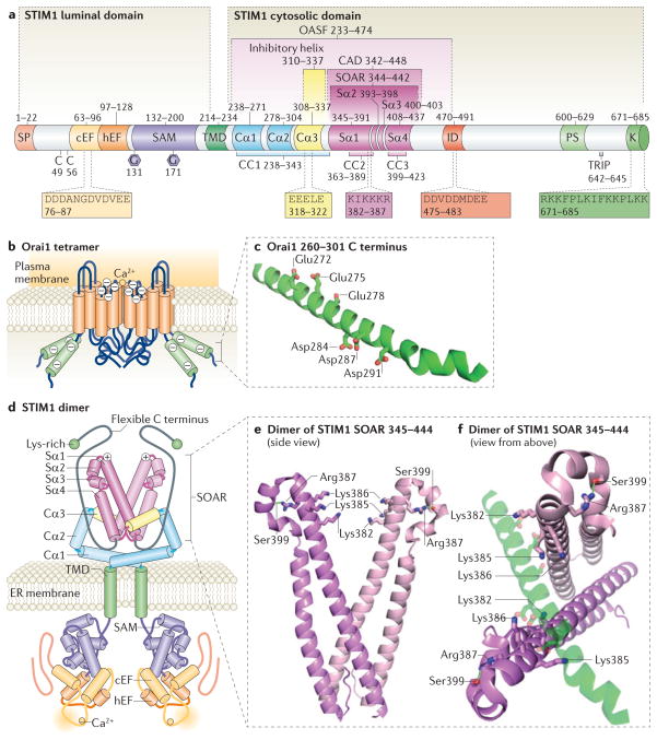
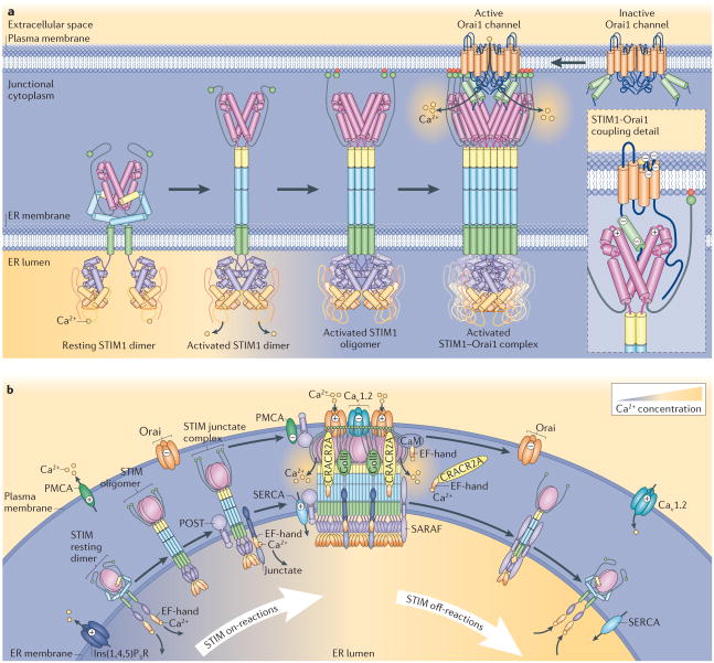
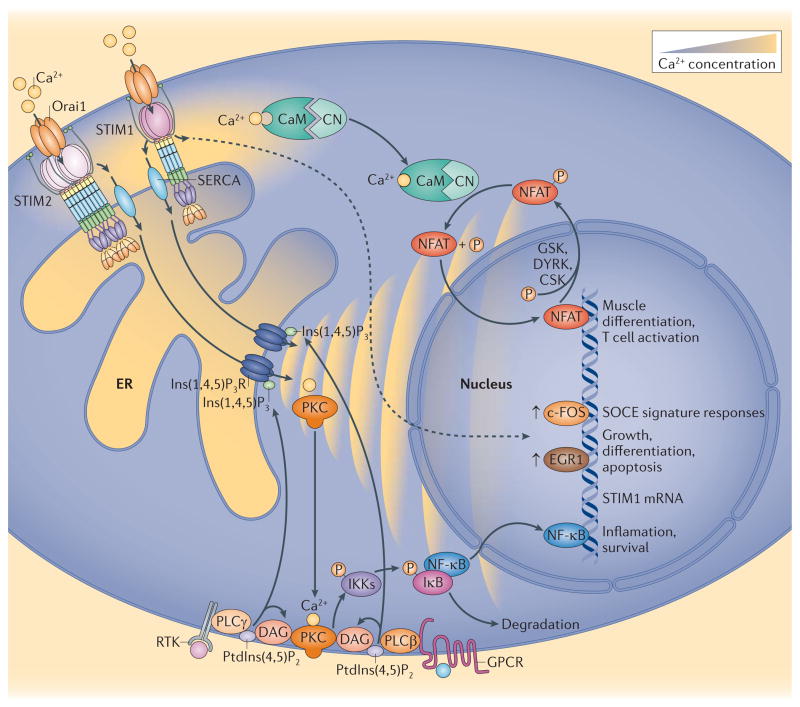
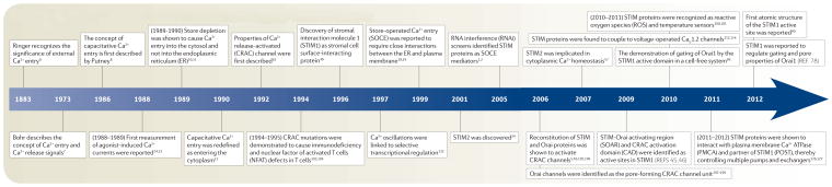
Stromal interaction molecule (STIM) proteins function in cells as dynamic coordinators of cellular calcium (Ca(2+)) signals. Spanning the endoplasmic reticulum (ER) membrane, they sense tiny changes in the levels of Ca(2+) stored within the ER lumen. As ER Ca(2+) is released to generate primary Ca(2+) signals, STIM proteins undergo an intricate activation reaction and rapidly translocate into junctions formed between the ER and the plasma membrane. There, STIM proteins tether and activate the highly Ca(2+)-selective Orai channels to mediate finely controlled Ca(2+) signals and to homeostatically balance cellular Ca(2+). Details are emerging on the remarkable organization within these STIM-induced junctional microdomains and the identification of new regulators and alternative target proteins for STIM.


Conflict of interest statement

Competing interests statement

The authors declare competing financial interests; see Web version for details.

Figures

Figure 1
Figure 1. Structure and activation of STIM1
a| The molecular domains of stromal interaction molecule 1 (STIM1). Endoplasmic reticulum (ER) STIM1 contains a luminal and a cytosolic domain. The amino-terminal signal peptide (SP) is cleaved during translation. The ER luminal N-terminal domain includes a conserved Cys pair, a Ca2+-binding canonical EF-hand domain, (cEF), a non-Ca2+-binding hidden EF-hand (hEF) domain, a sterile α-motif (SAM) with two Asn-linked glycosylation sites (shown as hexagons) and a single transmembrane domain (TMD). The cytosolic carboxy-terminal domain is considered to include three coiled-coil regions (called CC1, CC2 and CC3). CC1 is divided into three α-helices (termed Cα1, Cα2 and Cα3) on the basis of sequence analysis predictions by using JPred3 (REF. 188). The structure of Cα3 is also determined on the basis of homology with the recently solved Caenorhabditis elegans STIM structure. SOAR (STIM–Orai activating region) is the minimal sequence required for the activation of Orai1 (REF. 45). SOAR contains four α-helices, termed Sα1, Sα2, Sα3 and Sα4 (REF. 40). The segments CAD (Ca2+ release-activated Ca2+ (CRAC) activation domain) and OASF (Orai1-activating small fragment) are larger than SOAR, contain the CC1 region and also activate Orai1. SOAR includes the polybasic region, with the sequence KIKKKR (amino acids 382–387), which is crucial for the interaction with Orai1 (REFS 40,68,71). Cα3 contains an inhibitory helix that inhibits SOAR function,,. The acidic EEELE (residues 318–322) region is required for the action of the inhibitory helix,. Downstream of SOAR resides an acidic inhibitory domain (ID) that mediates fast Ca2+-dependent inactivation of Orai1 (REFS 89,92,93). The C-terminal tail contains a Pro/Ser-rich domain (PS), a microtubule interacting domain (TRIP) and a Lys-rich domain responsible for phospholipid interaction at the plasma membrane. b | The tetrameric structure of the Orai1 channel is shown, highlighting the TMDs (shown in orange), extracellular and intracellular sequences (blue) and C-terminal predicted α-helices (shown in green). Negatively charged residues are indicated in the C-terminal helices and in the Ca2+ selectivity filter at the predicted mouth of the pore. c | The predicted α-helical structure of the Orai1 C terminus and potential sites for SOAR binding are illustrated on the basis of secondary structure prediction by using JPred3 (REF. 188). Side chains from acidic residues Glu272, Glu275, Glu278, Asp284, Asp287 and Asp291 are shown as amphipathic helices that may electrostatically interact with basic residues in the SOAR dimer. d | Proposed structure of a resting STIM1 dimer. The predicted α-helices in CC1 are indicated in the figure and are shown in a folded configuration. The inhibitory Cα3 helix (highlighted in yellow) is bound to SOAR (shown in red), which comprises four α-helices as indicated. The SOAR polybasic region is shown (+). The STIM1 dimer is held together predominantly by interactions between the CC1 and SOAR regions,. The C-terminal flexible region (shown in grey) together with the C-terminal Lys-rich domain (shown in green) may provide some steric shielding of SOAR. α-helices are shown as cylinders; flexible regions as lines. e | The side view of the SOAR structure reveals that Lys382, Lys385 and Lys386 are located in the polybasic Orai-interacting region, and that these residues are oriented towards the centre of the cleft. Lys384 (not shown) is oriented in another direction. The Arg387 residue may be involved in hydrogen bonding with Ser399 (indicated by a green line). Coordinates were obtained from Protein data bank entry 3TEQ. f | SOAR structure shown in (e) rotated 90° to illustrate the potential sites of association with the Orai1 C terminus depicted in (c). The hypothetical electrostatic binding of the Orai1 C terminus within the cleft between the SOAR monomers is shown.
Figure 2
Figure 2. STIM activation and organization of the Ca2+ signalling junction
a | Hypothetical model of stromal interaction molecule 1 (STIM1) activation and coupling to Orai1. The resting STIM1 dimer in the Ca2+-replete endoplasmic reticulum (ER) is shown on the left. The activation of the STIM1 dimer is initiated by Ca2+ dissociation from the STIM1 dimer. This causes EF-hand–SAM domains within the STIM1 dimer to interact, which induces an extended configuration of the cytoplasmic coiled-coil domains, dissociation of the Cα3 inhibitory helix from SOAR (STIM–Orai activating region),,, and the carboxy-terminal flexible domains recede and expose SOAR. STIM1 continues to oligomerize and migrates into ER–plasma membrane junctions, and the polybasic C termini bind and anchor STIM1 to negatively charged phospholipids in the plasma membrane (shown in red) and active SOAR is fully exposed. Large aggregates of anchored STIM1 within ER–plasma membrane junctions are able to tether and activate Orai1 proteins. Each SOAR dimer interacts with one Orai1 protein, therefore eight STIM1 molecules form an active complex with one tetrameric Orai1 channel. b | Role of regulatory and target proteins in the STIM-activated Ca2+ signalling junction. In response to inositol-1,4,5-trisphosphate (Ins(1,4,5)P3) receptor (Ins(1,4,5)P3R)-mediated ER Ca2+ depletion, Ca2+ dissociates from STIM1, and STIM1 aggregates and translocates into ER–plasma membrane junctions. During activation, STIM1 initially interacts with plasma membrane lipids through its Lys-rich domain assisted by interaction with junctate in or near ER–plasma membrane junctions,. This interaction is stabilized by the Ca2+-free form of Ca2+ release-activated (CRAC) regulatory protein 2A (CRACR2A) and golli,. During targeting, STIM1 interacts with and activates Orai1 channels and inhibits CaV1.2 channels,. STIM1 recruits partner of STIM1 (POST) to the junction, and this adaptor protein recruits both plasma membrane Ca2+ ATPase (PMCA) and sarcoendoplasmic reticulum Ca2+ ATPase (SERCA) to the Ca2+ signalling junction. POST and STIM1 (REF. 126) inhibit PMCA-mediated Ca2+ efflux from the cell and thereby increase Ca2+ availability for signalling. SERCA recruited into junctions by POST may also assist ER Ca2+ loading. During STIM deactivation, increased cytosolic Ca2+-dependent loss of both CRACR2A and golli destabilizes the STIM complex. Ca2+-binding to calmodulin (CaM) promotes Orai channel inactivation. The STIM-binding protein store-operated Ca2+ entry (SOCE)-associated regulatory factor (SARAF) also mediates dissociation of STIM proteins, resulting in their configuration into a resting state.
Figure 3
Figure 3. STIM proteins control gene expression by generating spatiotemporally controlled Ca2+ signals
Activation of either receptor Tyr kinase-coupled or G protein-coupled receptors (RTK or GPCR, respectively) result in phospholipase C (PLC)-mediated production of inositol-1,4,5-trisphosphate (Ins(1,4,5)P3) and diacylglycerol (DAG). RTK-mediated PLCγ activation can be slower and more gradual than GPCR-mediated PLCβ responses. The rapid ER Ca2+ depletion mediated by GPCR leads to stromal interaction protein 1 (STIM1)-mediated but not STIM2-mediated Orai channel activation. By contrast, ‘shallow’ Ca2+ release through RTK may cause both STIM1- and STIM2-mediated Orai channel activation. Close apposition of sarcoplasmic reticulum Ca2+ ATPase (SERCA) with junctions allows the entry of Ca2+ to refill stores and maintain oscillations while contributing only minimally to global changes in cytosolic Ca2+ concentration. However, the local increase in Ca2+ near the STIM1–Orai junction is crucial for the activation of both c-FOS and nuclear factor of activated T cells (NFAT),. Hence, Ca2+-Calmodulin (CaM) activates calcineurin (CN), which dephosphorylates NFAT, resulting in nuclear translocation. Casein kinase (CK), dual specificity Tyr phosphorylationregulated kinase 2 (DYRK) and glycogen synthase kinase (GSK) phosphorylate NFAT, which results in a cycling of NFAT across the nuclear membrane as long as the Ca2+ signal continues. Store-operated Ca2+ entry (SOCE) can also lead to both nuclear factor-κB (NF-κB) activation, and early growth response protein 1 (EGR1) upregulation, both of which directly stimulate STIM1 transcription (not shown),. Ca2+-mediated NF-κB activation may be mediated by protein kinase C (PKC), which phosphorylates (P) inhibitor of NF-κB (IκB) kinases (IKKs). IKKs phosphorylate IκB, resulting in its dissociation from NF-κB.
Timeline
Timeline
Major events in the discovery and characterization of store-operated Ca2+ entry

Similar articles

Cited by

References

    1. Roos J, et al. STIM1, an essential and conserved component of store-operated Ca2+ channel function. J Cell Biol. 2005;169:435–445. - PMC - PubMed
    1. Liou J, et al. STIM is a Ca2+ sensor essential for Ca2+-store-depletion-triggered Ca2+ influx. Curr Biol. 2005;15:1235–1241. - PMC - PubMed
    1. Feske S, et al. A mutation in Orai1 causes immune deficiency by abrogating CRAC channel function. Nature. 2006;441:179–185. - PubMed
    1. Vig M, et al. CRACM1 is a plasma membrane protein essential for store-operated Ca2+ entry. Science. 2006;312:1220–1223. - PMC - PubMed
    1. Zhang SL, et al. Genome-wide RNAi screen of Ca2+ influx identifies genes that regulate Ca2+ release-activated Ca2+ channel activity. Proc Natl Acad Sci USA. 2006;103:9357–9362. - PMC - PubMed

Publication types