. 2012 Dec 1;21(23):5039-47.
doi: 10.1093/hmg/dds348. Epub 2012 Aug 21.

Fragile X premutation RNA is sufficient to cause primary ovarian insufficiency in mice

Affiliations

Fragile X premutation RNA is sufficient to cause primary ovarian insufficiency in mice

Cuiling Lu et al. Hum Mol Genet. .

Abstract

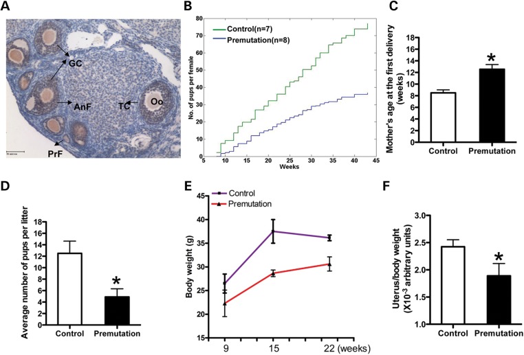
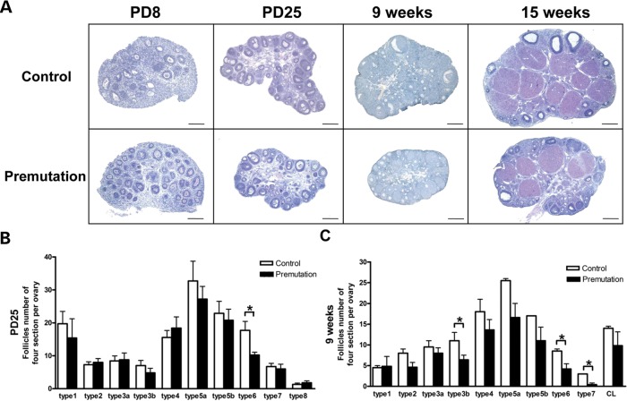
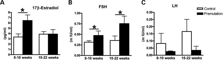
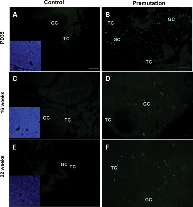
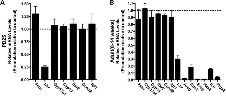
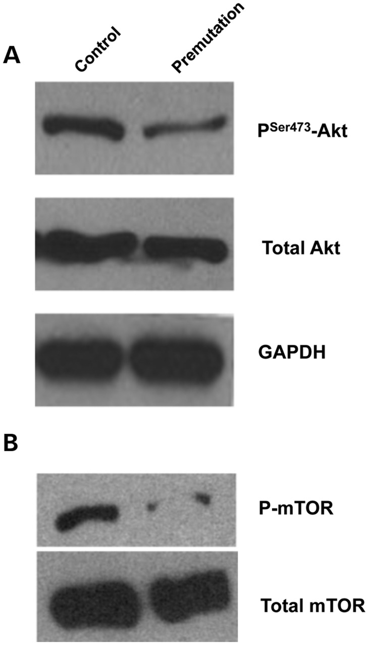
Spontaneous 46,XX primary ovarian insufficiency (POI), also known as 'premature menopause' or 'premature ovarian failure', refers to ovarian dysfunction that results in a range of abnormalities, from infertility to early menopause as the end stage. The most common known genetic cause of POI is the expansion of a CGG repeat to 55-199 copies (premutation) in the 5' untranslated region in the X-linked fragile X mental retardation 1 (FMR1) gene. POI associated with the FMR1 premutation is referred to as fragile X-associated POI (FXPOI). Here, we characterize a mouse model carrying the human FMR1 premutation allele and show that FMR1 premutation RNA can cause a reduction in the number of growing follicles in ovaries and is sufficient to impair female fertility. Alterations in selective serum hormone levels, including FSH, LH and 17β-estradiol, are seen in this mouse model, which mimics findings in humans. In addition, we also find that LH-induced ovulation-related gene expression is specifically altered. Finally, we show that the FMR1 premutation allele can lead to reduced phosphorylation of Akt and mTOR proteins. These results together suggest that FMR1 premutation RNA could cause the POI associated with FMR1 premutation carriers, and the Akt/mTOR pathway may serve as a therapeutic target for FXPOI.


Figures

Figure 1.
Figure 1.
Fragile X premutation RNA impairs female fertility. (A) immunohistochemistry staining of Fmrp in the ovary of a 9-week-old WT mouse. Brown staining indicates immunoreactivity and nuclei are counterstained blue. Bar: 50 µm. GC, granulosa cell; Oo, oocyte; TC, theca cell; AnF, antral follicle; PrF, primary follicle. (B) Lifespan breeding assays started at ∼8 weeks of age. Comparison of the cumulative number of pups per fragile X premutation (TG296-Premutation) female (blue line) and per WT littermate (green line), n = 7 for WT mice, n = 8 for TG296 mice. (C) the ages of mothers at first delivery (mean ± SD). (D) the litter sizes of WT and premutation females (mean ± SD). N = 13 for premutation mice, n = 9 for WT mice. (E) the body weights of females at different ages (mean ± SD). (F) Graph of the uterus/body weight ratio of females at 9 weeks of age (mean ± SD). Statistical significance (P < 0.05) was calculated by Student's t-test.
Figure 2.
Figure 2.
Reduced number of growing follicles in fragile X premutation ovaries. (A) Histology of ovarian sections from premutation and WT littermates of PD8, PD25, 9- and 15-week-old females stained with hematoxylin/eosin dye (bar: 200 µm). (B and C) the quantifications of numbers for each type of follicle cell at PD25 and 9 weeks. The experiments were repeated four times, and for each time and each age, ovaries from three mice of each genotype were used. Relative follicle counts at PD25 (B) and 9 weeks (C) of age for premutation mice and WT littermates. Numbers represent the four serial biggest sections from every fifth section per sectioned ovaries (n = 5 animals per genotype). For all panels, data are shown as mean ± SEM, and statistical significance (*P < 0.05) was calculated by Student's t-test.
Figure 3.
Figure 3.
Altered serum hormone levels in fragile X premutation mice. The elevated levels of estradiol (A) and FSH (B) and the decreased level of LH (C) in adult premutation versus WT littermates. On average, 8–11 mice were used for each time point and each genotype. Female mice of both genotypes were sacrificed at the proestrus stage, and sera were collected for measurement of 17β-estradiol, FSH and LH levels. Statistical significance (*P < 0.05) was calculated by Student's t-test.
Figure 4.
Figure 4.
Increased apoptosis of follicle cells in fragile X premutation ovaries. Follicular apoptosis was detected by TUNEL on serial sections. Atresic follicles and cells exhibited positive TUNEL staining, whereas healthy ones were TUNEL negative. Representatives of slides from six animals are shown. Bar: 50 µm. TC, theca cell; GC, granulosa cell; inset denotes nuclear staining by Hoechst.
Figure 5.
Figure 5.
Altered expression of the LH-induced ovulation-related genes in fragile X premutation ovaries. The ovaries from both premutation and WT littermates at PD25 and 10–14 weeks of age were collected for gene expression analyses. Results shown are the representative real-time PCR results (using two or three ovaries per genotype in each experiment) of three independent experiments. Data were analyzed by using Student's t test and are shown as mean ± SEM.
Figure 6.
Figure 6.
Reduced phosphorylation of Akt and mTOR in fragile X premutation ovaries. Ovaries were isolated from PD25 and adult (8–14 weeks) fragile X premutation mice and their WT littermates for western blot analyses. Levels of p-Akt (serine 473) and p-mTOR (serine 2448) as well as total Akt and mTOR are shown. GAPDH was used as a loading control. All experiments were repeated at least three times. Representative images are shown.

Similar articles

Cited by

References

    1. Nelson L.M. Clinical practice. Primary ovarian insufficiency. N. Engl. J. Med. 2009;360:606–614. doi:10.1056/NEJMcp0808697. - DOI - PMC - PubMed
    1. Gallagher J.C. Effect of early menopause on bone mineral density and fractures. Menopause. 2007;14:567–571. doi:10.1097/gme.0b013e31804c793d. - DOI - PubMed
    1. Jacobsen B.K., Knutsen S.F., Fraser G.E. Age at natural menopause and total mortality and mortality from ischemic heart disease: the Adventist Health Study. J. Clin. Epidemiol. 1999;52:303–307. doi:10.1016/S0895-4356(98)00170-X. - DOI - PubMed
    1. Mondul A.M., Rodriguez C., Jacobs E.J., Calle E.E. Age at natural menopause and cause-specific mortality. Am. J. Epidemiol. 2005;162:1089–1097. doi:10.1093/aje/kwi324. - DOI - PubMed
    1. Popat V.B., Calis K.A., Vanderhoof V.H., Cizza G., Reynolds J.C., Sebring N., Troendle J.F., Nelson L.M. Bone mineral density in estrogen-deficient young women. J. Clin. Endocrinol. Metab. 2009;94:2277–2283. doi:10.1210/jc.2008-1878. - DOI - PMC - PubMed

Publication types

MeSH terms