

Clinically relevant hormone treatments fail to induce spinogenesis in prefrontal cortex of aged female rhesus monkeys
- PMID: 22915112
- PMCID: PMC3657730
- DOI: 10.1523/JNEUROSCI.1881-12.2012
Clinically relevant hormone treatments fail to induce spinogenesis in prefrontal cortex of aged female rhesus monkeys
Abstract
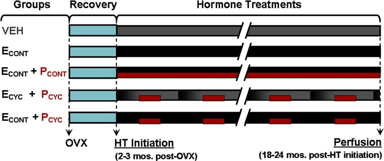
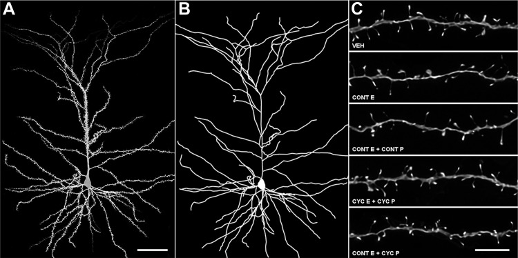
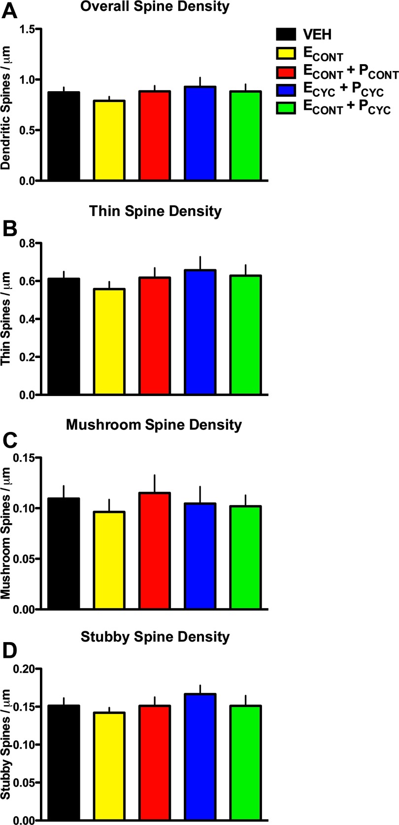
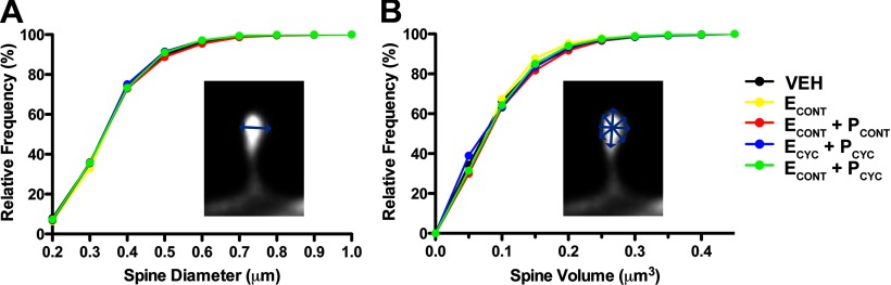
Preclinical animal models have provided strong evidence that estrogen (E) therapy (ET) enhances cognition and induces spinogenesis in neuronal circuits. However, clinical studies have been inconsistent, with some studies revealing adverse effects of ET, including an increased risk of dementia. In an effort to bridge this disconnect between the preclinical and clinical data, we have developed a nonhuman primate (NHP) model of ET combined with high-resolution dendritic spine analysis of dorsolateral prefrontal cortical (dlPFC) neurons. Previously, we reported cyclic ET in aged, ovariectomized NHPs increased spine density on dlPFC neurons. Here, we report that monkeys treated with cyclic E treatment paired with cyclic progesterone (P), continuous E combined with P (either cyclic or continuous), or unopposed continuous E failed to increase spines on dlPFC neurons. Given that the most prevalent form of ET prescribed to women is a combined and continuous E and P, these data bring into convergence the human neuropsychological findings and preclinical neurobiological evidence that standard hormone therapy in women is unlikely to yield the synaptic benefit presumed to underlie the cognitive enhancement reported in animal models.
Figures
Similar articles
-
Continuously delivered ovarian steroids do not alter dendritic spine density or morphology in macaque dorsolateral prefrontal cortical neurons.Neuroscience. 2013;255:219-25. doi: 10.1016/j.neuroscience.2013.09.062. Epub 2013 Oct 10. Neuroscience. 2013. PMID: 24120552 Free PMC article.
-
Interactive effects of age and estrogen on cortical neurons: implications for cognitive aging.Neuroscience. 2011 Sep 15;191:148-58. doi: 10.1016/j.neuroscience.2011.05.045. Epub 2011 Jun 2. Neuroscience. 2011. PMID: 21664255 Free PMC article. Review.
-
Estrogen Restores Multisynaptic Boutons in the Dorsolateral Prefrontal Cortex while Promoting Working Memory in Aged Rhesus Monkeys.J Neurosci. 2016 Jan 20;36(3):901-10. doi: 10.1523/JNEUROSCI.3480-13.2016. J Neurosci. 2016. PMID: 26791219 Free PMC article.
-
Estrogen alters spine number and morphology in prefrontal cortex of aged female rhesus monkeys.J Neurosci. 2006 Mar 1;26(9):2571-8. doi: 10.1523/JNEUROSCI.3440-05.2006. J Neurosci. 2006. PMID: 16510735 Free PMC article.
-
The evolving role of dendritic spines and memory: Interaction(s) with estradiol.Horm Behav. 2015 Aug;74:28-36. doi: 10.1016/j.yhbeh.2015.05.004. Epub 2015 May 17. Horm Behav. 2015. PMID: 25993604 Free PMC article. Review.
Cited by
-
Alzheimer's disease: review of hormone therapy trials and implications for treatment and prevention after menopause.J Steroid Biochem Mol Biol. 2014 Jul;142:99-106. doi: 10.1016/j.jsbmb.2013.05.010. Epub 2013 May 28. J Steroid Biochem Mol Biol. 2014. PMID: 23727128 Free PMC article. Review.
-
The scientific body of knowledge - Whose body does it serve? A spotlight on oral contraceptives and women's health factors in neuroimaging.Front Neuroendocrinol. 2021 Jan;60:100874. doi: 10.1016/j.yfrne.2020.100874. Epub 2020 Sep 28. Front Neuroendocrinol. 2021. PMID: 33002517 Free PMC article. Review.
-
Differential effects of aging on dendritic spines in visual cortex and prefrontal cortex of the rhesus monkey.Neuroscience. 2014 Aug 22;274:33-43. doi: 10.1016/j.neuroscience.2014.05.008. Epub 2014 May 20. Neuroscience. 2014. PMID: 24853052 Free PMC article.
-
Distinct cognitive effects of estrogen and progesterone in menopausal women.Psychoneuroendocrinology. 2015 Sep;59:25-36. doi: 10.1016/j.psyneuen.2015.04.020. Epub 2015 May 14. Psychoneuroendocrinology. 2015. PMID: 26010861 Free PMC article. Clinical Trial.
-
Continuously delivered ovarian steroids do not alter dendritic spine density or morphology in macaque dorsolateral prefrontal cortical neurons.Neuroscience. 2013;255:219-25. doi: 10.1016/j.neuroscience.2013.09.062. Epub 2013 Oct 10. Neuroscience. 2013. PMID: 24120552 Free PMC article.
References
-
- Bimonte-Nelson HA, Nelson ME, Granholm AC. Progesterone counteracts estrogen-induced increases in neurotrophins in the aged female rat brain. Neuroreport. 2004;15:2659–2663. - PubMed
-
- Bimonte-Nelson HA, Francis KR, Umphlet CD, Granholm AC. Progesterone reverses the spatial memory enhancements initiated by tonic and cyclic oestrogen therapy in middle-aged ovariectomized female rats. Eur J Neurosci. 2006;24:229–242. - PubMed
Publication types
MeSH terms
Substances
Grants and funding
LinkOut - more resources
Full Text Sources
Medical