. 2012 Aug 22;32(34):11706-15.
doi: 10.1523/JNEUROSCI.0730-12.2012.

Lipopolysaccharide-induced microglial activation and neuroprotection against experimental brain injury is independent of hematogenous TLR4

Affiliations

Lipopolysaccharide-induced microglial activation and neuroprotection against experimental brain injury is independent of hematogenous TLR4

Zhihong Chen et al. J Neurosci. .

Abstract

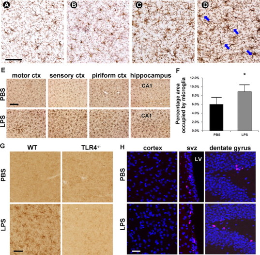
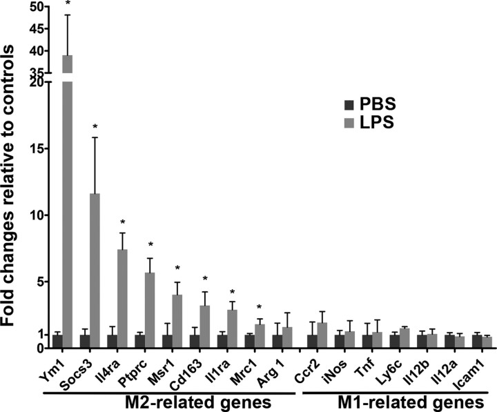
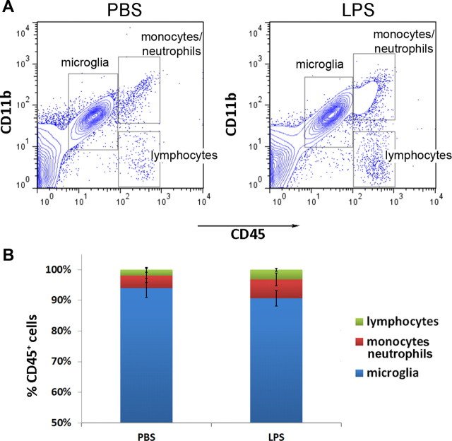
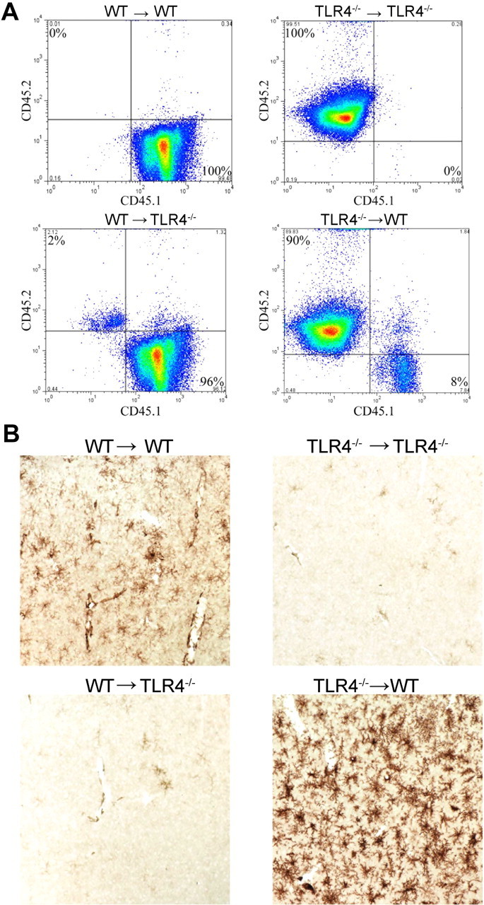
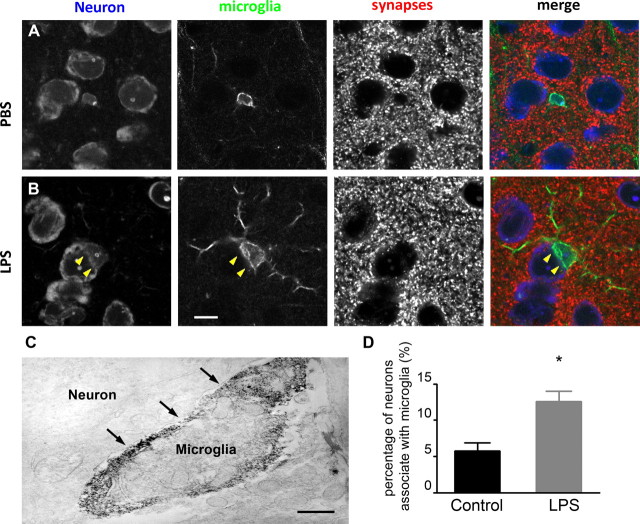
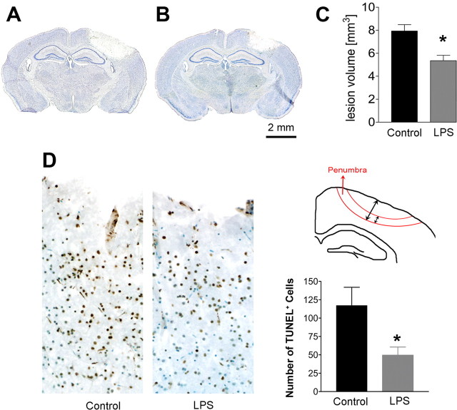
Intraperitoneal injection of the Gram-negative bacterial endotoxin lipopolysaccharide (LPS) elicits a rapid innate immune response. While this systemic inflammatory response can be destructive, tolerable low doses of LPS render the brain transiently resistant to subsequent injuries. However, the mechanism by which microglia respond to LPS stimulation and participate in subsequent neuroprotection has not been documented. In this study, we first established a novel LPS treatment paradigm where mice were injected intraperitoneally with 1.0 mg/kg LPS for four consecutive days to globally activate CNS microglia. By using a reciprocal bone marrow transplantation procedure between wild-type and Toll-like receptor 4 (TLR4) mutant mice, we demonstrated that the presence of LPS receptor (TLR4) is not required on hematogenous immune cells but is required on cells that are not replaced by bone marrow transplantation, such as vascular endothelia and microglia, to transduce microglial activation and neuroprotection. Furthermore, we showed that activated microglia physically ensheathe cortical projection neurons, which have reduced axosomatic inhibitory synapses from the neuronal perikarya. In line with previous reports that inhibitory synapse reduction protects neurons from degeneration and injury, we show here that neuronal cell death and lesion volumes are significantly reduced in LPS-treated animals following experimental brain injury. Together, our results suggest that activated microglia participate in neuroprotection and that this neuroprotection is likely achieved through reduction of inhibitory axosomatic synapses. The therapeutic significance of these findings rests not only in identifying neuroprotective functions of microglia, but also in establishing the CNS location of TLR4 activation.


Figures

Figure 1.
Figure 1.
Microglia in the brain parenchyma are globally activated by four daily intraperitoneal injections of 1.0 mg/kg LPS. A–E, Microglial morphology was visualized by DAB immunohistostaining with mouse-anti-Iba-1 antibody. The morphology of microglia was similar in LPS-injected animals 24 h after the first injection (B) to microglia in PBS-injected animals (A), both having small rounded cell bodies and long, fine processes. Twenty-four hours after 2 LPS injections, processes were retracted and thickened and cell bodies were expanded (C). Microglial activation was apparent 24 h after 4 LPS injections (D), as evidenced by thick cell bodies and asymmetric cell processes. Blue arrows denote cup-shaped microglia. Scale bar, 60 μm. E, The observed microglial activation was global throughout the brain parenchyma. Scale bar, 120 μm. F, Percentage of area occupied by microglia over the total area of interest was significantly higher in 4 daily LPS-treated mice than in PBS-treated mice (representative of three replicates). G, Microglial activation is further confirmed by F4/80 antibody, which stains activated but not resting microglia. The specificity of TLR4 in this process was confirmed by injecting LPS into TLR4-deficient mice. No microglial activation was detected in TLR4−/− mice, as indicted by a lack of F4/80 positivity. Scale bar, 60 μm. H, LPS treatment does not induce microglial proliferation. BrdU+ cells cannot be observed in the cerebral cortex. However, BrdU+ cells (red) are easily identifiable in the SVZ and dentate gyrus regions (images represent three replicates of two different approaches). Nuclei are counterstained in blue. Scale bar, 120 μm. ctx, Cortex; SVZ, subventricular zone; LV, lateral ventricle.
Figure 2.
Figure 2.
Multiple low-dose LPS treatment skews activated microglia toward an M2-like phenotype. Microarray analysis (n = 6/group) of cortical tissues shows that M2-related genes are significantly increased following LPS treatment, but that M1-related genes remain either unchanged or reduced when compared with PBS-treated controls.
Figure 3.
Figure 3.
Intraperitoneal LPS injections did not lead to infiltration of peripheral leukocytes into the CNS. A, Brain cells analyzed by flow cytometry displayed similar patterns of CD11b and CD45 expression between LPS- and saline-treated mice. B, The percentages of lymphocytes, monocytes/neutrophils, and microglia over total CD45+ cells were quantified and were not significantly different (n = 3/group).
Figure 4.
Figure 4.
Bone marrow chimera mice show that peripheral hematogenous TLR4 is not required for LPS-induced microglial activation. A, Blood cells taken from mice that underwent bone marrow transplant surgery were analyzed using flow cytometry and showed that most of cells in the WT→TLR4−/− chimera displayed the WT isoform of CD45 and vice versa. B, No microglial activation was seen in the TLR4−/−→TLR4−/− or the WT→TLR4−/− chimeras, as evidenced by the lack of DAB immunohistostaining using the anti-F4/80 antibody. Microglial activation was observed in the WT→WT chimera and was more robust in the TLR4−/−→WT chimera. Images represent at least 12 different chimerae.
Figure 5.
Figure 5.
Synapses cannot be seen where microglia appose neuronal cell bodies. A, In PBS-injected animals, microglia (green, Iba-1) are small and round with fine processes. Synaptic terminals (red, synaptophysin) are evenly distributed on the circumference of the neuronal perikarya (blue, Nissl). B, In LPS-injected animals, microglia have larger cell bodies, thicker processes, and can be observed partially ensheathing neuronal cell bodies. No synaptic staining is visible where microglia appose neuronal perikarya (arrowhead). Scale bar, 10 μm. C, Immunoelectron micrograph shows an Iba-1-positive microglia in direct contact with the plasma membrane of a neuronal cell body (arrows). Scale bar, 1 μm. D, The percentage of neurons in the cerebral cortex that are contacted by microglia is significantly increased following LPS injections compared with controls (n = 3/group). A–C represent at least three different experiments. *p < 0.05.
Figure 6.
Figure 6.
LPS-treated mice had a smaller lesion volume and reduced apoptotic cells following cryogenic injury. A–C, Mice treated with 4 daily injections of PBS or LPS underwent cryogenic injury and were killed 72 h postinjury. Serial sections were visualized with Giemsa stain (discoloration in A and B demarcates the lesions). LPS-treated animals (B) had a significantly smaller lesion volume than those treated daily with PBS (A) (n = 11/group). The lesion size was quantified in C. D, Significantly fewer TUNEL-positive cells (brown) were present in the penumbra region (illustrated) in LPS-treated animals compared with controls (n = 3/group). Data are expressed as mean ± SEM. *p < 0.05.

References

    1. Baughman VL, Hoffman WE, Miletich DJ, Albrecht RF, Thomas C. Neurologic outcome in rats following incomplete cerebral ischemia during halothane, isoflurane, or N2O. Anesthesiology. 1988;69:192–198. - PubMed
    1. Blinzinger K, Kreutzberg G. Displacement of synaptic terminals from regenerating motor neurons by microglial cells. Z Zellforsch Mikrosk Anat. 1968;85:145–157. - PubMed
    1. Chakravarty S, Herkenham M. Toll-like receptor 4 on nonhematopoietic cells sustains CNS inflammation during endotoxemia, independent of systemic cytokines. J Neurosci. 2005;25:1788–1796. - PMC - PubMed
    1. Chen J, Simon R. Ischemic tolerance in the brain. Neurology. 1997;48:306–311. - PubMed
    1. Ford AL, Goodsall AL, Hickey WF, Sedgwick JD. Normal adult ramified microglia separated from other central nervous system macrophages by flow cytometric sorting. Phenotypic differences defined and direct ex vivo antigen presentation to myelin basic protein-reactive CD4+ T cells compared. J Immunol. 1995;154:4309–4321. - PubMed

Publication types

MeSH terms

LinkOut - more resources