Review
. 2013 Mar;70(6):1067-79.
doi: 10.1007/s00018-012-1112-4. Epub 2012 Aug 23.

Modeling melanoblast development

Affiliations
Review

Modeling melanoblast development

Lionel Larue et al. Cell Mol Life Sci. 2013 Mar.

Abstract

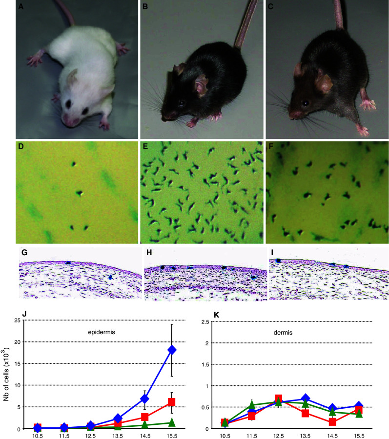
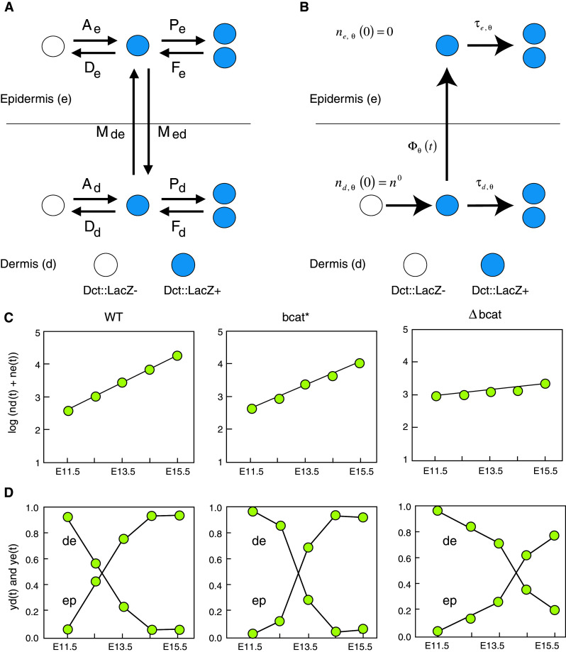
Melanoblasts are a particular type of cell that displays extensive cellular proliferation during development to contribute to the skin. There are only a few melanoblast founders, initially located just dorsal to the neural tube, and they sequentially colonize the dermis, epidermis, and hair follicles. In each compartment, melanoblasts are exposed to a wide variety of developmental cues that regulate their expansion. The colonization of the dermis and epidermis by melanoblasts involves substantial proliferation to generate thousands of cells or more from a few founders within a week of development. This review addresses the cellular and molecular events occurring during melanoblast development. We focus on intrinsic and extrinsic factors that control melanoblast proliferation. We also present a robust mathematical model for estimating the doubling-time of dermal and epidermal melanoblasts for all coat color phenotypes from black to white.


Figures

Fig. 1
Fig. 1
Melanoblast development in wt mice and β-catenin coat color mutants. ac β-catenin loss-of-function (Δbcat), wild-type (wt), and gain-of-function (bcat*) adult mice. (A, D, G: Δbcat), (B, E, H: wt) and (C, F, I: bcat*). Note that melanocyte-specific disruption of β-catenin (Δbcat) led to the complete absence of pigmentation, whereas a stabilized and nuclear form of β-catenin (bcat*) in melanocytes leads to a less intense coat color than controls (wt). df Macroscopic observations of the trunk region of Δbcat, wt, and bcat* E14.5 embryos. The melanoblasts, identified as X-gal-positive cells, are less abundant in both β-catenin mutants than in wt, and in Δbcat than in bcat*. gi Sections through the trunk of embryos at E13.5. Melanoblasts, the X-gal positive cells, are observed in both compartments of the skin: epidermis (ep) and dermis (de). jk The total number of epidermal and dermal melanoblasts in the trunk was estimated from E10.5 onwards. In the epidermis (j), the number of melanoblasts increased with time with the most substantial increase in wt mice and the smallest in Δbcat mice. In the dermis (k), the total number of melanoblasts remained low for all genotypes and was fairly constant from E11.5 onwards
Fig. 2
Fig. 2
Modeling melanoblast proliferation dynamics. a Theoretical scheme of melanoblast proliferation in the dermis and the epidermis. Melanoblasts, considered as Dct::LacZ-positive cells (Dct::LacZ+), may appear from Dct::LacZ-negative cells (Dct::LacZ−) in the dermis and the epidermis. The appearance (A) of Dct::LacZ-positive cells is associated with differentiation, transdifferentiation, or/and recruitment. The disappearance (D) of Dct::LacZ-positive cells is associated with transdifferentiation, loss of differentiation, and/or apoptosis. The cells may proliferate (P) or fuse from two cells to one cell (F). Finally, they can migrate from the dermis to the epidermis (M de) and in the opposite direction (M ed). b Mathematical scheme of melanoblast proliferation in the dermis and the epidermis based on biological evaluation. Melanoblasts (Dct::LacZ+) are determined from neural crest cells in the dermis and penetrate from the dermis into the epidermis (Φ(t) representing the flow of the melanoblasts crossing the basal membrane) and proliferate in the dermis and the epidermis (solid arrows). In theory, melanoblasts can lose their differentiation or transdifferentiate, undergo apoptosis, and therefore become Dct::LacZ− (white circle) in the epidermis and dermis, can migrate from the epidermis to the dermis and can fuse; however, these events seem unlikely and are not supported in any way by biological observation. The initial number of melanoblasts at E8.5 is represented by n d,θ(0) = n 0 for the dermis and n e,θ(0) = 0 for the epidermis. The mathematical model was designed to estimate melanoblast doubling times in the dermis τd(t) and epidermis τe(t) and the number of melanoblast founders at E8.5. cd Additional knowledge extracted from the data. The total number of melanoblasts n(t) = n d(t) + n e(t) increases exponentially and the progress of the fraction of melanoblasts in the dermis y d(t) through time is sigmoidal (or S-shaped). y e(t) represents the fraction of melanoblasts in the epidermis

References

    1. Lamoreux ML, Delmas V, Larue L, Bennett D. The colors of mice: a model genetic network. New York: Wiley; 2010.
    1. MacKenzie MA, Jordan SA, Budd PS, Jackson IJ. Activation of the receptor tyrosine kinase kit is required for the proliferation of melanoblasts in the mouse embryo. Dev Biol. 1997;192:99–107. doi: 10.1006/dbio.1997.8738. - DOI - PubMed
    1. Delmas V, Martinozzi S, Bourgeois Y, Holzenberger M, Larue L. Cre-mediated recombination in the skin melanocyte lineage. Genesis. 2003;36:73–80. doi: 10.1002/gene.10197. - DOI - PubMed
    1. Yajima I, Belloir E, Bourgeois Y, Kumasaka M, Delmas V, Larue L. Spatiotemporal gene control by the Cre-ERT2 system in melanocytes. Genesis. 2006;44:34–43. doi: 10.1002/gene.20182. - DOI - PubMed
    1. Le Douarin N, Kalcheim C. The neural crest. Cambridge: Cambridge University Press; 1999.

Publication types

LinkOut - more resources