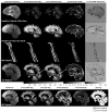
One component? Two components? Three? The effect of including a nonexchanging "free" water component in multicomponent driven equilibrium single pulse observation of T1 and T2
- PMID: 22915316
- PMCID: PMC3711852
- DOI: 10.1002/mrm.24429
One component? Two components? Three? The effect of including a nonexchanging "free" water component in multicomponent driven equilibrium single pulse observation of T1 and T2
Abstract
Quantitative myelin content imaging provides novel and pertinent information related to underlying pathogenetic mechanisms of myelin-related disease or disorders arising from aberrant connectivity. Multicomponent driven equilibrium single pulse observation of T1 and T2 is a time-efficient multicomponent relaxation analysis technique that provides estimates of the myelin water fraction, a surrogate measure of myelin volume. Unfortunately, multicomponent driven equilibrium single pulse observation of T1 and T2 relies on a two water-pool model (myelin-associated water and intra/extracellular water), which is inadequate within partial volume voxels, i.e., containing brain tissue and ventricle or meninges, resulting in myelin water fraction underestimation. To address this, a third, nonexchanging "free-water" component was introduced to the multicomponent driven equilibrium single pulse observation of T1 and T2 model. Numerical simulations and experimental in vivo data show that the model to perform advantageously within partial volume regions while providing robust and reproducible results. It is concluded that this model is preferable for future studies and analysis.
Copyright © 2012 Wiley Periodicals, Inc.
Figures


Similar articles
-
Comparison of myelin water fraction from multiecho T2 decay curve and steady-state methods.Magn Reson Med. 2015 Jan;73(1):223-32. doi: 10.1002/mrm.25125. Epub 2014 Feb 11. Magn Reson Med. 2015. PMID: 24515972
-
Investigating the stability of mcDESPOT myelin water fraction values derived using a stochastic region contraction approach.Magn Reson Med. 2015 Jan;73(1):161-9. doi: 10.1002/mrm.25108. Epub 2014 Jan 24. Magn Reson Med. 2015. PMID: 24464472 Free PMC article.
-
Brain and cord myelin water imaging: a progressive multiple sclerosis biomarker.Neuroimage Clin. 2015 Oct 3;9:574-80. doi: 10.1016/j.nicl.2015.10.002. eCollection 2015. Neuroimage Clin. 2015. PMID: 26594633 Free PMC article.
-
MRI-based myelin water imaging: A technical review.Magn Reson Med. 2015 Jan;73(1):70-81. doi: 10.1002/mrm.25198. Epub 2014 Mar 6. Magn Reson Med. 2015. PMID: 24604728 Review.
-
Multispectral quantitative magnetic resonance imaging of brain iron stores: a theoretical perspective.Top Magn Reson Imaging. 2006 Feb;17(1):19-30. doi: 10.1097/01.rmr.0000245460.82782.69. Top Magn Reson Imaging. 2006. PMID: 17179894 Review.
Cited by
-
Longitudinal white matter and cognitive development in pediatric carriers of the apolipoprotein ε4 allele.Neuroimage. 2020 Nov 15;222:117243. doi: 10.1016/j.neuroimage.2020.117243. Epub 2020 Aug 18. Neuroimage. 2020. PMID: 32822813 Free PMC article.
-
A Nutrient Formulation Affects Developmental Myelination in Term Infants: A Randomized Clinical Trial.Front Nutr. 2022 Feb 10;9:823893. doi: 10.3389/fnut.2022.823893. eCollection 2022. Front Nutr. 2022. PMID: 35242798 Free PMC article.
-
Longitudinal associations between white matter maturation and cognitive development across early childhood.Hum Brain Mapp. 2019 Oct 1;40(14):4130-4145. doi: 10.1002/hbm.24690. Epub 2019 Jun 12. Hum Brain Mapp. 2019. PMID: 31187920 Free PMC article.
-
Myelin water imaging using a short-TR adiabatic inversion-recovery (STAIR) sequence.Magn Reson Med. 2022 Sep;88(3):1156-1169. doi: 10.1002/mrm.29287. Epub 2022 May 25. Magn Reson Med. 2022. PMID: 35613378 Free PMC article.
-
High-resolution quantitative MRI of multiple sclerosis spinal cord lesions.Magn Reson Med. 2022 Jun;87(6):2914-2921. doi: 10.1002/mrm.29152. Epub 2022 Jan 11. Magn Reson Med. 2022. PMID: 35014736 Free PMC article.
References
-
- MacKay A, Laule C, Vavsour I, Bjarnason T, Kolling S, Madler B. Insights into Brain Microstructure from the T2 Distribution. Magn Reson Imag. 2006;24:515–525. - PubMed
-
- Laule C, Leung E, Lis DK, Traboulsee AL, Paty DW, MacKay AL, Moore GR. Myelin Water Imaging in Multiple Sclerosis: Quantitative Correlations with Histopathology. Mult Scler. 2006;12:747–753. - PubMed
-
- Webb S, Munro CA, Midha R, Stanisz GJ. Is Multicomponent T2 a Good Measure of Myelin Content in Peripheral Nerve? Magn Reson Med. 2003;49:638–645. - PubMed
-
- Laule C, Vavasour IM, Leung E, Li DK, Kozlowski P, Traboulsee AL, Oger J, MacKay AL, Moore GR. Pathological Basis of Diffusely Abnormal White Matter: Insights from Magnetic Resonance Imaging and Histology. Mult Scler. 2011;17:144–150. - PubMed
-
- Kolind SH, Laule C, Vavasour IM, Li DK, Traboulsee AL, Madler B, Moore GR, MacKay AL. Complementary Information from Multi-Exponential T2 Relaxation and Diffusion Tensor Imaging Reveals Differences Between Multiple Sclerosis Lesions. NeuroImage. 2008;40:77–85. - PubMed
Publication types
MeSH terms
Grants and funding
LinkOut - more resources
Full Text Sources
Medical

