. 2012 Oct 12;287(42):35382-35396.
doi: 10.1074/jbc.M112.409797. Epub 2012 Aug 22.

Anti-apoptotic role of caspase-cleaved GAB1 adaptor protein in hepatocyte growth factor/scatter factor-MET receptor protein signaling

Affiliations

Anti-apoptotic role of caspase-cleaved GAB1 adaptor protein in hepatocyte growth factor/scatter factor-MET receptor protein signaling

Arnaud Le Goff et al. J Biol Chem. .

Abstract

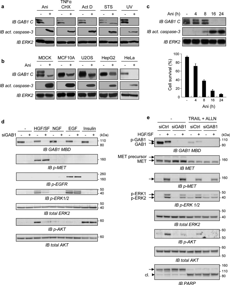
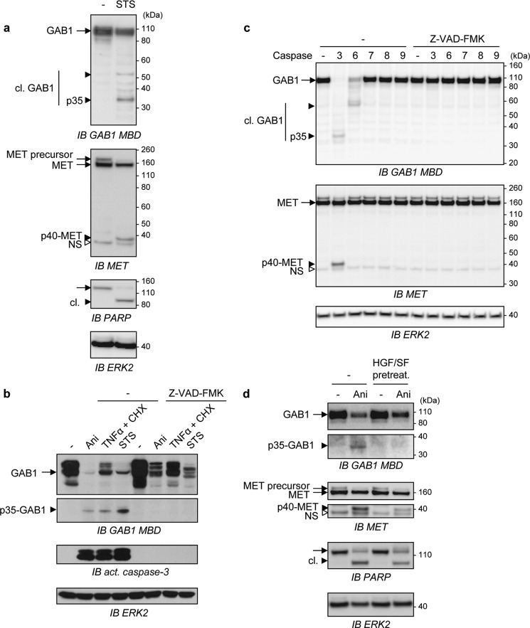
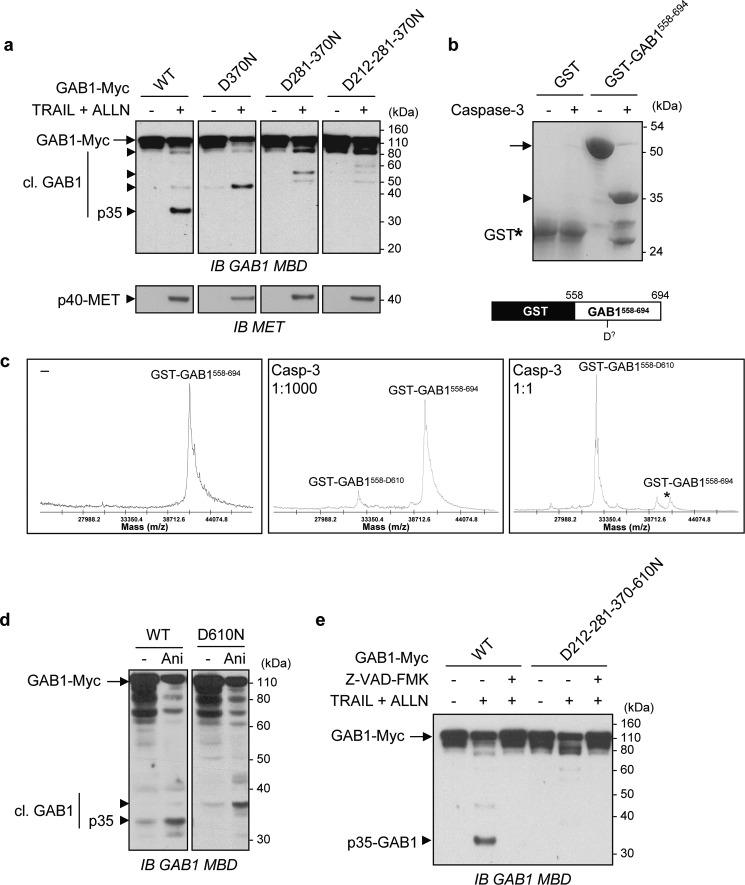
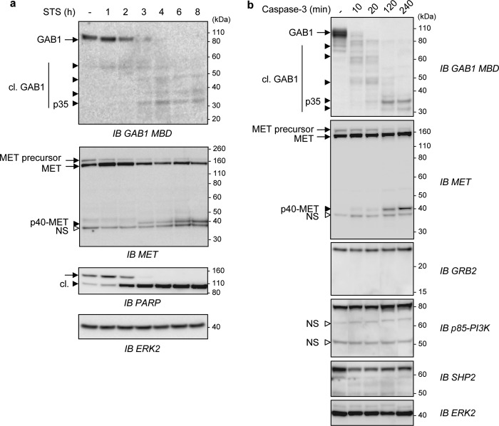
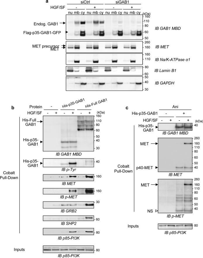
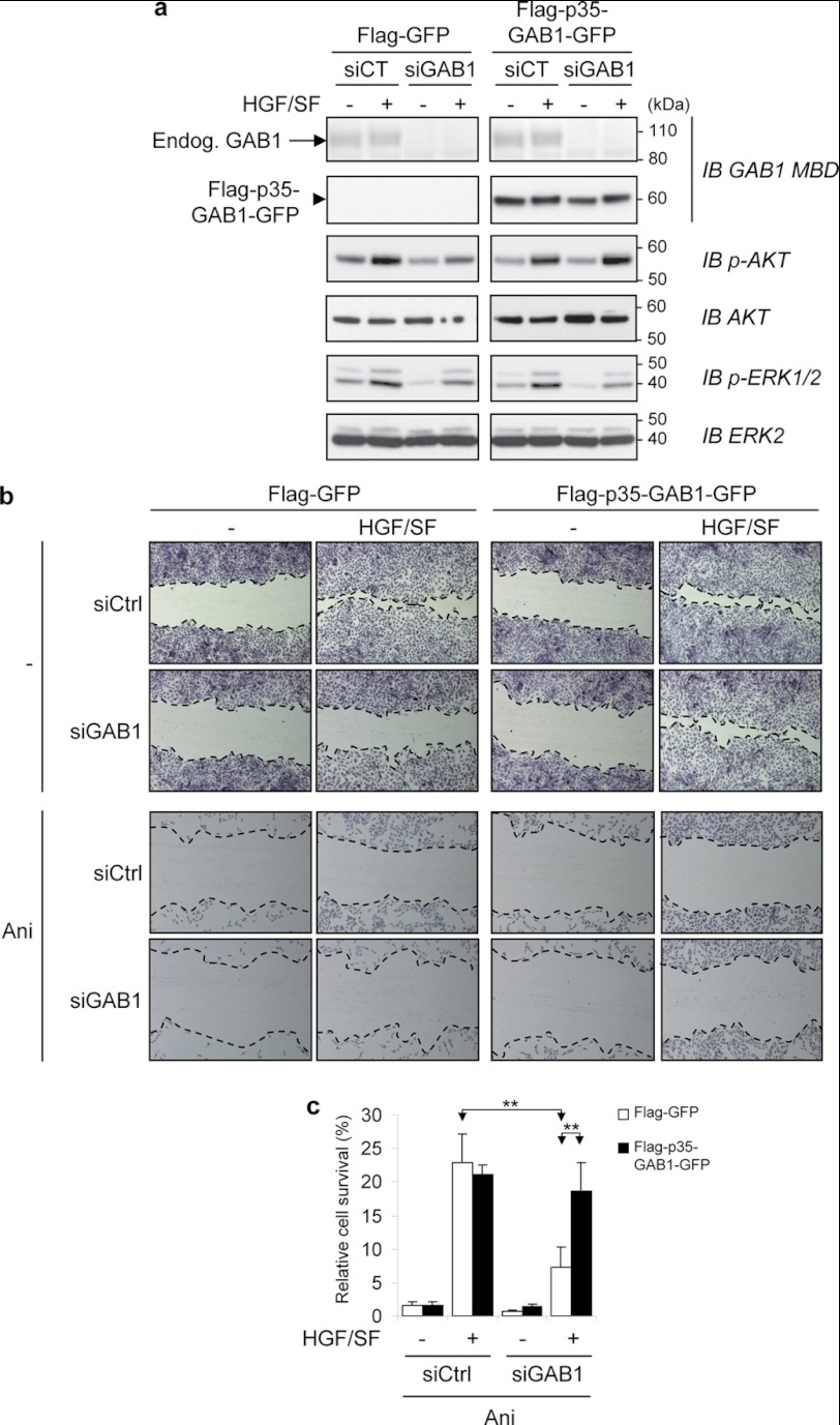
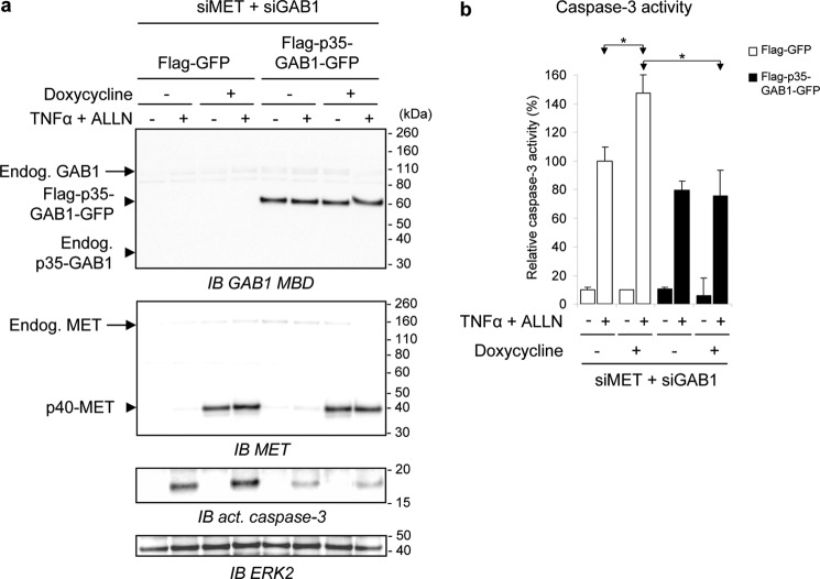
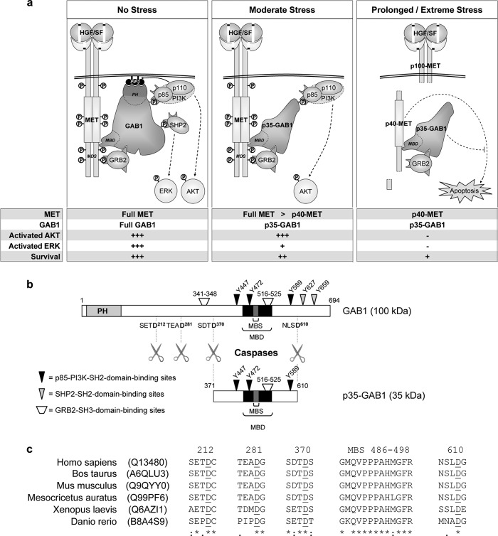
The GRB2-associated binder 1 (GAB1) docking/scaffold protein is a key mediator of the MET-tyrosine kinase receptor activated by hepatocyte growth factor/scatter factor (HGF/SF). Activated MET promotes recruitment and tyrosine phosphorylation of GAB1, which in turn recruits multiple proteins and mediates MET signaling leading to cell survival, motility, and morphogenesis. We previously reported that, without its ligand, MET is a functional caspase target during apoptosis, allowing the generation of a p40-MET fragment that amplifies apoptosis. In this study we established that GAB1 is also a functional caspase target by evidencing a caspase-cleaved p35-GAB1 fragment that contains the MET binding domain. GAB1 is cleaved by caspases before MET, and the resulting p35-GAB1 fragment is phosphorylated by MET upon HGF/SF binding and can interact with a subset of GAB1 partners, PI3K, and GRB2 but not with SHP2. This p35-GAB1 fragment favors cell survival by maintaining HGF/SF-induced MET activation of AKT and by hindering p40-MET pro-apoptotic function. These data demonstrate an anti-apoptotic role of caspase-cleaved GAB1 in HGF/SF-MET signaling.


Figures

FIGURE 1.
FIGURE 1.
GAB1 down-expression by either stress conditions or by siGAB1 knockdown contributes to inhibition of HGF/SF-MET downstream signaling. a, MDCK cells were cultured in medium, 0% FBS for 8 h in the absence (−) or presence (+) of different apoptotic inducers: anisomycin (Ani, 50 μm), a combination of TNFα (30 ng/ml) and cycloheximide (CHX, 10 μg/ml), actinomycin D (ActD, 5 μg/ml), or staurosporine (STS, 1 μm). For UV treatment, cells were exposed to UV-B light 400 J/m2 for 2.5 min and then cultured in medium-0% FBS for an additional 8 h. Whole-cell lysates were immunoblotted (IB) using the anti-C terminus GAB1 antibody (GAB1 C) and an anti-active caspase-3 antibody. The filter was stripped and reprobed using an anti-ERK2 antibody to verify comparable loading. GAB1 was detected as a doublet that may correspond to the known A and B isoforms of GAB1 produced by alternative splicing. b, MDCK, MCF10A, U2OS, HepG2, or HeLa cells were treated for 8 h without (−) or with (+) anisomycin (Ani, 50 μm) in medium-0% FBS. Whole-cell lysates were immunoblotted using antibodies, as described for panel a. c, upper panel, MDCK cells were treated with anisomycin (Ani) or left untreated as described above for the indicated times in hours. Cell lysates were collected and then analyzed by immunoblotting using the same antibodies as described for panel a (IB act. caspase-3). Lower panel, the percentage of cell survival was determined by trypan blue staining. d, HeLa cells were transiently transfected with either siRNA negative control (−) or siRNA targeting the endogenous Gab1 transcript (siGab1, +). The next day cells were cultured in medium, 0.1% FBS for 6 h and subsequently treated with either HGF (50 ng/ml, 10 min), NGF (100 ng/ml, 10 min), EGF (10 ng/ml, 5 min), or insulin (0.1 μm, 3 min) or left untreated for 10 min. Cell lysates were then subjected to immunoblot analysis using an anti-GAB1 middle antibody (GAB1 M) directed against the residues surrounding Tyr-472 of GAB1. The same blot was stripped and reprobed several times using the indicated antibodies. e, HeLa cells were transiently transfected with either siRNA negative control (siCtrl) or siRNA targeting the endogenous GAB1 transcript (siGAB1). The next day cells were cultured overnight in medium, 0.1% FBS and subsequently treated with TRAIL (30 ng/ml) in combination with the proteasome inhibitor ALLN (50 μm) or left untreated (−) for 4 h. Cells were then treated (+) or not (−) with HGF/SF (50 ng/ml) for 10 min. Cell lysates were subjected to immunoblot analysis using an antibody directed against the GAB1-MBD (IB GAB1-MBD)). The same blot was stripped and reprobed several times using the indicated antibodies. Detection of PARP cleavage (cl.) was assessed to monitor caspases activation.
FIGURE 2.
FIGURE 2.
Stress conditions cause the generation of a stable fragment of GAB1 and this caspase-dependent mechanism is hindered by HGF/SF. a, MDCK cells were treated with staurosporine (STS, 1 μm) or left untreated (−) for 8 h and then lysed. Immunoblot analysis (IB) was performed on cell lysates, and the membrane was probed using anti-GAB1 MBD or anti-MET antibodies. The filter was stripped and reprobed with anti-PARP antibody and anti-ERK2 antibody. b, MDCK cells were cultured in medium, 0% FBS and pretreated without (−) or with the general caspase inhibitor Z-VAD-FMK (20 μm) for 30 min followed by a treatment with either anisomycin (Ani, 50 μm), a combination of TNFα (30 ng/ml) and cycloheximide (CHX, 10 μg/ml), or staurosporine (STS, 2 μm) for 16 h. Cell lysates were analyzed by immunoblotting using the indicated antibodies. c, MDCK cells were lysed, and the same amount of protein for each sample was incubated without (−) or with active purified caspase-3, -6, -7, -8, or -9 at 37 °C for 4 h in the absence (−) or presence of the general caspase inhibitor Z-VAD-FMK (40 μm). Samples were then analyzed by immunoblotting using the anti-GAB1 MBD or the anti-MET antibodies. The filter was stripped and reprobed using an anti-ERK2 antibody to verify comparable loading. d, MDCK cells were pretreated with HGF/SF (HGF/SF pretreat., 10 ng/ml) or left untreated (−) for 1 h and incubated for an additional 10 h with or without (−) anisomycin (Ani, 50 μm). Cell lysates were analyzed by immunoblotting using the indicated antibodies. a-c, arrows point to full-length (black arrow) and cleaved (black arrowhead) proteins. The small white arrowhead corresponds to a nonspecific immunoreactive band (NS). Cleaved proteins are abbreviated as cl.
FIGURE 3.
FIGURE 3.
The p35-GAB1 fragment is surrounded by four caspase cleavage sites and contains the MET binding domain. a, after transient transfection for 24 h using Myc-tagged wild type (WT) GAB1 or its mutated versions D370N, D281N/D370N, or D212–281-370N, HeLa cells were cultured overnight in medium, 0.1% FBS. Cells were then treated (+) with TRAIL (30 ng/ml) in combination with the proteasome inhibitor ALLN (50 μm) or left untreated (−) for 5 h. For each condition, the same amount of whole-cell extracts was analyzed by immunoblotting (IB) using anti-GAB1 MBD or anti-MET antibodies. b, purified recombinant GST and GST-GAB1558–694 proteins were incubated with (+) or without (−) recombinant active caspase-3 at 37 °C for 1 h. Products were then separated by SDS-PAGE and stained using Coomassie Blue (upper panel). Schematic representation of the GST-GAB1558–694 structure is also shown on the lower panel. c, purified recombinant GST-GAB1558–694 protein was analyzed on a MALDI-TOF spectrometer before (left panel) and after cleavage with caspase-3 at a low (1:100 dilution, middle panel) or high concentration (1:1 dilution, right panel) at 37 °C for 1 h. Experimentally determined masses of GST-GAB1 and the main cleavage product are indicated. d, MDCK cells were transfected for 24 h with Myc-tagged wild type (WT) GAB1 or its mutant D610N. Cells were then treated with anisomycin (Ani, 50 μm) or left untreated (−) for 8 h. Cell lysates were collected and analyzed by immunoblotting using an anti-GAB1 MBD antibody. e, after transient transfection for 24 h with Myc-tagged wild type (WT) GAB1 or its mutated version D212N/D281N/D370N/D610N, HeLa cells were cultured overnight in medium, 0.1% FBS. Cells were then left untreated (−) or pretreated (+) with Z-VAD-FMK (20 μm) for 30 min immediately followed by an additional 5-h incubation with (+) or without (−) TRAIL and ALLN (as described in panel a). Immunoblot analysis was then performed on whole-cell lysates using an anti-GAB1 MBD antibody. a–e, arrows point to full-length (black arrow) and cleaved (black arrowhead) proteins. Cleaved GAB1 is abbreviated as cl. GAB1.
FIGURE 4.
FIGURE 4.
Both MET and GAB1 are degraded and fragmented by stress or by caspase 3, with the GAB1 adaptor being caspase-cleaved faster and more progressively than the MET receptor. a, HeLa cells were left untreated (−) or treated with staurosporine (STS, 1 μm) for the indicated times in hours. Cell lysates were then immunoblotted (IB) using the indicated antibodies. b, lysates from MDCK cells were incubated without (−) or with active purified caspase-3 at 37 °C for the indicated times in minutes. Samples were then analyzed by immunoblotting using the indicated antibodies. a and b, arrows point to full-length (black arrow) and cleaved (black arrowhead) proteins. The small white arrowhead corresponds to a nonspecific immunoreactive band (NS). Cleaved proteins are abbreviated as cl.
FIGURE 5.
FIGURE 5.
Subcellular localization and association capacities of the p35-GAB1 fragment. a, MDCK cells stably expressing FLAG-p35-GAB1-GFP proteins were transiently transfected with siRNA control (siCtrl) or targeting endogenous GAB1 transcripts (siGAB1). The next day cells were grown overnight in medium, 0.1% FBS and subsequently treated with HGF/SF (50 ng/ml) for 10 min (+) or left untreated (−). Cell lysates were sequentially fractionated in subcellular compartments (nu, nucleus; mb, membrane; cy, cytosol) and subjected to immunoblot (IB) analysis using GAB1 or MET antibodies as well as antibodies directed against classical compartment markers (Na+/K+-ATPase, Lamin B1, and GAPDH). b and c, MDCK cells were cultured overnight in medium, 0.1% FBS in the absence (b) or presence (c) of anisomycin (Ani, 50 μm) and were then left untreated (−) or treated (+) with HGF/SF (100 ng/ml) for 5 min. Cell lysates were prepared and incubated without (−) or with purified recombinant His-tagged p35-GAB1 (His-p35-GAB1) or full-length GAB1 (His-Full GAB1) proteins. Pulldown assays were performed, and eluates were analyzed by immunoblotting using the indicated antibodies. Total cell lysates before pull down (Inputs) were also loaded and analyzed (supplemental Fig. S4, b and c). In c, the white arrowhead corresponds to a nonspecific immunoreactive band (NS).
FIGURE 6.
FIGURE 6.
In response to HGF/SF, the p35-GAB1 fragment maintains AKT activation and cell migration and allows resistance to apoptotic stress. a, MDCK cells stably expressing FLAG-GFP or FLAG-p35-GAB1-GFP proteins were transiently transfected with either siRNA negative control (siCT) or siRNA targeting the endogenous GAB1 transcript (siGAB1). The next day cells were grown overnight in medium, 0.1% FBS and subsequently treated with HGF/SF (50 ng/ml) for 10 min (+) or left untreated (−). Cell lysates were subjected to immunoblot (IB) analysis using the indicated antibodies. Note that expression of endogenous GAB1 (Endog. GAB1) was faint compared with FLAG-p35-GAB1-GFP. b, MDCK cells stably expressing FLAG-GFP or FLAG-p35-GAB1-GFP protein were transiently transfected with either siRNA negative control (siCtrl) or siRNA targeting the endogenous GAB1 transcript (siGAB1). The next day a scratch wound-healing assay was performed. After 24 h of incubation without (−) or with HGF/SF (30 ng/ml) in medium, 0.1% FBS, cells were stained using Hemacolor kit, and the wound closure areas were visualized under an inverted microscope (×40). Dotted lines were added to delineate the migrating cell front. Note that the width of scratched wounds was comparable between samples just before incubation of HGF/SF. c, MDCK cells stably expressing FLAG-GFP or FLAG-p35-GAB1-GFP proteins were transiently transfected with siRNA as described above. The next day cells were treated for 16 h with or without anisomycin (Ani, 1 μm) in medium-0.1% FBS in the absence (−) or presence (+) of HGF/SF (30 ng/ml). An MTT assay was performed to evaluate cell survival. Results are expressed as a percentage of untreated control cells. All values are shown as the mean ± S.D. of three independent MTT assays, each condition measured in triplicate (**, p < 0.01 using Student's t test with Bonferroni correction).
FIGURE 7.
FIGURE 7.
The p35-GAB1 fragment hinders amplification of caspase 3 activity by the p40-MET fragment. MDCK-p40-MET-Tet-On cells stably expressing FLAG-GFP or FLAG-p35-GAB1-GFP proteins were transiently transfected with siRNAs targeting both endogenous GAB1 and MET transcripts (siMET + siGAB1). After 48 h of cell culture in a complete medium, cells were starved overnight in medium, 0.1% FBS with (+) or without (−) doxycycline (0.2 μg/ml) and subsequently treated with TNFα (30 ng/ml) and ALLN (50 μm) for 4 h (+) or left untreated (−). Cell lysates were subjected to immunoblot analysis (IB) using indicated antibodies (a), or caspase-3 activity assay was performed from an independent experiment (b) (*, p < 0.05 using Student's t test with Bonferroni correction). Similar results were obtained in distinct experiments.
FIGURE 8.
FIGURE 8.
The GAB1 adaptor is a checkpoint regulator of HGF/SF-MET signaling under stress conditions. a, shown is a model for the regulatory role of the GAB1 adaptor and its caspase-cleaved p35-GAB1 fragment in HGF/SF-MET signaling under distinct stress conditions (see “Discussion”). Full MET or Full GAB1 stands for full-length MET or full-length GAB1, respectively; p40-MET and p35-GAB1 stands for the caspase-dependent fragment of MET and GAB1, respectively; +++, strong response to HGF/SF; +, decreased response to HGF/SF; −, no response. b, shown is a schematic representation of the presently identified four caspase cleavage sites in the GAB1 adaptor and main signaling partners binding sites for either full-length GAB1 protein or the p35-GAB1 fragment. Specific aa in GAB1 are indicated corresponding in particular to phosphorylated tyrosine residues known to be involved in binding of SH2 domain-containing proteins. The apparent molecular weights of GAB1 and p35-GAB1 in SDS gels are indicated. c, sequence alignment shows conservation of the GAB1-MBS and the presently identified caspase cleavage sites in GAB1 from various species. All sequences were retrieved from the UniProt database, and UniProt entries are indicated. Numbering is indicated according to the human sequence. Aspartate sites are underlined. See also the full sequence alignment of various species in supplemental Fig. S5.

Similar articles

Cited by

References

    1. Schlessinger J. (2000) Cell signaling by receptor-tyrosine kinases. Cell 103, 211–225 - PubMed
    1. Pawson T., Scott J. D. (1997) Signaling through scaffold, anchoring, and adaptor proteins. Science 278, 2075–2080 - PubMed
    1. Buday L., Tompa P. (2010) Functional classification of scaffold proteins and related molecules. FEBS J. 277, 4348–4355 - PubMed
    1. Holgado-Madruga M., Emlet D. R., Moscatello D. K., Godwin A. K., Wong A. J. (1996) A Grb2-associated docking protein in EGF and insulin receptor signaling. Nature 379, 560–564 - PubMed
    1. Weidner K. M., Di Cesare S., Sachs M., Brinkmann V., Behrens J., Birchmeier W. (1996) Interaction between Gab1 and the c-Met receptor-tyrosine kinase is responsible for epithelial morphogenesis. Nature 384, 173–176 - PubMed

Publication types

MeSH terms