. 2012 Oct 15;72(20):5209-18.
doi: 10.1158/0008-5472.CAN-12-1187. Epub 2012 Aug 20.

PD-1 blockade enhances T-cell migration to tumors by elevating IFN-γ inducible chemokines

Affiliations

PD-1 blockade enhances T-cell migration to tumors by elevating IFN-γ inducible chemokines

Weiyi Peng et al. Cancer Res. .

Abstract

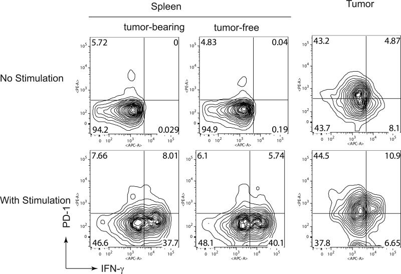
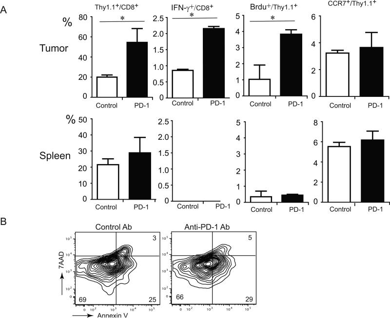
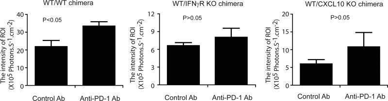
Adoptive cell transfer (ACT) is considered a promising modality for cancer treatment, but despite ongoing improvements, many patients do not experience clinical benefits. The tumor microenvironment is an important limiting factor in immunotherapy that has not been addressed fully in ACT treatments. In this study, we report that upregualtion of the immunosuppressive receptor programmed cell death-1 (PD-1) expressed on transferred T cells at the tumor site, in a murine model of ACT, compared with its expression on transferred T cells present in the peripheral blood and spleen. As PD-1 can attenuate T-cell-mediated antitumor responses, we tested whether its blockade with an anti-PD-1 antibody could enhance the antitumor activity of ACT in this model. Cotreatment with both agents increased the number of transferred T cells at the tumor site and also enhanced tumor regressions, compared with treatments with either agent alone. While anti-PD-1 did not reduce the number of immunosuppressive regulatory T cells and myeloid-derived suppressor cells present in tumor-bearing mice, we found that it increased expression of IFN-γ and CXCL10 at the tumor site. Bone marrow-transplant experiments using IFN-γR-/- mice implicated IFN-γ as a crucial nexus for controlling PD-1-mediated tumor infiltration by T cells. Taken together, our results imply that blocking the PD-1 pathway can increase IFN-γ at the tumor site, thereby increasing chemokine-dependent trafficking of immune cells into malignant disease sites.


Figures

Figure 1
Figure 1
The expression of PD-1 by transferred T cells at the tumor and spleen of mice receiving ACT treatment. Mice with or without 7-day established B16 tumor were transferred with cultured T cells from pmel-1 TCR/Thy1.1 transgenic mice. Six days after T-cell transfer, single cell suspensions were obtained from spleens and tumors and stimulated with gp100-peptide pulsed DC in the presence of Golgi stop for 4 hours. Lymphocytes with or without stimulation were evaluated by flow cytometry for PD-1 versus IFN-γ after gating on CD8+ and Thy1.1+. Number indicates the percentage of cells showing in each quadrant. The flow data was obtained from pooled lymphocyte samples from 5 mice in each group. Two independent studies showed similar results.
Figure 2
Figure 2
Increased accumulation of pmel-1 T cells to tumor sites and enhanced anti-tumor immune response in mice receiving ACT combined with anti-PD-1 antibody treatment. (A) Schematic representation of treatment schedule. (B) In vivo trafficking of transferred pmel-1 T cells. Luciferase-expressing pmel-1 T cells (1 × 10⁶) were transferred into mice bearing established 7-day MC38/gp100 tumors. DC vaccine and IL-2 treatment were performed as previously described. Mice were intraperitoneally injected with 250μg either control antibody or anti-PD-1 Ab on days 0, 2 and 4 after T cell transfer. Imaging was performed on day 6 after T cell transfer. Data shown were from representative mice. (C) Quantitative imaging analysis of transferred T cells in tumor-bearing mice. Intensities of the luciferase signal at tumor sites in all tumor-bearing mice are depicted(N=4 per group). (D) Tumor growth curve of MC38/gp100 tumor-bearing mice receiving anti-PD-1 Ab with or without adoptive T cell transfer (N=5 per group). (E) Tumor growth curve of B16 tumor-bearing mice receiving anti-PD-1 Ab with or without adoptive T cell transfer (N=5 per group).
Figure 3
Figure 3
The phenotype and function of transferred pmel-1 T cells in mice receiving ACT and anti-PD-1 Ab treatment. (A) Change of frequency and function of transferred pmel-1 T cells within tumor and spleen in response to PD-1 blockade. Six-days after T cell transfer, B16-bearing mice were intraperitoneally treated with Brdu solution. 24-hours later, mice were sacrificed to harvest tumor tissue and spleen (N=3 per group). Single cell suspension was made from tumor and spleen and stained with anti-CD8, anti-Thy1.1, anti-CCR7 anti-IFN-γ, and anti-Brdu. (B) Apoptosis of transferred pmel-1 T cells at the tumor site. Six-days after T cell transfer, lymphocytes from tumor tissues were stained with anti-CD8, anti-Thy1.1, Annexin V and 7AAD. Representative contour plots for one tissue sample from mice treated with ACT and anti-PD-1, as well as one tissue sample from mice treated with ACT with control antibody, were shown after gating with anti-Thy1.1+ and anti-CD8+ subsets. Values in each quadrant indicate the percentage of cells in the corresponding quadrant. (* indicates P<0.05)
Figure 4
Figure 4
Change in chemokine and cytokine expression within tumors from mice receiving ACT in response to PD-1 blockade. (A) Chemokine mRNA levels within the tumor site from mice infused with pmel-1 T cells with or without PD-1 blockade. Mice challenged with 5×10⁵ B16 cells received pmel-1 T cells on day 7, then treated with either anti-PD-1 antibody or control antibody on days 7, 9 and 11 and sacrificed on day 13. RNA was isolated from the tumor tissues of mice (N=3 for each group). The expression levels of chemokine within tumors were analyzed by realtime PCR. (B) Representative plot of CXCL10-producing cells within tumor tissue. (C) Representative histogram plots of CXCL10 production within tumor tissue from mice treated with ACT and anti-PD-1, as well as from mice treated with ACT with control antibody, were shown after gating with CD11b+ subsets. (D) Percentage of CXCL10-producing cells in CD11b+ cells within tumor tissue from mice treated with ACT and anti-PD-1, as well as from mice treated with ACT with control antibody (N=3-5 per group). (E) Cytokine mRNA levels within the tumor site from mice infused with pmel-1 T cells with or without PD-1 blockade. (** indicates P<0.01)
Figure 5
Figure 5
Failure to enhance anti-tumor immune response by anti-PD-1 antibody in mice with IFN-γ receptor deficient or CXCL10 deficient in BM derived cells receiving ACT therapy. (A) Frequency of Thy1.1+ in CD8+ cells within the tumor site in IFN-γ receptor deficient mice treated with ACT. Wild-type or IFN-γ receptor deficient mice were challenged with 5×10⁵ tumor cells and infused with pmel-1 T cells on day 7, treated with either anti-PD-1 antibody or control antibody on days 7, 9 and 11, and then sacrificed on day 13. Single cell suspensions were made from tumor and stained with anti-CD8, anti-Thy1.1. Data shown were from representative mice. (B) Frequency of Thy1.1+ in CD8+ cells within the tumor site in BM chimera mice treated with ACT. IFN-γ receptor deficient mice received bone marrow either from wild-type mice or IFN-γ receptor deficient mice 16-hours after 1000 rad irradiation. Eight weeks after BM transfer, the BM chimera mice were challenged with tumor and received pmel-1 T cell as previously described. Anti-PD-1 or control antibody was intraperitoneally injected on days 0, 2 and 4 after T cell transfer. The percentage of transferred pmel-1 T cells at the tumor site was analyzed on day 6 after T cell transfer. (C) Tumor growth curve of MC38/gp100 tumor-bearing BM chimera mice receiving ACT with control Ab or anti-PD-1 antibody(N=3 for each group). Wild-type female mice received bone marrow from wild-type mice, IFN-γ receptor deficient mice, or CXCL10-deficient mice. Eight weeks after BM transfer, the BM chimera mice were challenged with MC38/gp100 tumor and received pmel-1 T cell as previously described.
Figure 6
Figure 6
Failure to increase accumulation of pmel-1 T cells to tumor sites by anti-PD-1 in mice with IFN-γ receptor deficient or CXCL10 deficient in BM-derived cells receiving ACT therapy. BM chimera mice (N=3 for each group) were challenged with MC38/gp100 tumor and transferred with luciferase expressing pmel-1 T cells. Mice were intraperitoneally injected with 250μg either control antibody or anti-PD-1 Ab on days 0, 2 and 4 after T cell transfer. In vivo luciferase imaging was performed on day 6 after T cell transfer. Intensities of the luciferase signal at tumor sites in all tumor-bearing mice are depicted .

References

    1. Peddareddigari VR, Miller PW, Nebiyou BB, Overwijk WW, Ross MI, Lee JE, et al. Effect of clinical status and previous treatment of melanoma patients on expansion of TIL for adoptive T-cell therapy. ASCO Meeting Abstracts. 2009;27:3021.
    1. Pule MA, Savoldo B, Myers GD, Rossig C, Russell HV, Dotti G, et al. Virus-specific T cells engineered to coexpress tumor-specific receptors: persistence and antitumor activity in individuals with neuroblastoma. Nat Med. 2008;14:1264–70. - PMC - PubMed
    1. Porter DL, Levine BL, Kalos M, Bagg A, June CH. Chimeric antigen receptor-modified T cells in chronic lymphoid leukemia. N Engl J Med. 365:725–33. - PMC - PubMed
    1. Laszlo G, Radvanyi CB, Minying Zhang, Priscilla Miller MG, Nicholas Papadopoulos, Patrick Hwu. Adoptive T Cell Therapy for Metastatic Melanoma: The MD Anderson Experience. Journal of Immunotherapy. 2010;33:863.
    1. Fisher B, Packard BS, Read EJ, Carrasquillo JA, Carter CS, Topalian SL, et al. Tumor localization of adoptively transferred indium-111 labeled tumor infiltrating lymphocytes in patients with metastatic melanoma. J Clin Oncol. 1989;7:250–61. - PubMed

Publication types

MeSH terms