. 2012 Nov;343(2):479-88.
doi: 10.1124/jpet.112.195446. Epub 2012 Aug 22.

Vinpocetine suppresses pathological vascular remodeling by inhibiting vascular smooth muscle cell proliferation and migration

Affiliations

Vinpocetine suppresses pathological vascular remodeling by inhibiting vascular smooth muscle cell proliferation and migration

Yujun Cai et al. J Pharmacol Exp Ther. 2012 Nov.

Abstract

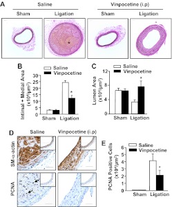
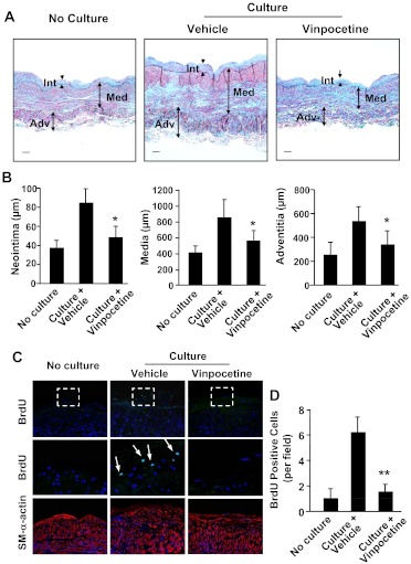
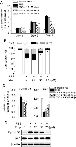
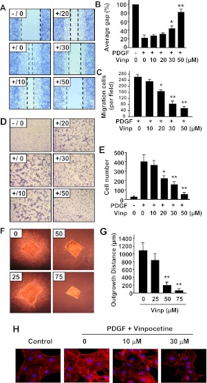
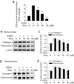
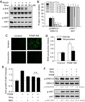
Abnormal vascular smooth muscle cell (SMC) activation is associated with various vascular disorders such as atherosclerosis, in-stent restenosis, vein graft disease, and transplantation-associated vasculopathy. Vinpocetine, a derivative of the alkaloid vincamine, has long been used as a cerebral blood flow enhancer for treating cognitive impairment. However, its role in pathological vascular remodeling remains unexplored. Herein, we show that systemic administration of vinpocetine significantly reduced neointimal formation in carotid arteries after ligation injury. Vinpocetine also markedly decreased spontaneous remodeling of human saphenous vein explants in ex vivo culture. In cultured SMCs, vinpocetine dose-dependently suppressed cell proliferation and caused G1-phase cell cycle arrest, which is associated with a decrease in cyclin D1 and an increase in p27Kip1 levels. In addition, vinpocetine dose-dependently inhibited platelet-derived growth factor (PDGF)-stimulated SMC migration as determined by the two-dimensional migration assays and three-dimensional aortic medial explant invasive assay. Moreover, vinpocetine significantly reduced PDGF-induced type I collagen and fibronectin expression. It is noteworthy that PDGF-stimulated phosphorylation of extracellular signal-regulated kinases 1/2 (ERK1/2), but not protein kinase B, was specifically inhibited by vinpocetine. Vinpocetine powerfully attenuated intracellular reactive oxidative species (ROS) production, which largely mediates the inhibitory effects of vinpocetine on ERK1/2 activation and SMC growth. Taken together, our results reveal a novel function of vinpocetine in attenuating neointimal hyperplasia and pathological vascular remodeling, at least partially through suppressing ROS production and ERK1/2 activation in SMCs. Given the safety profile of vinpocetine, this study provides insight into the therapeutic potential of vinpocetine in proliferative vascular disorders.


Figures

Fig. 1.
Fig. 1.
Effects of systemic administration of vinpocetine on ligation injury-induced neointima formation. A, representative histological images of left common carotid arteries stained with Verhoeff-van Gieson from mice subjected to complete carotid ligation or sham operation for 14 days with systemic administration of vinpocetine (5 mg/kg/day) or vehicle saline intraperitoneally. Bar, 100 μm. Blue-black indicates elastic fibers. * indicates yellowish intraplaque hemorrhage. B and C, morphometric analyses show that vinpocetine decreases intimal + medial areas (B) and increases luminal areas (C) at 14 days after ligation. Values are means ± S.E.M. (n = 8 for saline and 10 for vinpocetine). *, P < 0.05 compared with saline under ligation. D, immunohistochemical analyses showing vinpocetine decreases cell proliferation as stained with PCNA. Insets are corresponding images of sham-operated samples. Brown staining represents smooth muscle SM-α-actin or PCNA. Blue represents counterstaining with hematoxylin. Bar, 100 μm. E, quantitative data of PCNA-positive cells. *, P < 0.05 compared with saline under ligation.
Fig. 2.
Fig. 2.
Effects of vinpocetine on human saphenous vein remodeling in ex vivo organ culture. A, representative images of sections with Verhoeff Masson trichrome combination staining. Sections are from human saphenous vein before or after culture for 7 days in the presence of vehicle or 100 μM vinpocetine. Smooth muscles are stained with red, collagen with light blue or blue-green, and elastin with dark blue. Bar, 50 μm. Int, intima; Med, media; Adv, adventitia. B, quantification of thickness of intima, media, and adventitia. Values are means ± S.E.M. (n = 6). C, immunofluorescent images of human saphenous vein sections immunostained with BrdU or smooth muscle-α-actin. Middle panels are magnified images corresponding to the dotted-line squares in upper panels. The proliferative cells were stained with BrdU. Arrows point to BrdU-positive cells. D, quantification of BrdU-positive cells. **, P < 0.01 compared with control.
Fig. 3.
Fig. 3.
Effects of vinpocetine (Vinp) on VSMC proliferation and cell cycle regulation. A, vinpocetine dose-dependently inhibited 5% FBS-induced proliferation of VSMCs. The cell proliferation was measured by SRB assay as described under Materials and Methods. B, vinpocetine promotes G1 cell cycle arrest of VSMCs in a dose-dependent manner. Cell cycle was measured by flow cytometric DNA analysis as described under Materials and Methods. C, vinpocetine down-regulates cyclin D1 and up-regulates and p27Kip1. VSMCs were serum-starved for 48 h and pretreated with vehicle or vinpocetine at the indicated doses, followed by stimulation with 5% FBS for 6 h. The expression of cyclin D1 and p27Kip1 was analyzed by real-time PCR. Values are means ± S.D. from at least three independent experiments. *, P < 0.05; **, P < 0.01 versus FBS with zero vinpocetine. D, vinpocetine down-regulates cyclin D1 and up-regulates p27Kip1 protein measured by Western blotting.
Fig. 4.
Fig. 4.
Effects of vinpocetine on VSMC migration. A to C, scratch wound assay. A, representative images show that vinpocetine dose-dependently inhibited PDGF-induced migration of VSMCs by scratch wound assay. Dotted lines show the edges of cell migration. Confluent VSMCs were starved and scratched with a pipette tip, then treated with vinpocetine at the indicated doses and stimulated with 25 ng/ml of PDGF-BB for 16 h. The cells were fixed with 4% paraformaldehyde and stained with hematoxylin. B and C, quantitative data of the scratch wound assay were analyzed by the percentage of gap area (B) or migrating cell numbers (C). D and E, Boyden chamber assay. D, representative images show that vinpocetine dose-dependently inhibited PDGF-induced transmigration of VSMCs by Boyden chamber assay. One hundred microliters of VSMC suspension was placed in the upper microchemotaxis chamber, and 600 μl of DMEM containing 25 ng/ml of PDGF-BB was placed in the lower polycarbonate filter chamber. The chamber was incubated at 37°C and 5% CO2 for 6 h. E, the transmigrated cells on the filter membrane were fixed and stained with hematoxylin and quantified. F and G, ex vivo aortic medial explant migration assay. F, representative images show that vinpocetine dose-dependently inhibited PDGF-induced VSMC outgrowth in 3D collagen I gel. Media explants of mouse aorta were embedded in 3D gel containing collagen type I, and the migration of VSMCs was initiated by addition of PDGF-BB/FGF2. G, migration was quantified by measuring the distance migrated by the leading front of VSMCs from the explanted tissue. Values are means ± S.D. from at least three independent experiments. *, P < 0.05; **, P < 0.01 versus PDGF with no vinpocetine. H, effects of vinpocetine on actin cytoskeleton polymerization in VSMCs. VSMCs were seeded in six-well plates overnight in DMEM supplemented with 10% FBS, serum-starved for 48 h, and treated with or without vinpocetine for 0.5 h, then stimulated with 20 ng/ml of PDGF-BB for 24 h. F-actin was stained by Alexa Fluor 546 phalloidin (red), and nucleus was stained by DAPI (blue).
Fig. 5.
Fig. 5.
Effects of vinpocetine on ECM production in VSMCs. A, vinpocetine dose-dependently inhibited PDGF-induced total collagen synthesis measured by [3H]proline incorporation. VSMCs were serum-starved for 48 h, then treated with vinpocetine at the indicated doses and stimulated with 20 ng/ml PDGF-BB for 48 h. Total collagen synthesis was determined by [3H]proline incorporation. B and C, vinpocetine dose-dependently inhibited PDGF-induced expression of collagen I and fibronectin in VSMCs (C) determined by Western blot (B). D and E, vinpocetine inhibited PDGF-induced secretion of collagen I and fibronectin. VSMCs were serum-starved for 48 h, then treated with vinpocetine at the indicated doses and stimulated with 20 ng/ml of PDGF-BB for 48 h (E). The culture media were collected and filtered, then subjected to Western blot (D). *, P < 0.05; **, P < 0.01 versus PDGF with no vinpocetine (for collagen I). †, P < 0.05 versus PDGF with no vinpocetine (for fibronectin).
Fig. 6.
Fig. 6.
Effects of vinpocetine on PDGF-induced ERK1/2 and AKT phosphorylation and ROS production in VSMCs. A and B, VSMCs were serum-starved for 48 h, then treated with vinpocetine at the indicated doses and stimulated with 20 ng/ml of PDGF-BB for 15 min. ERK1/2 (A) and AKT (B) phosphorylation and total ERK1/2 and AKT levels were measured by Western blotting with phospho-specific antibodies and total antibodies, respectively. **, P < 0.01 versus PDGF with no vinpocetine. C and D, serum-starved VSMCs were labeled with 10 μM DCFH2-DA, then treated with vinpocetine and stimulated with 20 ng/ml of PDGF-BB for 1 h. C, the photographs were taken by using an Olympus (BX-51) fluorescent microscope. D, intracellular ROS was quantified by flow cytometry. *, P < 0.05 versus PDGF with vehicle. E, serum-starved VSMCs were pretreated with 30 μM vinpocetine, 5 mM NAC, or both, followed by PDGF-BB (50 ng/ml) stimulation for 48 h. Cell growth was measured by SRB assay. n.s., not significant. F, serum-starved VSMCs were pretreated with 30 μM vinpocetine, 5 mM NAC, or both, followed by PDGF-BB (50 ng/ml) stimulation for 15 min. ERK1/2 and AKT phosphorylation and total ERK1/2 and AKT levels were measured by Western blotting. The blots were analyzed by densitometry. Fold changes normalized to the left lane are shown below the blots (n = 2–3).

Similar articles

Cited by

References

    1. Adiguzel E, Ahmad PJ, Franco C, Bendeck MP. (2009) Collagens in the progression and complications of atherosclerosis. Vasc Med 14:73–89 - PubMed
    1. Andrae J, Gallini R, Betsholtz C. (2008) Role of platelet-derived growth factors in physiology and medicine. Genes Dev 22:1276–1312 - PMC - PubMed
    1. Bagoly E, Fehér G, Szapáry L. (2007) [The role of vinpocetine in the treatment of cerebrovascular diseases based in human studies]. Orv Hetil 148:1353–1358 - PubMed
    1. Balestreri R, Fontana L, Astengo F. (1987) A double-blind placebo controlled evaluation of the safety and efficacy of vinpocetine in the treatment of patients with chronic vascular senile cerebral dysfunction. J Am Geriatr Soc 35:425–430 - PubMed
    1. Bhardwaj S, Roy H, Ylä-Herttuala S. (2008) Gene therapy to prevent occlusion of venous bypass grafts. Expert Rev Cardiovasc Ther 6:641–652 - PubMed

Publication types

MeSH terms