. 2012 Nov;86(21):11763-78.
doi: 10.1128/JVI.01353-12. Epub 2012 Aug 22.

Isolation of novel synthetic prion strains by amplification in transgenic mice coexpressing wild-type and anchorless prion proteins

Affiliations

Isolation of novel synthetic prion strains by amplification in transgenic mice coexpressing wild-type and anchorless prion proteins

Gregory J Raymond et al. J Virol. 2012 Nov.

Abstract

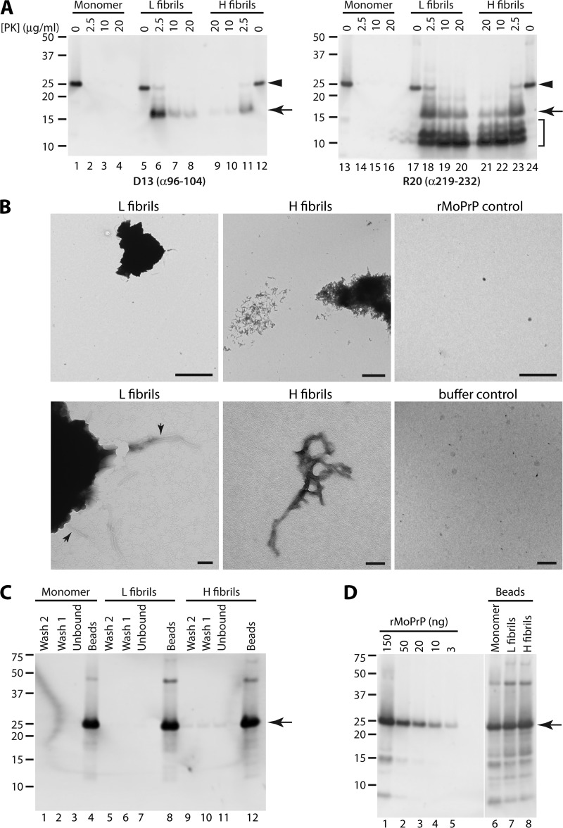
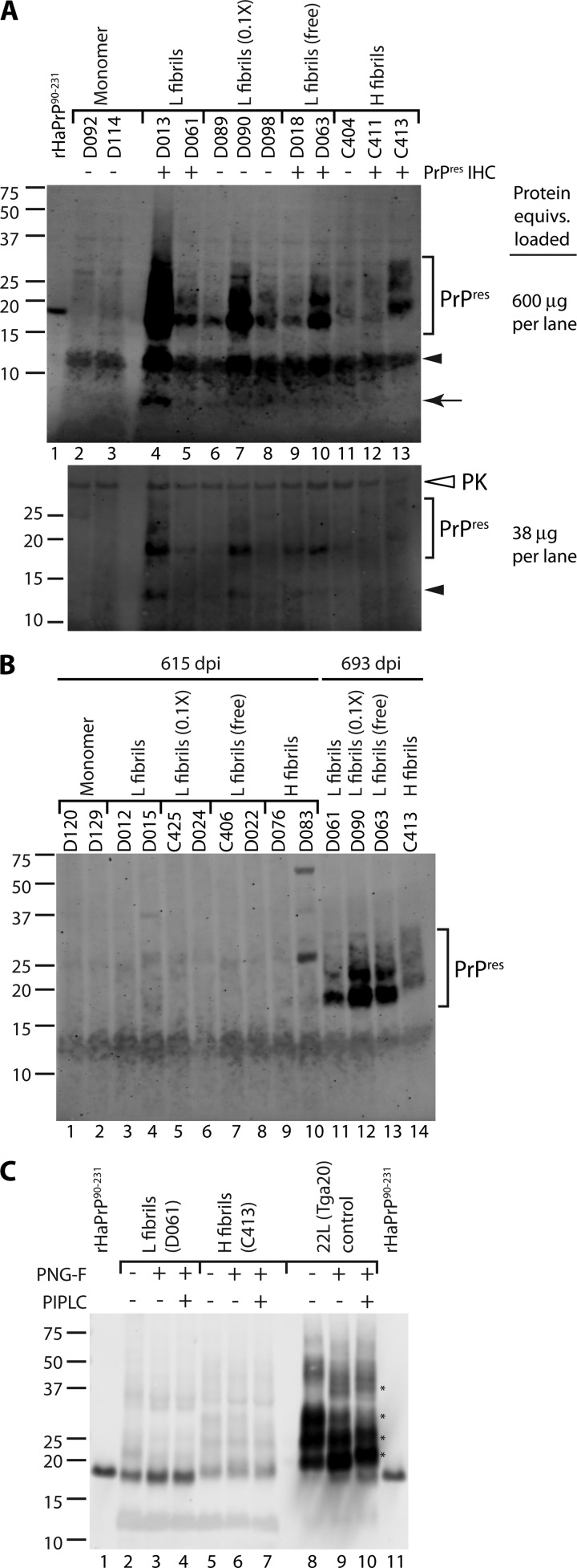
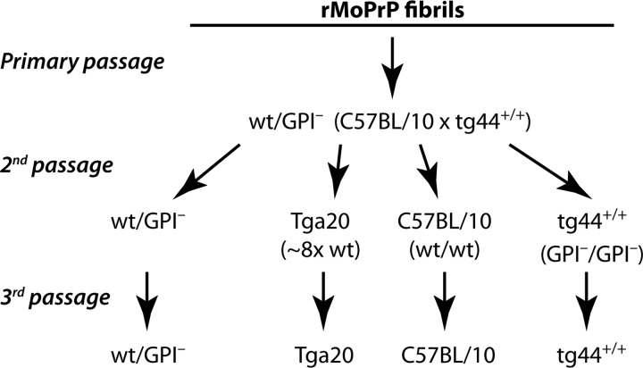
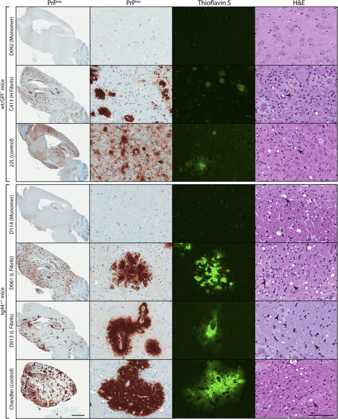
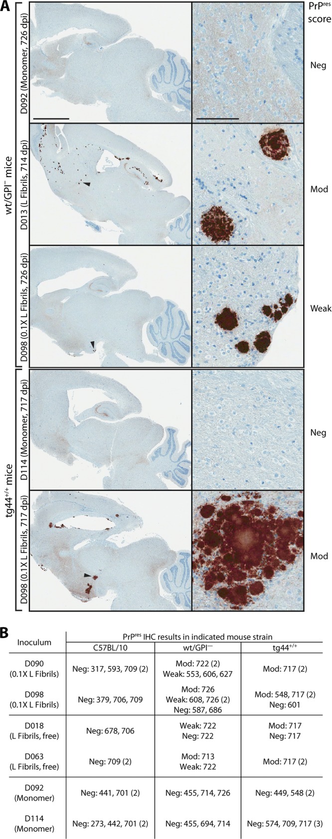
Mammalian prions are thought to consist of misfolded aggregates (protease-resistant isoform of the prion protein [PrP(res)]) of the cellular prion protein (PrP(C)). Transmissible spongiform encephalopathy (TSE) can be induced in animals inoculated with recombinant PrP (rPrP) amyloid fibrils lacking mammalian posttranslational modifications, but this induction is inefficient in hamsters or transgenic mice overexpressing glycosylphosphatidylinositol (GPI)-anchored PrP(C). Here we show that TSE can be initiated by inoculation of misfolded rPrP into mice that express wild-type (wt) levels of PrP(C) and that synthetic prion strain propagation and selection can be affected by GPI anchoring of the host's PrP(C). To create prions de novo, we fibrillized mouse rPrP in the absence of molecular cofactors, generating fibrils with a PrP(res)-like protease-resistant banding profile. These fibrils induced the formation of PrP(res) deposits in transgenic mice coexpressing wt and GPI-anchorless PrP(C) (wt/GPI(-)) at a combined level comparable to that of PrP(C) expression in wt mice. Secondary passage into mice expressing wt, GPI(-), or wt plus GPI(-) PrP(C) induced TSE disease with novel clinical, histopathological, and biochemical phenotypes. Contrary to laboratory-adapted mouse scrapie strains, the synthetic prion agents exhibited a preference for conversion of GPI(-) PrP(C) and, in one case, caused disease only in GPI(-) mice. Our data show that novel TSE agents can be generated de novo solely from purified mouse rPrP after amplification in mice coexpressing normal levels of wt and anchorless PrP(C). These observations provide insight into the minimal elements required to create prions in vitro and suggest that the PrP(C) GPI anchor can modulate the propagation of synthetic TSE strains.


Figures

Fig 1
Fig 1
Characterization of rMoPrP fibrils. (A) Immunoblot analysis of rMoPrP fibrils or monomer control treated with the indicated concentrations of PK. PrP was detected using either D13 (epitope hamster residues 96 to 104; left) or R20 (epitope hamster residues 219 to 232; right) antibody. Arrowhead, full-length rMoPrP; arrow, ∼17-kDa, PK-truncated fragment; bracket, low-MW PK-resistant bands detected with R20 antibody. Numbers on the left indicate apparent molecular masses (in kDa). (B) Ultrastructure of rMoPrP fibrils. Aliquots of H or L fibril reaction mixtures were directly spotted onto EM grids, washed, and stained with ammonium molybdate. Control grids were prepared using purified rMoPrP monomer (rMoPrP control) or reaction buffer used to form H fibrils (buffer control). Bars, 100 nm (lower panels of H and L fibrils) and 2 μm (all other panels). (C) Conjugation of rMoPrP preparations to magnetic beads. Aliquots (1/100 equivalents) from the unbound fractions (lanes 3, 7, and 11), first bead wash fractions (lanes 2, 6 and 10), and second bead wash fractions (lanes 1, 5, and 9) were analyzed. Bead-associated rMoPrP was eluted by boiling an aliquot of beads (1/100 equivalents) in sample buffer (lanes 4, 8, and 12). (D) Immunoblot quantitation of bead-associated rMoPrP. Bead-bound rMoPrP was eluted by boiling in sample buffer (lanes 6 to 8). Various amounts of purified rMoPrP (lanes 1 to 5) were loaded as standards. The vertical white line indicates the removal of irrelevant lanes. For panels C and D, rMoPrP was detected by immunoblotting with D13 anti-PrP antibody. Arrows, full-length rMoPrP monomer.
Fig 2
Fig 2
rMoPrP fibrils induce PrPres formation in mice coexpressing wt and GPI PrPC. (A) Immunoblot assay for PrPres in rMoPrP fibril-inoculated mice at 693 dpi. Lanes 2 to 13, brain homogenates from wt/GPI mice inoculated with the indicated rMoPrP preparations. L fibrils (0.1×), animals received 1/10 dose of bead-conjugated L fibrils; L fibrils (free), L fibrils mixed with preblocked beads immediately prior to inoculation. Brain homogenates were PK digested and precipitated with PTA (top) or methanol (bottom). Each lane contains 600 μg (top) or 38 μg (bottom) total protein equivalents. The 90 to 231 fragment of purified recombinant hamster PrP (0.2 ng) was loaded on the upper gel for comparison (lane 1). Closed arrowhead and arrow, low-molecular-mass PK-resistant bands in fibril-inoculated animals; open arrowhead, detection of methanol-precipitated PK due to antibody cross-reactivity. PrPres IHC, detection of PrPres by immunohistochemistry (see Fig. S2 in the supplemental material; data not shown). (B) Immunoblot assay for PrPres in rMoPrP fibril-inoculated mice at 615 dpi. Brain homogenates from wt/GPI mice inoculated with the indicated rMoPrP preparations and euthanized at 615 dpi (lanes 1 to 10) or 693 dpi (lanes 11 to 14). Each lane contains 600 μg total protein equivalents. Bands in lane 10 are likely due to residual mouse IgG in the brain homogenate. (C) rMoPrP fibril-induced PrPres lacks a GPI anchor. After PK digestion and PTA precipitation, brain homogenate samples (lanes 2 to 10) were denatured and treated with PNGase F (PNG-F) and PIPLC, as indicated. 22L-infected Tga20 mouse brain was included as a wt PrPres control (lanes 8 to 10). Lanes 2 to 7, 600 μg total protein equivalents per lane; lanes 8 to 10, 15 μg total protein equivalents per lane. The 90 to 231 fragment of purified recombinant hamster PrP (1 ng) was loaded for comparison (lanes 1 and 11). Asterisks, bands shifted by PIPLC treatment (lane 9 versus lane 10). For panels A to C (except where indicated), all samples were PK treated, PTA precipitated, and probed with anti-PrP mouse monoclonal antibody 6D11.
Fig 3
Fig 3
Overview of serial passage studies of rMoPrP fibrils in various mouse strains. After primary passage of rMoPrP fibrils in mice coexpressing wt and wt/GPI, subsequent passages were conducted in wt/GPI, Tga20 (which overexpresses wt MoPrPC 8-fold) (23), C57BL/10, and tg44+/+ (homozygous for GPI Prnp transgene on C57BL/10 [wt] PrP-knockout background) mice. Incubation time and attack rate data corresponding to secondary and tertiary passage studies are shown in Table 1.
Fig 4
Fig 4
Histopathology of C57BL/10 mice infected with rMoPrP fibril agents from wt/GPI PrP mice. Brain homogenates (1%) from 693 dpi wt/GPI mice treated with rMoPrP fibril preparations or monomer control were passaged into C57BL/10 mice. Brain sagittal sections were stained for PrPres with D13 antibody (PrPres), GFAP, or H&E, as indicated. Small panels correspond to the thalamus. All fibril agent-treated animals that developed TSE-like neurological disease showed similar PrPres deposition, astrocytosis, and spongiform change (arrows). A C413 agent-treated animal is shown as a representative example. Note the differences in distribution (large panels) and nature (punctate for fibril versus diffuse for 22L; small panels) of PrPres deposits induced by the fibril agent compared with that induced by the 22L scrapie strain control. A Monomer control-inoculated animal (D114, 441 dpi) is shown for comparison. Bars, 2 mm (large panels) and 50 μm (small panels).
Fig 5
Fig 5
Histopathology of rMoPrP fibril agent passaged in wt/GPI and tg44+/+ mice. Brain homogenates (1%) from 693 dpi wt/GPI mice treated with rMoPrP fibril preparations or Monomer control were passaged into wt/GPI or tg44+/+ mice. Brain sagittal sections were stained for PrPres with D13 antibody (PrPres), thioflavin S, or H&E, as indicated. Images for wt/GPI mice correspond to the thalamus. In wt/GPI mice, note the diffuse PrPres deposits (arrow) in the 22L-infected control mouse that are absent from the C413-infected mouse. Thioflavin S-positive amyloid plaques and spongiform pathology (arrows) are present in C413- and 22L-infected control mice. tg44+/+ images correspond to cerebral cortex (columns 1 to 3) or corpus callosum (column 4). Note differences in the size, morphology (the D061 agent is multifocal), and distribution of plaques in D061 agent- and D013 agent-infected tg44+/+ mice versus Chandler-infected control mice. Although aged tg44+/+ mice (D114, 574 dpi and uninfected controls [data not shown]) show some white matter spongiosis, vacuoles (arrows) were more abundant in D061 agent-, D013 agent-, and Chandler-infected mice, especially around plaques (arrowheads). Bars, 2 mm (column 1) and 100 μm (columns 2 to 4).
Fig 6
Fig 6
Histopathology of subclinical wt/GPI mice infected with secondary-passage rMoPrP fibril agents. Brain homogenates (1%) from 693 dpi wt/GPI mice treated with rMoPrP fibril preparations or Monomer control underwent secondary passage in wt/GPI or tg44+/+ mice. (A) Brain sagittal sections were stained for PrPres with D13 antibody. Representative examples are shown. Arrowheads, PrPres plaques shown at a higher magnification on the right. PrPres plaque score: Neg, negative, Weak, weak positive (≤8 plaques); Mod, moderate positive (>8 plaques). Bars, 2 mm (column 1) and 100 μm (column 2). (B) Summary of PrPres immunohistochemistry. Numbers indicate the dpi on which individual animals were euthanized. Values in parentheses indicate the number of animals with the same dpi of euthanization.
Fig 7
Fig 7
Transmission of PrPres propagation on secondary passage. PrPres profiles in brain homogenates from inoculation of Tga20 (A), wt/GPI (B), or tg44+/+ (C) mice with the indicated inocula from primary passage in wt/GPI mice in Fig. 2A. Monomer control samples were derived from age-matched (usually older) mice (Table 1). Where available, samples from Tga20, wt/GPI, and tg44+/+ mice infected with established mouse-adapted scrapie strains (22L, Chandler, ME7) were included for comparison, as indicated. Brackets indicate bands corresponding to PrPres monomers. SDS-resistant oligomers are also visible. Darker exposure panels are a darker exposure of the corresponding panel above. Arrowhead and arrow, ∼12-kDa and ∼8-kDa PK-resistant bands, respectively; vertical white lines, removal of irrelevant lanes. All samples were PK treated, PTA precipitated, and probed with 6D11 antibody. The total protein equivalents loaded per lane were as follows: 30 μg in lanes 1 to 9 and 300 μg in lanes 10 to 22 in panel A; 150 μg in lanes 1 to 7 and 15 μg in lanes 8 to 14 in panel B; 200 μg in panel C.

Similar articles

Cited by

References

    1. Atarashi R, et al. 2007. Ultrasensitive detection of scrapie prion protein using seeded conversion of recombinant prion protein. Nat. Methods 4:645–650 - PubMed
    1. Ayers JI, Kincaid AE, Bartz JC. 2009. Prion strain targeting independent of strain-specific neuronal tropism. J. Virol. 83:81–87 - PMC - PubMed
    1. Ayers JI, et al. 2011. The strain-encoded relationship between PrP replication, stability and processing in neurons is predictive of the incubation period of disease. PLoS Pathog. 7:e1001317 doi:10.1371/journal.ppat.1001317 - DOI - PMC - PubMed
    1. Baron GS, et al. 2011. Effect of glycans and the glycophosphatidylinositol anchor on strain dependent conformations of scrapie prion protein: improved purifications and infrared spectra. Biochemistry 50:4479–4490 - PMC - PubMed
    1. Barria MA, Mukherjee A, Gonzalez-Romero D, Morales R, Soto C. 2009. De novo generation of infectious prions in vitro produces a new disease phenotype. PLoS Pathog. 5:e1000421 doi:10.1371/journal.ppat.1000421 - DOI - PMC - PubMed

Publication types

LinkOut - more resources