. 2012 Nov;86(21):11886-98.
doi: 10.1128/JVI.01879-12. Epub 2012 Aug 22.

Regulated interaction of tegument proteins UL16 and UL11 from herpes simplex virus

Affiliations

Regulated interaction of tegument proteins UL16 and UL11 from herpes simplex virus

Pooja Chadha et al. J Virol. 2012 Nov.

Abstract

It is well known that proteins in the tegument (located between the viral capsid and envelope proteins) play critical roles in the assembly and budding of herpesviruses. Tegument proteins UL16 and UL11 of herpes simplex virus (HSV) are conserved among all the Herpesviridae. Although these proteins directly interact in vitro, UL16 was found to colocalize poorly with UL11 in cotransfected cells. To explain this discrepancy, we hypothesized that UL16 is initially made in an inactive form and is artificially transformed to the binding-competent state when cells are disrupted. Consistent with a regulated interaction, UL16 was able to fully colocalize with UL11 when a large C-terminal segment of UL16 was removed, creating mutant UL16(1-155). Moreover, membrane flotation assays revealed a massive movement of this mutant to the top of sucrose gradients in the presence of UL11, whereas both the full-length UL16 and the C-terminal fragment (residues 156 to 373) remained at the bottom. Further evidence for the presence of a C-terminal regulatory domain was provided by single-amino-acid substitutions at conserved cysteines (C269S, C271S, and C357S), which enabled the efficient interaction of full-length UL16 with UL11. Lastly, the binding site for UL11 was further mapped to residues 81 to 155, and to our surprise, the 5 Cys residues within UL16(1-155) are not required, even though the modification of free cysteines in UL16 with N-ethylmaleimide does in fact prevent binding. Collectively, these results reveal a regulatory function within the C-terminal region of UL16 that controls an N-terminal UL11-binding activity.


Figures

Fig 1
Fig 1
Interaction of UL16(1-155) with UL11 in vitro. (A) The His-tagged UL16 constructs used in the GST pulldown assay. They were expressed in E. coli, and cell lysates were either treated with NEM or left untreated. (B) As demonstrated by Ponceau S staining, equal amounts of GST and GST-UL11 beads were added to the lysates. (C) After incubating and washing the beads, they were resuspended in sample buffer prior to the analysis of bound proteins via Western blotting with UL16-specific antibodies. (D) Direct loading and Western blotting of input lysate samples show the amounts of wild-type and mutant UL16 that were initially present and the shift in migration that results from NEM modification.
Fig 2
Fig 2
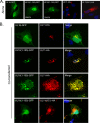
Coexpression of UL16(1-155)-GFP with UL11 in mammalian cells. (A) Cells were singly transfected with vectors that express UL16-GFP, UL16(1-155)-GFP, or UL11-HA to show the sites where these proteins accumulate on their own. (B) UL16-GFP and UL16(1-155)-GFP were coexpressed with UL11-HA to look for changes in subcellular localization. In all cases, the cells were fixed at 16 to 20 h posttransfection and stained with DAPI (4′,6-diamidino-2-phenylindole) to reveal nuclei (blue). UL16 and UL16(1-155) were revealed by the fluorescence of their GFP tags (green). The position of UL11 was revealed by a monoclonal antibody specific for the HA peptide (red).
Fig 3
Fig 3
Membrane flotation analyses of UL16 derivatives in the presence or absence of UL11. (A) Vero cells were transfected with the indicated UL16 constructs, either alone or with UL11-HA or its acidic-cluster mutant (AC−). At 16 to 20 h posttransfection, the cells were osmotically disrupted, and the ability of each protein to float to the upper fractions of sucrose step gradients was examined. Six equal fractions were collected, and detergent was added to solubilize the membranes. Proteins were concentrated by immunoprecipitation with antibodies specific for either GFP or UL11 and analyzed by Western blotting with the indicated antibodies. The tops and bottoms (Bot.) of the gradients are indicated. (B) Densitometry was used to quantitate the immunoblots from three independent experiments. The results are shown as the percentage of floating protein (top three fractions) relative to the total protein (all fractions). The error bars indicate standard deviations.
Fig 4
Fig 4
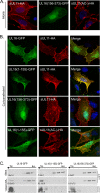
Relocalization of UL16(1-155)-GFP by sUL11. (A) Vero cells were singly transfected with expression vectors to show where the indicated proteins accumulate when expressed alone. (B) The indicated derivatives of UL16 were coexpressed with either sUL11-HA or sUL11(AC−)-HA to see which could be relocated. UL11 constructs were detected with a monoclonal antibody specific for the HA peptide (red), while the UL16 constructs were detected by the fluorescence of their GFP tags (green). DAPI was used to stain nuclei (blue). The images were captured with a confocal microscope. (C) Sucrose step gradients were used to examine the ability of the UL16 derivatives to associate with coexpressed, membrane-bound sUL11 (as described in the legend to Fig. 3). Representative immunoblots are shown. The number at the bottom left of each panel is the percentage of floating protein (top three fractions) relative to the total protein (all fractions) from three independent experiments.
Fig 5
Fig 5
Positions of cysteines and putative domain organization of UL16. The 20 cysteines contained within the 373 amino acids of UL16 are indicated, along with their residue numbers. Five are found in the N-terminal fragment (NTD) (residues 1 to 155), which binds to UL11 and gE. The eight conserved cysteines (boldface) are located in the putative C-terminal regulatory domain (CTD). Amino acid substitutions at 3 of the 20 cysteines (indicated with asterisks) enabled the full-length protein to bind to UL11 in vivo. The position of the CXXC-X21-CXC motif that resembles the unusual zinc finger of E. coli chaperone Hsp33 is shown (see Discussion).
Fig 6
Fig 6
Substitutions in the putative C-terminal regulatory domain activate sUL11 binding in cells. (A) Cells were singly transfected to express either UL16-GFP or the three cysteine substitution mutants that enable binding to UL11. Confocal microscopy revealed heterogeneous phenotypes for the mutants, examples of which are shown. (B) Cells were cotransfected with the indicated UL16-GFP derivatives and sUL11-HA. After 16 to 20 h of transfection, the cells were fixed, stained for UL11 with monoclonal anti-HA antibodies, and visualized by confocal microscopy.
Fig 7
Fig 7
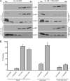
Membrane flotation analyses of cysteine mutants in the presence or absence of sUL11. Cells were transfected to express either UL16-GFP, the full-length cysteine substitution mutants, or a C-terminal fragment (CT-GFP.C269S) by themselves or with sUL11. The abilities of the UL16 derivatives to float to the top fractions in sucrose step gradients were measured as described in the legend to Fig. 3. (A) Representative immunoblots. (B) Data from three independent experiments are shown as the percentage of protein in the top three fractions relative to the total protein. The error bars indicate standard deviations.
Fig 8
Fig 8
Cysteines are not required for interaction with UL11. Mutant UL16(1-155)-GFP or its derivative that lacks all five cysteines, (5C−)-GFP, was either expressed alone or coexpressed with sUL11-HA or its acidic-cluster derivative (AC−), as indicated. (A) Cells were fixed and stained for sUL11 with anti-HA antibodies (red) and viewed by confocal microscopy. (B) The ability of the cysteine mutant to float when coexpressed with sUL11-HA, but not with the acidic cluster mutant, is shown in immunoblots from a membrane flotation assay. (C) The combined results from three independent flotation experiments are shown. The error bars indicate standard deviations.
Fig 9
Fig 9
Mapping of the UL11-binding region within the N-terminal fragment. (A) Diagrams of UL16(1-155) mutants used. WT, wild type. (B and C) Cells were transfected with the indicated GFP-tagged plasmids, either alone (B) or cotransfected with sUL11-HA (C), and examined by confocal microscopy. (D) The interactions of these deletion mutants with sUL11-HA were analyzed by membrane flotation assays; representative immunoblots are shown.

Similar articles

Cited by

References

    1. Baines JD, Jacob RJ, Simmerman L, Roizman B. 1995. The herpes simplex virus 1 UL11 proteins are associated with cytoplasmic and nuclear membranes and with nuclear bodies of infected cells. J. Virol. 69:825–833 - PMC - PubMed
    1. Baines JD, Koyama AH, Huang T, Roizman B. 1994. The UL21 gene products of herpes simplex virus 1 are dispensable for growth in cultured cells. J. Virol. 68:2929–2936 - PMC - PubMed
    1. Baines JD, Roizman B. 1991. The open reading frames UL3, UL4, UL10, and UL16 are dispensable for the replication of herpes simplex virus 1 in cell culture. J. Virol. 65:938. - PMC - PubMed
    1. Baines JD, Roizman B. 1992. The UL11 gene of herpes simplex virus 1 encodes a function that facilitates nucleocapsid envelopment and egress from cells. J. Virol. 66:5168–5174 - PMC - PubMed
    1. Baird NL, Starkey JL, Hughes DJ, Wills JW. 2010. Myristylation and palmitylation of HSV-1 UL11 are not essential for its function. Virology 397:80–88 - PMC - PubMed

Publication types

LinkOut - more resources