. 2012 Nov;86(21):11779-88.
doi: 10.1128/JVI.01525-12. Epub 2012 Aug 22.

Inclusion bodies are a site of ebolavirus replication

Affiliations

Inclusion bodies are a site of ebolavirus replication

Thomas Hoenen et al. J Virol. 2012 Nov.

Abstract

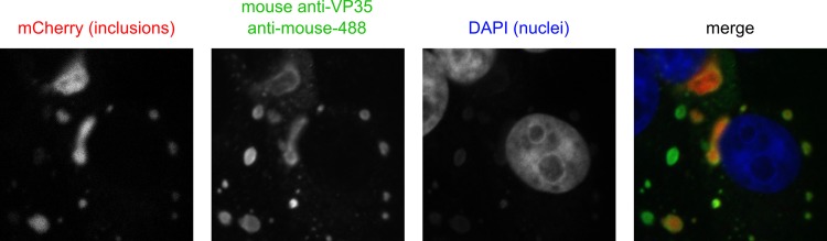
Inclusion bodies are a characteristic feature of ebolavirus infections in cells. They contain large numbers of preformed nucleocapsids, but their biological significance has been debated, and they have been suggested to be aggregates of viral proteins without any further biological function. However, recent data for other viruses that produce similar structures have suggested that inclusion bodies might be involved in genome replication and transcription. In order to study filovirus inclusion bodies, we fused mCherry to the ebolavirus polymerase L, which is found in inclusion bodies. The resulting L-mCherry fusion protein was functional in minigenome assays and incorporated into virus-like particles. Importantly, L-mCherry fluorescence in transfected cells was readily detectable and distributed in a punctate pattern characteristic for inclusion bodies. A recombinant ebolavirus encoding L-mCherry instead of L was rescued and showed virtually identical growth kinetics and endpoint titers to those for wild-type virus. Using this virus, we showed that the onset of inclusion body formation corresponds to the onset of viral genome replication, but that viral transcription occurs prior to inclusion body formation. Live-cell imaging further showed that inclusion bodies are highly dynamic structures and that they can undergo dramatic reorganization during cell division. Finally, by labeling nascent RNAs using click technology we showed that inclusion bodies are indeed the site of viral RNA synthesis. Based on these data we conclude that, rather than being inert aggregates of nucleocapsids, ebolavirus inclusion bodies are in fact complex and dynamic structures and an important site at which viral RNA replication takes place.


Figures

Fig 1
Fig 1
Cloning and expression of L-mCherry. (A) Schematic representation of L-mCherry. Shown are both the wild-type ZEBOV L protein and the L-mCherry fusion protein. The putative linker region is shown in green, and the arrow marks the site where mCherry (shown in purple) is inserted. Numbers indicate amino acid positions. (B) Fluorescence analysis of Huh-7 cells expressing L-mCherry. Cells were transfected with expression plasmids encoding L-mCherry or wild-type L and NP, VP35, and VP30, as indicated. Cells were visualized 18 h posttransfection. Shown are phase-contrast, mCherry signal, and a merged image. (C) Western blot analysis of 293T cells expressing L-mCherry. Cells were transfected with expression plasmids encoding L-mCherry or wild-type L and NP, VP35, and VP30, as indicated. At 48 h posttransfection, cell lysates were subjected to SDS-PAGE and Western blotting with an antibody against mCherry.
Fig 2
Fig 2
Functional analysis of L-mCherry. (A) Minigenome assay. 293T cells were transfected with expression plasmids encoding all ZEBOV RNP proteins, including the indicated amounts of either wild-type L or L-mCherry, a T7-driven minigenome, and a T7 RNA polymerase. At 48 h posttransfection, cells were lysed, and reporter activity, reflecting viral genome replication and transcription, was measured. (B) trVLP assay results. 293T producer cells were transfected as described for panel A, but in addition, expression plasmids for VP40 and GP were cotransfected. At 72 h posttransfection, naive Vero E6 target cells were infected with trVLP-containing supernatants of these cells. At 48 h postinfection, reporter activity in target cells, reflecting viral genome replication and production of trVLPs containing RNP complexes including L-mCherry, entry of trVLPs into target cells, and primary transcription in these target cells was measured. As negative controls, either the expression plasmid for L or VP40 was omitted when transfecting producer cells. (C) Growth kinetics of a recombinant ZEBOV expressing L-mCherry. Vero E6 cells were infected with either a recombinant WT or a ZEBOV expressing L-mCherry instead of L. Supernatants were collected at the times indicated and titers were determined based on TCID50 analysis. In all panels, means and standard errors from three independent experiments are shown.
Fig 3
Fig 3
Time course analysis of inclusion body formation. (A) Onset of inclusion bodies. Cells were infected with rg-ZEBOV-L-mCherry. At 2, 4, 6, 8, 10, 12, 14, 16, 18, 20, 22, and 24 h postinfection, cells were fixed, removed from the BSL-4 laboratory, counterstained with DAPI, and analyzed by confocal microscopy. Shown are the percentages of cells showing inclusion bodies from at least 5 random fields per time point. (B) Sizes of inclusion bodies. Images obtained as described for panel A were analyzed with ImageJ, and the cross-sectional areas of inclusion bodies at different time points were measured. Bars indicate the mean sizes of inclusion bodies at a given time. (C) Live-cell imaging of inclusion body formation. Cells were infected with rgZEBOV-L-mCherry and continuously monitored by live-cell fluorescence microscopy. Pictures show the same cell at the indicated time points after infection.
Fig 4
Fig 4
Fusion of smaller inclusion bodies into larger inclusions. Cells were infected with rgZEBOV-L-mCherry. After formation of early inclusion bodies, these were monitored by live-cell imaging. Fusion events are marked with arrows.
Fig 5
Fig 5
Colocalization of inclusion bodies with VP35. Vero E6 cells were infected with rgZEBOV-L-mCherry. At 18 h postinfection, cells were fixed and stained with a mouse anti-VP35 antibody and an anti-mouse Alexa Fluor 488-coupled secondary antibody. Nuclei were counterstained with DAPI.
Fig 6
Fig 6
Analysis of mRNA and vRNA production in infected cells. Vero E6 cells were infected with ZEBOV-L-mCherry. At 0, 1, 2, 4, 6, 8, 10, 12, 16, 20, and 24 h postinfection, cells were lysed and total RNA was purified. mRNA was reverse transcribed using an oligo(dT) primer, and vRNA was reverse transcribed using a vRNA specific primer, followed by quantitative real-time PCR using NP-specific primers. Shown are the means and standard errors from three independent experiments.
Fig 7
Fig 7
Colocalization of nascent RNA and inclusion bodies. Vero E6 cells were infected with rg-ZEBOV-L-mCherry. At 17.5 h postinfection, actinomycin (ActD) was added to the cells to inhibit cellular RNA synthesis. At 18 h postinfection, ethynyl-uridine (EU) was added to the medium for 1 h before cells were fixed, permeabilized, and the EU incorporated into nascent RNAs was detected using Alexa Fluor 488-azide. Nuclei were stained with DAPI, and cells were visualized by confocal microscopy.

Similar articles

Cited by

References

    1. Abramoff MD, Magalhaes PJ. 2004. Image processing with ImageJ. Biophotonics Int. 11:36–42
    1. Baskerville A, Fisher-Hoch SP, Neild GH, Dowsett AB. 1985. Ultrastructural pathology of experimental Ebola haemorrhagic fever virus infection. J. Pathol. 147:199–209 - PubMed
    1. Beniac DR, et al. 2012. The organisation of Ebola virus reveals a capacity for extensive, modular polyploidy. PLoS One 7:e29608 doi:10.1371/journal.pone.0029608 - DOI - PMC - PubMed
    1. Bharat TA, et al. 2012. Structural dissection of Ebola virus and its assembly determinants using cryo-electron tomography. Proc. Natl. Acad. Sci. U. S. A. 109:4275–4280 - PMC - PubMed
    1. Boehmann Y, Enterlein S, Randolf A, Muhlberger E. 2005. A reconstituted replication and transcription system for Ebola virus Reston and comparison with Ebola virus Zaire. Virology 332:406–417 - PubMed

Publication types