. 2012 Nov;86(21):11735-44.
doi: 10.1128/JVI.01084-12. Epub 2012 Aug 22.

Glycan-dependent immunogenicity of recombinant soluble trimeric hemagglutinin

Affiliations

Glycan-dependent immunogenicity of recombinant soluble trimeric hemagglutinin

Robert P de Vries et al. J Virol. 2012 Nov.

Abstract

Recombinant soluble trimeric influenza A virus (IAV) hemagglutinin (sHA(3)) has proven an effective vaccine antigen against IAV. Here, we investigate to what extent the glycosylation status of the sHA(3) glycoprotein affects its immunogenicity. Different glycosylation forms of subtype H5 trimeric HA protein (sH5(3)) were produced by expression in insect cells and different mammalian cells in the absence and presence of inhibitors of N-glycan-modifying enzymes or by enzymatic removal of the oligosaccharides. The following sH5(3) preparations were evaluated: (i) HA proteins carrying complex glycans produced in HEK293T cells; (ii) HA proteins carrying Man(9)GlcNAc(2) moieties, expressed in HEK293T cells treated with kifunensine; (iii) HA proteins containing Man(5)GlcNAc(2) moieties derived from HEK293S GnTI(-) cells; (iv) insect cell-produced HA proteins carrying paucimannosidic N-glycans; and (v) HEK293S GnTI(-) cell-produced HA proteins treated with endoglycosidase H, thus carrying side chains composed of only a single N-acetylglucosamine each. The different HA glycosylation states were confirmed by comparative electrophoretic analysis and by mass spectrometric analysis of released glycans. The immunogenicity of the HA preparations was studied in chickens and mice. The results demonstrate that HA proteins carrying terminal mannose moieties induce significantly lower hemagglutination inhibition antibody titers than HA proteins carrying complex glycans or single N-acetylglucosamine side chains. However, the glycosylation state of the HA proteins did not affect the breadth of the antibody response as measured by an HA1 antigen microarray. We conclude that the glycosylation state of recombinant antigens is a factor of significant importance when developing glycoprotein-based vaccines, such as recombinant HA proteins.


Figures

Fig 1
Fig 1
HA-glycan-dependent induction of antibodies against sH53N1. (A) Chickens (10 per group) were immunized twice with purified sH53N1 protein preparations produced in HEK293T cells (293T) or HEK293S GnTI(−) cells (GnTI). As a control, chickens were mock treated (PBS). Blood samples were taken 3 weeks after the first and after the second vaccinations. HI titers against 8 HAU sH53N1 in serum for each bird are shown. (B) Mice (6 per group) were immunized twice with sH53N1 protein preparations produced in HEK293T cells (293T), in HEK293T cells treated with 100 μM KIF (KIF), or in HEK293S GnTI(−) cells (GnTI). Blood samples were taken 3 weeks after the second immunization. HI titers against 8 HAU sH53N1 in serum for each mouse are shown. (A and B) The horizontal lines represent the geometric means per group. Significant differences between groups (panel A, Student's t test; panel B, ANOVA) are indicated as follows: *, P < 0.05; **, P < 0.01; ***, P < 0.001. (C) Purified sH53N1 protein preparations were analyzed by SDS-PAGE, followed by Western blotting. Where indicated, the proteins were treated with EndoH or PNGaseF prior to electrophoresis. The recombinant proteins were detected using a mouse anti-Strep tag antibody (29).
Fig 2
Fig 2
Structure representation of H5 derived from H5N1 with complex N-glycans attached at its N-glycosylation sites. The protein structure (black) was created with Protein Data Bank ID code 2IBX, and the N-linked glycans (gray) were modeled with Glyprot (3). For comparison, a model (labeled H5N1) is shown in which the N-linked glycan side chain that is missing in H5 from H5N7, compared to HA derived from H5N1, has been deleted.
Fig 3
Fig 3
Analysis of different sH53N7 preparations. sH53N7 proteins produced in HEK293T cells (293T), HEK293T cells treated with KIF (KIF), or HEK293S GnTI(−) cells (GnTI) or in insect S2 cells (S2), and HEK293S GnTI(−) cell-produced sH53N7 treated with EndoH (EndoH) were purified and analyzed. (A) The HA proteins were analyzed by SDS-PAGE, followed by Western blotting. The proteins were detected using a mouse anti-Strep tag antibody (29). Where indicated, samples were treated with PNGaseF or EndoH prior to electrophoresis. (B) Binding of Fc-tagged DC-SIGN (2.5 μg/well) to wells coated with the different HA preparations (5 μg/well) or with no HA protein (Mock). Binding of DC-SIGN was detected using HRP-labeled goat-anti-human IgG. (C) Blue Native-PAGE analysis of the HA protein preparations. The positions in the gel of the trimeric and monomeric protein species are indicated, as well as samples that were denatured by heating them for 1 min at 95°C prior to electrophoresis. (D) sH53 proteins were complexed with HRP-conjugated mouse antibody directed against the Strep tag prior to their application in a fetuin binding assay. HA-fetuin binding is shown at an HA concentration of 2.5 μg/ml. Standard deviations are indicated by the error bars.
Fig 4
Fig 4
Mass spectrometric glycan analysis of different sH53N7 preparations. Shown are MALDI-TOF mass spectra of the N-glycans of purified sH53N7 protein preparations produced in HEK293T cells (A and B), HEK293T cells treated with KIF (C), HEK293S GnTI(−) cells (D), and insect S2 cells (E). N-glycans were released by PNGaseF, labeled with anthranilic acid, and analyzed in negative-ion reflectron mode. All signals were labeled with monoisotopic masses, and the structures were deduced from the composition indicated by this mass. In the case of the HEK293T sample (A), only the 10 signals with the highest abundance were labeled with assigned structures; these structures were confirmed by beta-galactosidase treatment of the HEK293T sample (B), and in each case, the number of terminal galactose residues present is indicated. Compared to the spectrum before treatment (A), a clear loss of all galactoses not carrying N-acetylneuraminic acid is visible, indicating a high abundance of terminal galactose. The double-headed arrows indicate a difference in fucose content. Red triangles, fucose; purple diamonds, N-acetylneuraminic acid; yellow circles, galactose; blue squares, N-acetylglucosamine; green circles, mannose; white square, N-acetylhexosamine.
Fig 5
Fig 5
HA-glycan-dependent induction of antibodies against sH53N7. (A) Mice (six per group) were immunized twice with sH53N7 protein preparations produced in HEK293T cells (293T), HEK293T cells treated with KIF (KIF), HEK293S GnTI(−) cells (GnTI), or insect S2 cells or with HEK293S GnTI(−) cell-produced sH53N7 treated with EndoH (EndoH). Blood samples were taken 3 weeks after the second immunization. HI titers against 4 HAU sH53N7 in serum for each mouse are shown. (B) HA-specific IgG1 levels (μg per ml serum) were determined by ELISA. (C) Graph displaying the data shown in panel A pooled on the basis of the absence [−man; HEK293T cell-produced HA and EndoH-treated HA produced in HEK293S GnTI(−) cells] or presence [+man; HA produced in HEK293T cells treated with KIF, HEK293S GnTI(−) cell-produced HA, and S2 cell-produced HA] of terminal mannose residues in the sH53N7 protein preparations for which data are shown. (D) Similar to panel C, but showing the HA-specific IgG1 levels. The horizontal lines in the scatter dot plots represent geometric means per group. Significant differences between groups (panels A and B, ANOVA; panels C and D, Student's t test) are indicated as follows: *, P < 0.05; **, P < 0.01; ***, P < 0.001.
Fig 6
Fig 6
Inhibition of CpG-induced IFN-α production by HA. Freshly isolated pDCs were treated with sH53N7, produced in HEK293T, KIF-treated HEK293T, HEK293S GnTI(−), and S2 cells, in the presence of CpG. The culture supernatants were collected after 18 h for IFN-α quantification by ELISA. The data shown are from one representative experiment out of four independent experiments with pDCs from four different donors. Significant differences between groups treated with the different HA protein preparations are indicated: ***, P < 0.001.
Fig 7
Fig 7
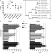
Profiling HA-glycan-dependent induction of antibodies by using HA1 microarrays. (A) Mice (nine per group) were immunized twice with sH53N1 or sH53N7 protein preparations produced in HEK293T cells (293T) or insect S2 cells (S2) or with HEK293S GnTI(−) cell-produced proteins treated with EndoH (EndoH). Blood samples were taken 3 weeks after the second immunization. HI titers in serum against 4 HAU of sH53 for each mouse are shown, corresponding to the immunogen used. The horizontal lines represent the geometric means per group. Significant differences between groups are indicated as follows: *, P < 0.05; **, P < 0.01; ***, P < 0.001. (B) An unrooted protein tree (neighbor joining) was generated from a sequence alignment (ClustalX 1.8; standard settings) of the HA1 domains, comprising the sequence from the signal cleavage site to the HA1-HA2 proteolytic cleavage site of the 12 indicated virus strains (numbers on right). Bootstrap values (1,000 replicates) are indicated on the branches. The HA proteins that were used as immunogens are boxed; the HA1 domains of the other HA proteins were spotted on the HA microarray. (C and D) A panel of recombinant HA1 domains of HA proteins derived from different viruses (see panel B for strain information) spotted in an HA1 antigen microarray format was probed with sera of mice immunized with sH53N1 (C) or sH53N7 (D). The titers were defined as the interpolated serum concentration that provoked a response halfway on a concentration-response curve between the minimum and maximum signals. Titers lower than the smallest serum dilution (1:20) were set to 20. The error bars indicate standard deviations.

Similar articles

Cited by

References

    1. Banerjee K, et al. 2009. Enzymatic removal of mannose moieties can increase the immune response to HIV-1 gp120 in vivo. Virology 389:108–121 - PMC - PubMed
    1. Banerjee K, et al. 2012. Occluding the mannose moieties on human immunodeficiency virus type 1 gp120 with griffithsin improves the antibody responses to both proteins in mice. AIDS Res. Hum. Retroviruses 28:206–214 - PMC - PubMed
    1. Bohne-Lang A, von der Lieth CW. 2005. GlyProt: in silico glycosylation of proteins. Nucleic Acids Res. 33:W214–W219 - PMC - PubMed
    1. Bosch BJ, et al. 2010. Recombinant soluble, multimeric HA and NA exhibit distinctive types of protection against pandemic swine-origin 2009 A(H1N1) influenza virus infection in ferrets. J. Virol. 84:10366–10374 - PMC - PubMed
    1. Cornelissen LA, et al. 2010. A single immunization with soluble recombinant trimeric hemagglutinin protects chickens against highly pathogenic avian influenza virus H5N1. PLoS One 5:e10645 doi:10.1371/journal.pone.0010645 - DOI - PMC - PubMed

Publication types