The assembly domain of the small capsid protein of Kaposi's sarcoma-associated herpesvirus
- PMID: 22915821
- PMCID: PMC3486306
- DOI: 10.1128/JVI.01430-12
The assembly domain of the small capsid protein of Kaposi's sarcoma-associated herpesvirus
Abstract
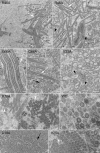
Self-assembly of Kaposi's sarcoma-associated herpesvirus capsids occurs when six proteins are coexpressed in insect cells using recombinant baculoviruses; however, if the small capsid protein (SCP) is omitted from the coinfection, assembly does not occur. Herein we delineate and identify precisely the assembly domain and the residues of SCP required for assembly. Hence, six residues, R14, D18, V25, R46, G66, and R70 in the assembly domain, when changed to alanine, completely abolish or reduce capsid assembly.
Figures


Similar articles
-
A hydrophobic domain within the small capsid protein of Kaposi's sarcoma-associated herpesvirus is required for assembly.J Gen Virol. 2014 Aug;95(Pt 8):1755-1769. doi: 10.1099/vir.0.064303-0. Epub 2014 May 13. J Gen Virol. 2014. PMID: 24824860 Free PMC article.
-
Small capsid protein pORF65 is essential for assembly of Kaposi's sarcoma-associated herpesvirus capsids.J Virol. 2008 Jul;82(14):7201-11. doi: 10.1128/JVI.00423-08. Epub 2008 May 7. J Virol. 2008. PMID: 18463150 Free PMC article.
-
Incorporation of the Kaposi's sarcoma-associated herpesvirus capsid vertex-specific component (CVSC) into self-assembled capsids.Virus Res. 2017 May 15;236:9-13. doi: 10.1016/j.virusres.2017.04.016. Epub 2017 Apr 26. Virus Res. 2017. PMID: 28456575 Free PMC article.
-
Kaposi's sarcoma-associated herpesvirus and Kaposi's sarcoma.Microbes Infect. 2000 May;2(6):671-80. doi: 10.1016/s1286-4579(00)00358-0. Microbes Infect. 2000. PMID: 10884618 Review.
-
Pathological Features of Kaposi's Sarcoma-Associated Herpesvirus Infection.Adv Exp Med Biol. 2018;1045:357-376. doi: 10.1007/978-981-10-7230-7_16. Adv Exp Med Biol. 2018. PMID: 29896675 Review.
Cited by
-
A hydrophobic domain within the small capsid protein of Kaposi's sarcoma-associated herpesvirus is required for assembly.J Gen Virol. 2014 Aug;95(Pt 8):1755-1769. doi: 10.1099/vir.0.064303-0. Epub 2014 May 13. J Gen Virol. 2014. PMID: 24824860 Free PMC article.
-
CryoEM and mutagenesis reveal that the smallest capsid protein cements and stabilizes Kaposi's sarcoma-associated herpesvirus capsid.Proc Natl Acad Sci U S A. 2015 Feb 17;112(7):E649-56. doi: 10.1073/pnas.1420317112. Epub 2015 Feb 2. Proc Natl Acad Sci U S A. 2015. PMID: 25646489 Free PMC article.
References
-
- Germi R, et al. 2012. Three-dimensional structure of the Epstein-Barr virus capsid. J. Gen. Virol. 93:1769–1773 - PubMed
Publication types
MeSH terms
Substances
Grants and funding
LinkOut - more resources
Full Text Sources

