. 2012;2(7):734-45.
doi: 10.7150/thno.4290. Epub 2012 Aug 1.

The Invasion and Reproductive Toxicity of QDs-Transferrin Bioconjugates on Preantral Follicle in vitro

Affiliations

The Invasion and Reproductive Toxicity of QDs-Transferrin Bioconjugates on Preantral Follicle in vitro

Gaixia Xu et al. Theranostics. 2012.

Abstract

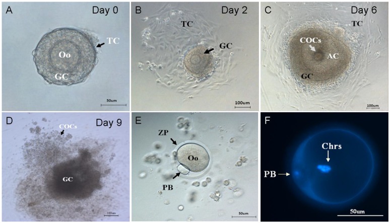
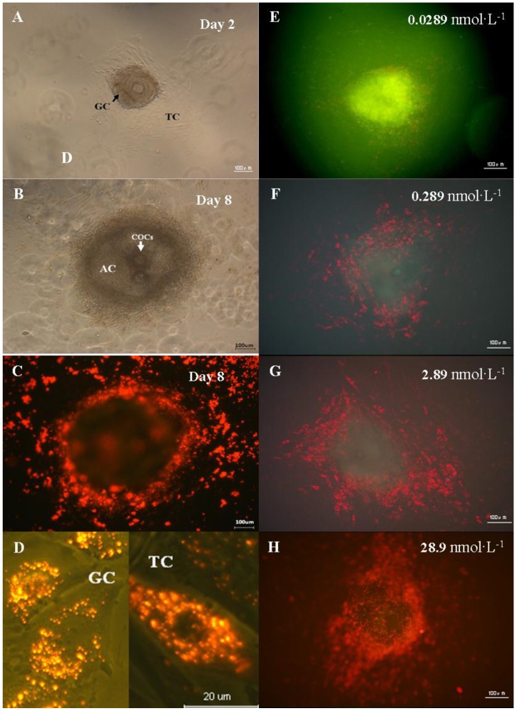
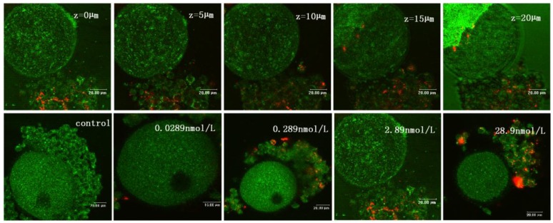
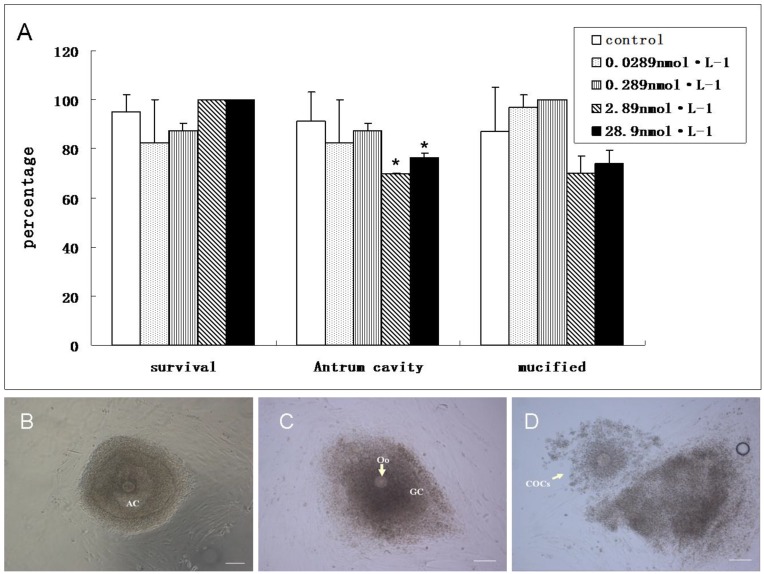
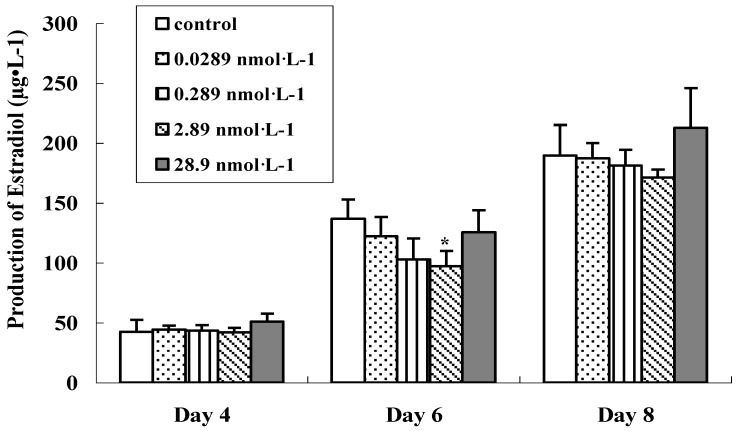
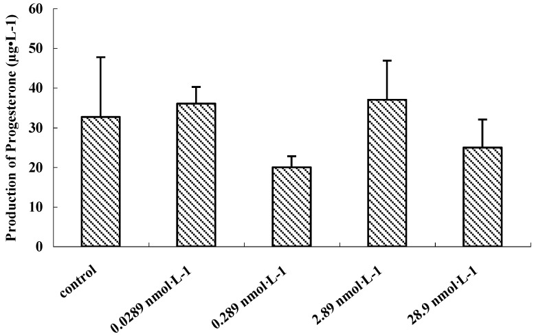
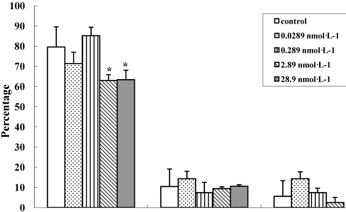
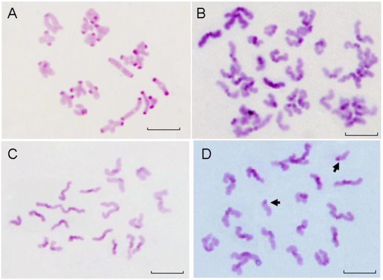
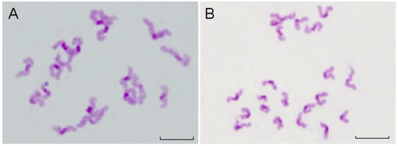
The toxicity of QD has been extensively studied over the past decade. However, the potential toxicity of QDs impedes its use for clinical research. In this work, we established a preantral follicle in vitro culture system to investigate the effects of QD-Transferrin (QDs-Tf) bioconjugates on follicle development and oocyte maturation. The preantral follicles were cultured and exposed to CdTe/ZnTe QDs-Tf bioconjugates with various concentrations and the reproductive toxicity was assessed at different time points post-treatment. The invasion of QDs-Tf for oocytes was verified by laser scanning confocal microscope. Steroid production was evaluated by immunoassay. C-band Giemsa staining was performed to observe the chromosome abnormality of oocytes. The results showed that the QDs-Tf bioconjugates could permeate into granulosa cells and theca cells, but not into oocyte. There are no obvious changes of oocyte diameter, the mucification of cumulus-oocyte-complexes and the occurrence of aneulpoidy as compared with the control group. However, delay in the antrum formation and decrease in the ratio of oocytes with first polar body were observed in QDs-Tf-treated groups. The matured oocytes with first polar body decreased significantly by ~16% (from 79.6±10 % to 63±2.9 %) when the concentration of QDs-Tf bioconjugates exceeded 2.89 nmol·L(-1) (P < 0.05). Our results implied that the CdTe/ZnTe QDs-Tf bioconjugates were reproductive toxic for follicle development, and thus also revealed that this in vitro culture system of preantral follicle is a highly sensitive tool for study on the reproductive toxicity of nanoparticles.

Keywords: QDs-transferrin bioconjugate.; in vitro culture system; invasion; preantral follicle; reproductive toxicity.


Conflict of interest statement

Competing Interests: The authors have declared that no competing interest exists.

Figures

Figure 1
Figure 1
Morphology of ovary and preantral follicles. (A) Mouse ovary, H&E staining. (B) Preantral follicles dissected mechanically. (C) Fixed single preantral follicle, H&E staining, Oo: oocyte; GC: granulosa cell; TC: theca cell.
Figure 2
Figure 2
Stages of folliculogenesis and oocyte maturation. (A) On the isolation day, intact preantral follicle was transferred into dishes, surrounded by granulosa cells and theca cells. (B) On the 2nd day, the theca cells attached to the bottom of culture dishes. (C) On the 6th day, the granulosa cells proliferated and differentiated, the antrum and COCs were formed. (D) On the 9th day, COCs was released under the stimulation of HCG and EGF. (E) On the 9th day, the granulosa cells were removed and the single oocytes with first polar body was denuded. (F) On the 9th day, two sets of chromosomes of oocyte stained by DAPI. Oo:oocyte; GC: granulosa cell; TC: Theca cell; COCs: Cumulus-Oocyte-Complexes; AC: Antrum cavity; PB: polar body; ZP: Zona pellucida; Chrs: Chromosome.
Figure 3
Figure 3
Distribution of QDs in the preantral follicles in vitro. (A) Preantral follicles cultured in vitro under phase-contrast microscope on the 2nd day. (B) Follicles with antrum cavity cultured in vitro under phase-contrast microscope on the 8th day. (C) Fluorescence microscopic image of follicles cultured in vitro on the 8th day. (D) Magnified fluorescence microscopic image of theca cells and granulosa cells on the 8th day. (E)-(H) Fluorescence microscopic image of follicle treated with QDs-Tf at different concentrations on the 8th day. The green pseudo-color represented emission from autofluorescence of samples and the red ones from QDs.
Figure 4
Figure 4
The confocal microscopic images of oocyte treated with QDs-Tf for 8 days. Upper panel: optical sectioning images of the oocyte treated with QDs-Tf (2.89nmol·L-1). Lower panel: the oocytes treated with QDs-Tf at different concentrations. Red pseudo-color represented fluorescence from QDs and green ones from reflecting signal.
Figure 5
Figure 5
(A)Comparison about the ratio of survival follicle, antrum cavity formation and COCs mucification between control and treated groups. (mean±SD, *P<0.05 vs control, Chi-Square test). (B) Follicle with antrum cavity. (C) Follicle without antrum cavity. (D) Mucified COCs. Scale bar: 100 µm.
Figure 6
Figure 6
Effect of QDs-Tf for production of 17 β-estradiol on the 4th, 6th and 8th days. (mean±SD, *P<0.05, ANOVA test)
Figure 7
Figure 7
Effect of QDs-Tf for production of progesterone on the 9th day (mean±SD, ANOVA test)
Figure 8
Figure 8
Comparision of cytoplasmic maturation of oocytes (mean±SD, *P<0.05 vs control, Chi-Square test).
Figure 9
Figure 9
The chromosomes of GVBD and PB oocytes treated with QDs-Tf (2.89 nmol·L-1). (A) Bivalent chromosomes in meiosis I oocyte. (B) Chromosomes in anaphase I of meiosis, with two sets of dyads. (C) Normal Chromosomes in metaphase II with one set of dyads. (D) Hyperploid oocyte with 19 dyads and 2 single chromatids (arrows). Scale bar: 10µm.
Figure 10
Figure 10
The chromosomes of PB oocytes treated with QDs-Tf (2.89 nmol·L-1). (A) Hypoploid with 19 dyads in oocyte. (B) Hyperploid with 21 dyds in oocyte. Scale bar: 10 µm.

References

    1. Alivisatos AP, Gu WW, Larabell C. Quantum dots as cellular probes. Annual Review of Biomedical Engineering. 2005;7:55–76. - PubMed
    1. Gao XH, Yang LL, Petros JA, Marshal FF, Simons JW, Nie SM. In vivo molecular and cellular imaging with quantum dots. Current Opinion in Biotechnology. 2005;16:63–72. - PubMed
    1. Ye YP, Chen XY. Integrin targeting for tumor optical imaging. Theranostics. 2011;1:102–126. - PMC - PubMed
    1. Chan WCW, Nie SM. Quantum dot bioconjugates for ultrasensitive nonisotopic detection. Science. 1998;281:2016–2018. - PubMed
    1. Michalet X, Pinaud F F, Bentolila LA, Tsay JM, Doose S, Li JJ. et al. Quantum dots for live cells, in vivo imaging, and diagnostics. Science. 2005;307:538–544. - PMC - PubMed