Coronary artery remodeling in a model of left ventricular pressure overload is influenced by platelets and inflammatory cells
- PMID: 22916095
- PMCID: PMC3423413
- DOI: 10.1371/journal.pone.0040196
Coronary artery remodeling in a model of left ventricular pressure overload is influenced by platelets and inflammatory cells
Abstract
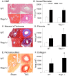
Left ventricular hypertrophy (LVH) is usually accompanied by intensive interstitial and perivascular fibrosis, which may contribute to arrhythmogenic sudden cardiac death. The mechanisms underlying the development of cardiac fibrosis are incompletely understood. To investigate the role of perivascular inflammation in coronary artery remodeling and cardiac fibrosis during hypertrophic ventricular remodeling, we used a well-established mouse model of LVH (transverse aortic constriction [TAC]). Three days after pressure overload, macrophages and T lymphocytes accumulated around and along left coronary arteries in association with luminal platelet deposition. Consistent with these histological findings, cardiac expression of IL-10 was upregulated and in the systemic circulation, platelet white blood cell aggregates tended to be higher in TAC animals compared to sham controls. Since platelets can dynamically modulate perivascular inflammation, we investigated the impact of thrombocytopenia on the response to TAC. Immunodepletion of platelets decreased early perivascular T lymphocytes' accumulation and altered subsequent coronary artery remodeling. The contribution of lymphocytes were examined in Rag1(-/-) mice, which displayed significantly more intimal hyperplasia and perivascular fibrosis compared to wild-type mice following TAC. Collectively, our studies support a role of early perivascular accumulation of platelets and T lymphocytes in pressure overload-induced inflammation.
Conflict of interest statement
Figures


Similar articles
-
Dapagliflozin Attenuates Cardiac Remodeling in Mice Model of Cardiac Pressure Overload.Am J Hypertens. 2019 Apr 22;32(5):452-459. doi: 10.1093/ajh/hpz016. Am J Hypertens. 2019. PMID: 30689697
-
Recombinant Human ADAMTS13 Treatment Improves Myocardial Remodeling and Functionality After Pressure Overload Injury in Mice.J Am Heart Assoc. 2018 Jan 24;7(3):e007004. doi: 10.1161/JAHA.117.007004. J Am Heart Assoc. 2018. PMID: 29367415 Free PMC article.
-
Intercellular Adhesion Molecule 1 Regulates Left Ventricular Leukocyte Infiltration, Cardiac Remodeling, and Function in Pressure Overload-Induced Heart Failure.J Am Heart Assoc. 2016 Mar 15;5(3):e003126. doi: 10.1161/JAHA.115.003126. J Am Heart Assoc. 2016. PMID: 27068635 Free PMC article.
-
Alpha-calcitonin gene-related peptide is protective against pressure overload-induced heart failure.Regul Pept. 2013 Aug 10;185:20-8. doi: 10.1016/j.regpep.2013.06.008. Epub 2013 Jun 28. Regul Pept. 2013. PMID: 23816470
-
The disruption of invariant natural killer T cells exacerbates cardiac hypertrophy and failure caused by pressure overload in mice.Exp Physiol. 2020 Mar;105(3):489-501. doi: 10.1113/EP087652. Epub 2020 Feb 9. Exp Physiol. 2020. PMID: 31957919
Cited by
-
Angiogenic Endothelial Cell Signaling in Cardiac Hypertrophy and Heart Failure.Front Cardiovasc Med. 2019 Mar 6;6:20. doi: 10.3389/fcvm.2019.00020. eCollection 2019. Front Cardiovasc Med. 2019. PMID: 30895179 Free PMC article. Review.
-
Mineralocorticoid Receptor in Smooth Muscle Contributes to Pressure Overload-Induced Heart Failure.Circ Heart Fail. 2021 Feb;14(2):e007279. doi: 10.1161/CIRCHEARTFAILURE.120.007279. Epub 2021 Feb 1. Circ Heart Fail. 2021. PMID: 33517669 Free PMC article.
-
Cardiac Hypertrophy: An Introduction to Molecular and Cellular Basis.Med Sci Monit Basic Res. 2016 Jul 23;22:75-9. doi: 10.12659/MSMBR.900437. Med Sci Monit Basic Res. 2016. PMID: 27450399 Free PMC article. Review.
-
The E3 ubiquitin ligase HectD3 attenuates cardiac hypertrophy and inflammation in mice.Commun Biol. 2020 Oct 9;3(1):562. doi: 10.1038/s42003-020-01289-2. Commun Biol. 2020. PMID: 33037313 Free PMC article.
-
Direct thrombin inhibition with dabigatran attenuates pressure overload-induced cardiac fibrosis and dysfunction in mice.Thromb Res. 2017 Nov;159:58-64. doi: 10.1016/j.thromres.2017.09.016. Epub 2017 Sep 21. Thromb Res. 2017. PMID: 28982031 Free PMC article.
References
-
- Rohr S (2009) Myofibroblasts in diseased hearts: new players in cardiac arrhythmias? Heart Rhythm 6: 848–856. - PubMed
-
- Nicoletti A, Michel JB (1999) Cardiac fibrosis and inflammation: interaction with hemodynamic and hormonal factors. Cardiovasc Res 41: 532–543. - PubMed
-
- Kwon DH, Smedira NG, Rodriguez ER, Tan C, Setser R, et al. (2009) Cardiac magnetic resonance detection of myocardial scarring in hypertrophic cardiomyopathy: correlation with histopathology and prevalence of ventricular tachycardia. J Am Coll Cardiol 54: 242–249. - PubMed
-
- Dellsperger KC, Marcus ML (1990) Effects of left ventricular hypertrophy on the coronary circulation. Am J Cardiol 65: 1504–1510. - PubMed
-
- Bache RJ (1988) Effects of hypertrophy on the coronary circulation. Prog Cardiovasc Dis 30: 403–440. - PubMed
Publication types
MeSH terms
Grants and funding
LinkOut - more resources
Full Text Sources
Other Literature Sources
Molecular Biology Databases

