. 2012;7(8):e43304.
doi: 10.1371/journal.pone.0043304. Epub 2012 Aug 16.

Luteolin induces microRNA-132 expression and modulates neurite outgrowth in PC12 cells

Affiliations

Luteolin induces microRNA-132 expression and modulates neurite outgrowth in PC12 cells

Lian-Fang Lin et al. PLoS One. 2012.

Abstract

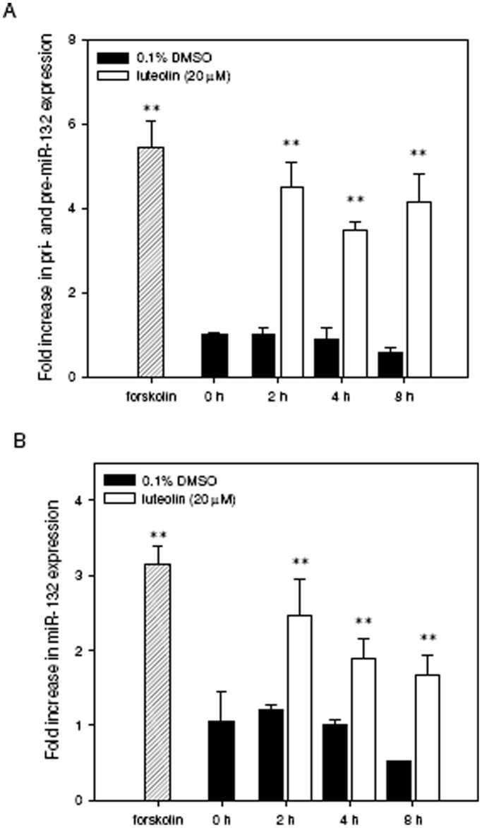
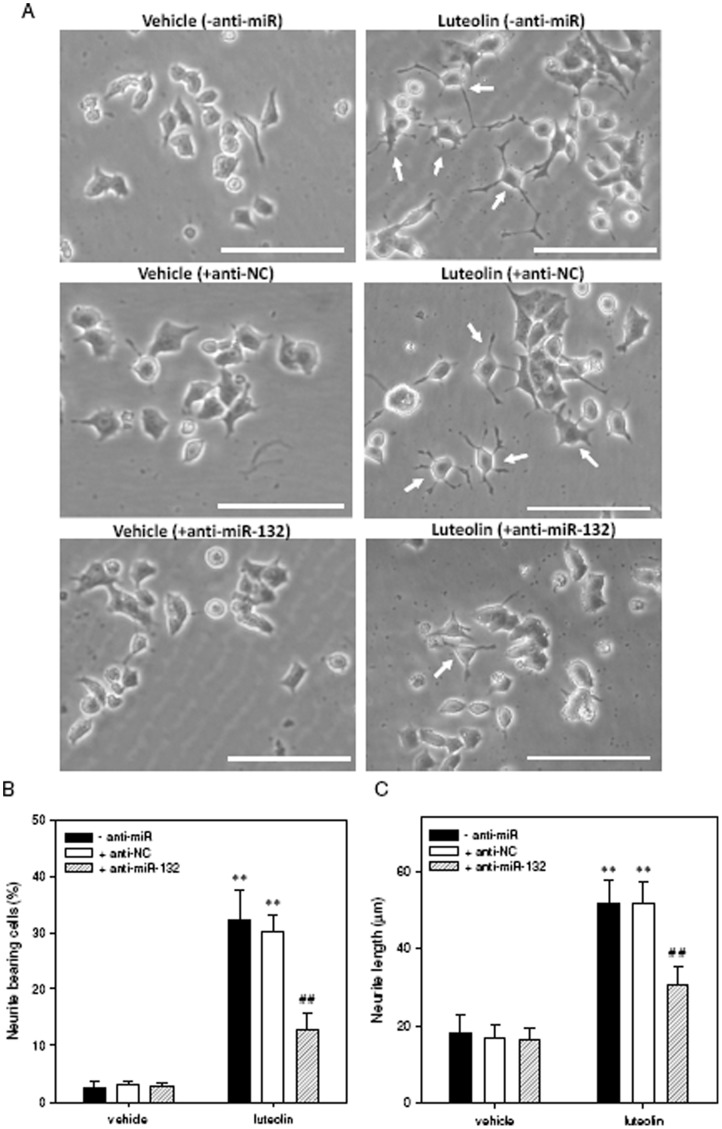
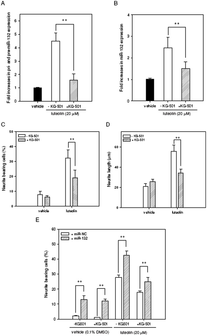
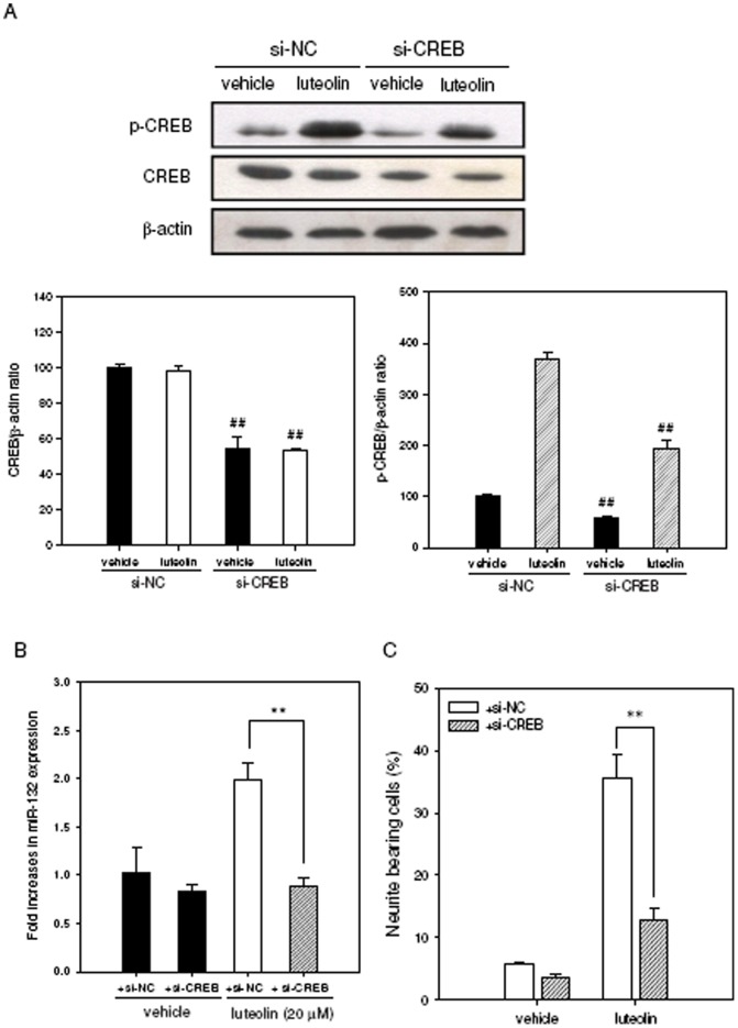
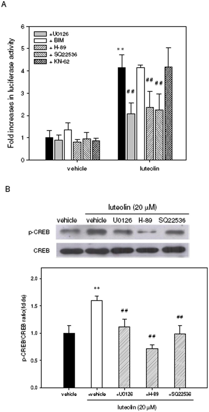
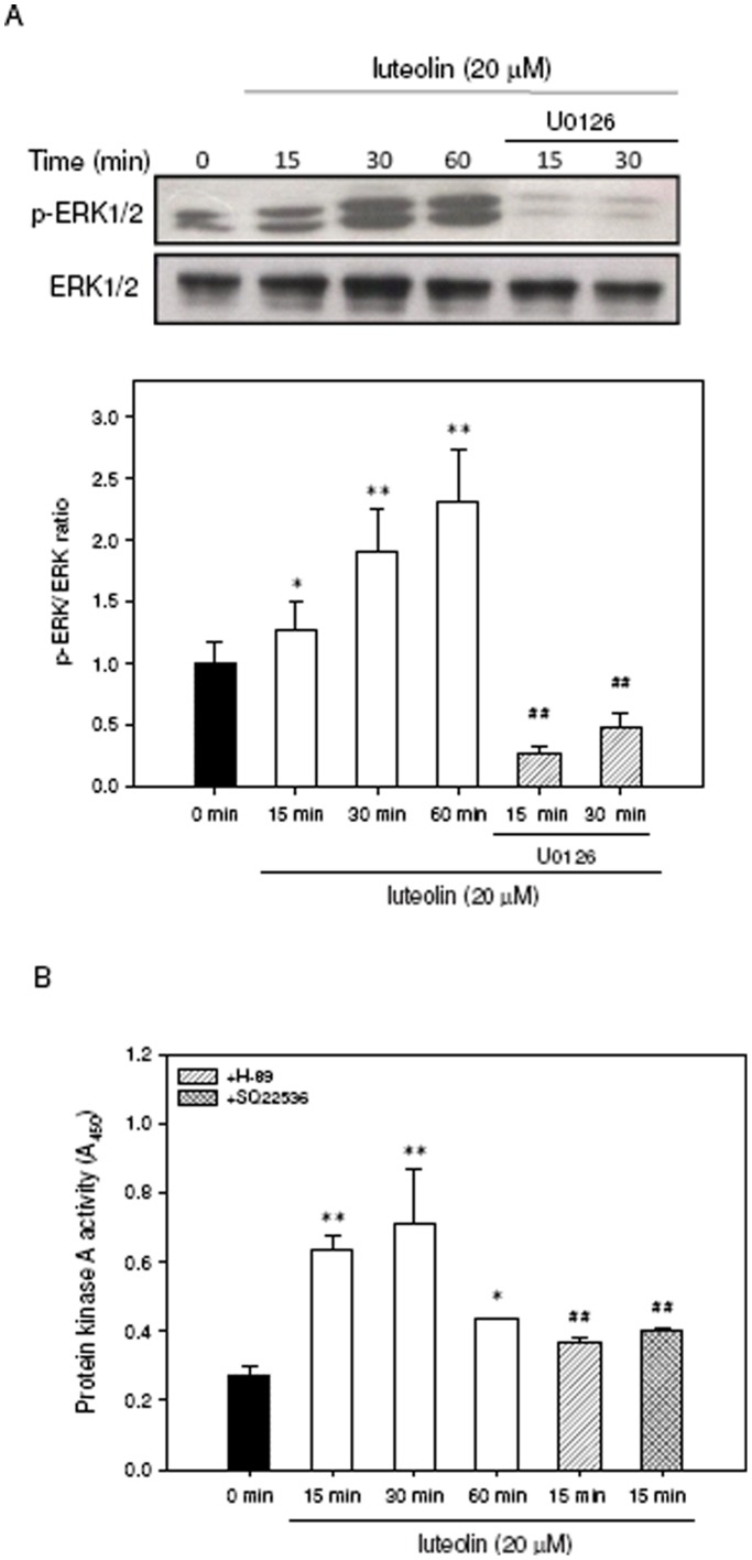
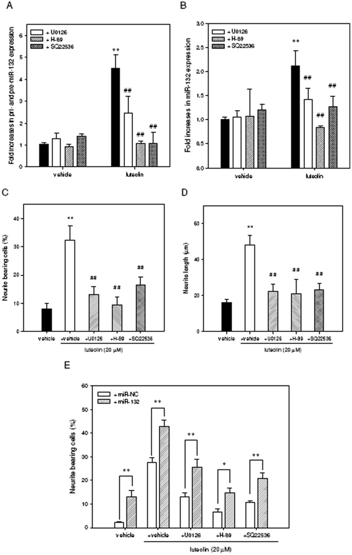
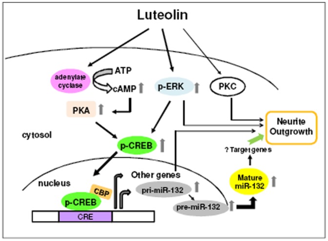
Luteolin (3',4',5,7-tetrahydroxyflavone), a food-derived flavonoid, has been reported to exert neurotrophic properties that are associated with its capacity to promote neuronal survival and neurite outgrowth. In this study, we report for the first time that luteolin induces the persistent expression of microRNA-132 (miR-132) in PC12 cells. The correlation between miR-132 knockdown and a decrease in luteolin-mediated neurite outgrowth may indicate a mechanistic link by which miR-132 functions as a mediator for neuritogenesis. Furthermore, we find that luteolin led to the phosphorylation and activation of cAMP response element binding protein (CREB), which is associated with the up-regulation of miR-132 and neurite outgrowth. Moreover, luteolin-induced CREB activation, miR-132 expression and neurite outgrowth were inhibited by adenylate cyclase, protein kinase A (PKA) and MAPK/ERK kinase 1/2 (MEK1/2) inhibitors but not by protein kinase C (PKC) or calcium/calmodulin-dependent protein kinase II (CaMK II) inhibitors. Consistently, we find that luteolin treatment increases ERK phosphorylation and PKA activity in PC12 cells. These results show that luteolin induces the up-regulation of miR-132, which serves as an important regulator for neurotrophic actions, mainly acting through the activation of cAMP/PKA- and ERK-dependent CREB signaling pathways in PC12 cells.


Conflict of interest statement

Competing Interests: The authors have declared that no competing interests exist.

Figures

Figure 1
Figure 1. Luteolin induces miR-132 expression in PC12 cells.
PC12 cells were seeded on poly-L-lysine-coated 6-well plates in low-serum medium (1% horse serum and 0.5% FBS) for 24 h prior to exposure to forskolin (10 µM) for 2 h, vehicle (0.1% DMSO) or luteolin (20 µM) for an additional 2–8 h. Cellular RNA was then prepared, and the levels of immature miR-132 (pri- and pre-miR-132) (A) and mature miR-132 (B) were detected by reverse transcription quantitative PCR as described in Materials and Methods. Data represent the mean ± SD from three independent experiments. **p<0.01 represents significant differences compared to vehicle-treated cells.
Figure 2
Figure 2. Knockdown of miR-132 expression attenuates luteolin-induced neurite outgrowth in PC12 cells.
PC12 cells were seeded on poly-L-lysine-coated 6-well plates in normal-serum medium for 24 h. Cells were then transfected with miR-132 antisense oligonucleotides (anti-miR-132, 150 pmol) or scramble antisense negative control (anti-NC, 150 pmol) for 24 h as described in Materials and Methods. After transfection, the PC12 cells were shifted to low-serum medium (1% horse serum and 0.5% FBS) and exposed to vehicle (0.1% DMSO) or luteolin (20 µM) for an additional 72 h. (A) Representative images of neurite outgrowth in PC12 cells. Cell morphology was observed using phase-contrast microscopy and photographed by the digital camera. Arrowheads indicate the neurite-bearing cells. Scale bar, 100 µm. (B) Neurite-bearing cells were analyzed as described in Materials and Methods. (C) The average maximal neurite length for each of the differentiated cells was analyzed by Image J software. Data represent the mean ± SD from three independent experiments. **p<0.01 represents significant differences compared to vehicle-treated cells. ##p<0.01 represents significant differences compared to antisense-untreated group (- anti-miR).
Figure 3
Figure 3. Luteolin increases the phosphorylation and activity of CREB in PC12 cells.
(A) PC12 cells were seeded on poly-L-lysine-coated 100 mm dishes in normal medium for 24 h and then shifted to low-serum medium (1% HS and 0.5% FBS) for 24 h prior to their exposure to the indicated agents. Adherent PC12 cells were treated with luteolin (20 µM) for 0–240 min. Phospho-CREB-Ser133 (p-CREB) and CREB proteins were analyzed by Western blotting as described in Materials and Methods. The immunoblot experiments were replicated at least three times, and a representative blot is shown. The normalized intensity of p-CREB versus CREB is presented as the mean ± SD of three independent experiments. *p<0.05 and **p<0.01 represents significant differences compared to the 0 min group. (B) PC12 cells were seeded on poly-L-lysine-coated 24-well plates in DMEM containing 10% horse serum and 5% FBS for 24 h. Cells were then transfected with a CRE-mediated luciferase reporter construct and Renilla luciferase control plasmid as described in Materials and Methods. After transfection, PC12 cells were treated with vehicle (0.1% DMSO) or luteolin (10 or 20 µM) for 8 h. Cells were harvested, and the luciferase activities were determined as described in Materials and Methods. The intensities of the luciferase reactions measured in the lysates of the transfectants were normalized to their Renilla luciferase control activity. Data represent the mean ± SD from three independent experiments. *p<0.05 and **p<0.01 represent significant differences compared to vehicle-treated cells.
Figure 4
Figure 4. Contribution of CREB activity to miR-132 up-regulation and neurite outgrowth in response to luteolin.
PC12 cells were seeded on poly-L-lysine-coated 6-well plates in normal medium for 24 h. Cells were then shifted to low-serum medium (1% horse serum and 0.5% FBS) and were pretreated with the CREB inhibitor KG-501 (10 µM) for 30 min. Cells were then exposed to vehicle (0.1% DMSO) or luteolin (20 µM) for 2 h. Cellular RNA was then prepared, and the levels of immature miR-132 (pri- and pre-miR-132) (A) and mature miR-132 (B) were detected by reverse transcription quantitative PCR as described in Materials and Methods. Data represent the mean ± SD from three independent experiments. **p<0.01 represents significant differences compared KG-501-non-treated cells. (C) PC12 cells were seeded on poly-L-lysine-coated 6-well plates in normal medium for 24 h and then shifted to low-serum medium (1% horse serum and 0.5% FBS) for 24 h prior to exposure to vehicle (0.1% DMSO) or luteolin (20 µM) for an additional 72 h. For the treatment of cells with the inhibitor, adherent cells were pre-incubated with KG-501 (10 µM) for 30 min and then exposed to vehicle (0.1% DMSO) or luteolin (20 µM) for an additional 72 h. Neurite-bearing cells were analyzed as described in Materials and Methods. (D) The average maximal neurite length for each of the differentiated cells was analyzed by Image J software. Data represent the mean ± SD from three independent experiments. **p<0.01 represents significant differences compared to the KG-501-non-treated group. (E) PC12 cells were transfected with miR-132 mimics (miR-132) or miRNA mimics negative control (miR-NC) for 24 h as described in Materials and Methods. For the treatment of cells with the inhibitor, transfected cells were pre-incubated with KG-501 (10 µM) for 30 min and then exposed to vehicle (0.1% DMSO) or luteolin (20 µM) for an additional 72 h. Neurite-bearing cells were analyzed as described in Materials and Methods. Data represent the mean ± SD from three independent experiments. **p<0.01 represents significant differences compared to miR-NC transfected group.
Figure 5
Figure 5. Effects of the CREB protein knockdown on the luteolin-mediated miR-132 induction and neurite outgrowth in PC12 cells.
PC12 cells were transfected transiently with siRNA negative control (si-NC) or with CREB-specific siRNA (si-CREB) before vehicle and luteolin (20 µM) treatment. (A) CREB and Phospho-CREB-Ser133 (p-CREB) proteins were determined by Western blotting analysis after luteolin treatment for 60 min. β-actin protein is as an internal control. The immunoblot experiments were replicated at least three times, and a representative blot is shown. The normalized intensity of CREB or p-CREB versus β-actin is presented as the mean ± SD of three independent experiments. ##p<0.01 represents significant differences compared to the siRNA negative control-transfected group. (B) Effect of CREB knockdown on miR-132 levels after luteolin treatment for 2 h. The levels of miR-132 were determined by RT-Q-PCR as described in Materials and Methods. (C) Effect of CREB knockdown on neurite outgrowth of PC12 cells after luteolin treatment for 72 h. Neurite-bearing cells were analyzed as described in Materials and Methods. Data represent the mean ± SD from three independent experiments. **p<0.01 represents significant differences compared to the siRNA negative control-transfected group.
Figure 6
Figure 6. Involvement of ERK, PKC, CaMK and cAMP-dependent PKA signaling in luteoln-mediated CREB activation.
(A) PC12 cells were seeded on poly-L-lysine-coated 24-well plates in DMEM containing 10% horse serum and 5% FBS for 24 h. Cells were then transfected with a CRE-mediated luciferase reporter construct and Renilla luciferase control plasmid as described in Materials and Methods. After transfection, the cells were pre-treated for 30 min with the following inhibitors: 10 µM U0126, 2.5 µM BIM, 10 µM H-89, 500 µM SQ22536, 10 µM KN-62 or vehicle (0.1% DMSO) followed by exposure to luteolin (20 µM) for 8 h. The intensities of the luciferase reactions measured in the lysates of the transfectants were normalized to their Renilla luciferase control activity. (B) PC12 cells were seeded on poly-L-lysine-coated 100 mm dishes in normal medium for 24 h and then shifted to low-serum medium (1% horse serum and 0.5% FBS) for an additional 24 h of culture. Cells were treated with the inhibitors U0126, H-89 or SQ22536 for 30 min prior to their exposure to vehicle (0.1% DMSO) or luteolin (20 µM) for 60 min. Phospho-CREB-Ser133 (p-CREB) and CREB were analyzed by Western blotting as described in Materials and Methods. The immunoblot experiments were replicated at least three times, and a representative blot is shown. The normalized intensity of p-CREB versus CREB is presented as the mean ± SD of three independent experiments. **p<0.01 represents significant differences compared to vehicle-treated cells. ##p<0.01 represents significant differences compared to the respective inhibitor-non-treated group.
Figure 7
Figure 7. Effects of luteolin on the ERK phosphorylation and PKA activity.
PC12 cells were seeded on poly-L-lysine-coated 100 mm dishes in normal medium for 24 h and then shifted to low serum medium (1% HS and 0.5% FBS) for 24 h prior to exposure to indicated agents. (A) Cells were treated with luteolin (20 µM) for 0 min, 15 min, 30 min and 60 min. For inhibitor treatment, cells were treated with the inhibitors U0126 for 30 min prior to exposure to luteolin (20 µM) for 15 min and 30 min. Phospho-ERK1/2 (p-ERK1/2) and total ERK1/2 proteins were analyzed by Western blotting as described in Materials and Methods. The experiments were replicated at least three times and a representative blot was shown. (B) Cells were incubated with luteolin (20 µM) for indicated period and PKA activity was detected using ELISA kit as described in Materials and Methods. For inhibitor treatment, cells were treated with the inhibitors H-89 (10 µM) and SQ22536 (500 µM) for 30 min prior to exposure to luteolin (20 µM) for 15 min. Data represent the mean ± SD of three independent experiments. *p<0.05 and **p<0.01 represents significant differences compared with 0 min group. ##p<0.01 represents significant differences compared to the respective inhibitor-non-treated group.
Figure 8
Figure 8. Contribution of ERK- and cAMP-dependent PKA signaling pathways to luteolin–induced miR-132 up-regulation and neurite outgrowth in PC12 cells.
PC12 cells were seeded on poly-L-lysine-coated 6-well plates in normal medium for 24 h. Cells were then shifted to low-serum medium (1% horse serum and 0.5% FBS) and then pretreated with the inhibitors U0126 (10 µM), H-89 (10 µM) or SQ22536 (500 µM) for 30 min followed by exposure to vehicle or luteolin (20 µM) for 2 h. Cellular RNA was then prepared, and the levels of immature miR-132 (pri- and pre-miR-132) (A) and mature miR-132 (B) was detected by reverse transcription quantitative PCR as described in Materials and Methods. Data represent the mean ± SD from three independent experiments. **p<0.01 represents significant differences compared to vehicle-treated cells. ##p<0.01 represents significant differences compared to the respective inhibitor-non-treated group. (C) PC12 cells were seeded on poly-L-lysine-coated 6-well plates in normal medium for 24 h. Cells were then shifted to low-serum medium (1% horse serum and 0.5% FBS) for 24 h and then pre-treated for 30 min with the indicated inhibitors followed by exposure to vehicle or luteolin (20 µM) for 72 h. Neurite-bearing cells were analyzed as described in the Materials and Methods. (D) The average maximal neurite length for each of the differentiated cells was analyzed by Image J software. Data represent the mean ± SD from three independent experiments. **p<0.01 represents significant differences compared to vehicle-treated cells. ##p<0.01 represents significant differences compared to the respective inhibitor-non-treated group. (E) PC12 cells were transfected with miR-132 mimics (miR-132) or miRNA mimics negative control (miR-NC) for 24 h as described in Materials and Methods. For the treatment of cells with the inhibitor, transfected cells were pre-incubated with indicated inhibitors for 30 min and then exposed to luteolin (20 µM) for an additional 72 h. Neurite-bearing cells were analyzed as described in Materials and Methods. Data represent the mean ± SD from three independent experiments. *p<0.05 and **p<0.01 represent significant differences compared to miR-NC transfected group.
Figure 9
Figure 9. Effects of TrkA and EGFR signaling pathways on the luteolin-induced neurite outgrowth in PC12 cells.
PC12 cells were seeded on poly-L-lysine-coated 6-well plates in normal medium for 24 h. Cells were then shifted to low-serum medium (1% horse serum and 0.5% FBS) for 24 h and then pre-treated for 30 min with the TrkA antagonist K252a (100 nM) or EGFR inhibitor AG1478 (2 µM) and then exposed to vehicle (0.1% DMSO) or luteolin (20 µM) for an additional 72 h. Neurite-bearing cells were analyzed as described in Materials and Methods. Data represent the mean ± SD from three independent experiments. **p<0.01 represents significant differences compared to vehicle-treated cells.
Figure 10
Figure 10. Hypothetical mechanism of luteolin mediation of neurite outgrowth in PC12 cells.
Luteolin induces the up-regulation of miR-132, which may serve as a mediator for neurite outgrowth through the activation of cAMP/PKA- and ERK-dependent CREB signaling pathways in PC12 cells. In addition, ERK- or PKC-dependent but CREB/miR-132-independent pathways may also partially contribute to the mediation of neurite outgrowth by luteolin in PC12 cells.

Similar articles

Cited by

References

    1. Ambros V (2008) The evolution of our thinking about microRNAs. Nat Med 14: 1036–1040. - PubMed
    1. Baek D, Villen J, Shin C, Camargo FD, Gygi SP, et al. (2008) The impact of microRNAs on protein output. Nature 455: 64–71. - PMC - PubMed
    1. Friedman RC, Farh KK, Burge CB, Bartel DP (2009) Most mammalian mRNAs are conserved targets of microRNAs. Genome Res 19: 92–105. - PMC - PubMed
    1. Lim LP, Lau NC, Garrett-Engele P, Grimson A, Schelter JM, et al. (2005) Microarray analysis shows that some microRNAs downregulate large numbers of target mRNAs. Nature 433: 769–773. - PubMed
    1. Bartel DP (2004) MicroRNAs: genomics, biogenesis, mechanism, and function. Cell 116: 281–297. - PubMed

Publication types

MeSH terms