. 2012;7(8):e43685.
doi: 10.1371/journal.pone.0043685. Epub 2012 Aug 20.

Acetylcholine-binding protein in the hemolymph of the planorbid snail Biomphalaria glabrata is a pentagonal dodecahedron (60 subunits)

Affiliations

Acetylcholine-binding protein in the hemolymph of the planorbid snail Biomphalaria glabrata is a pentagonal dodecahedron (60 subunits)

Michael Saur et al. PLoS One. 2012.

Abstract

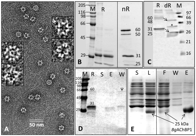
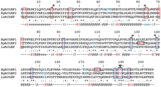
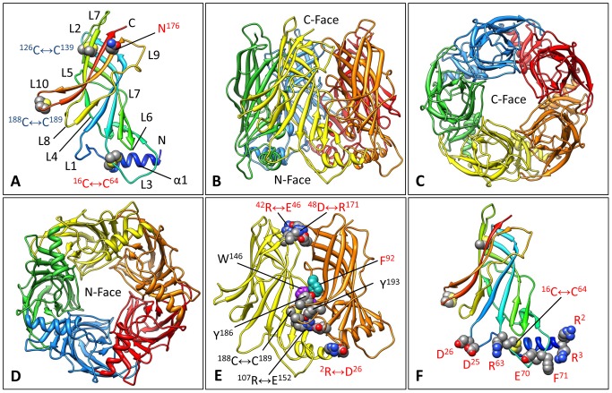
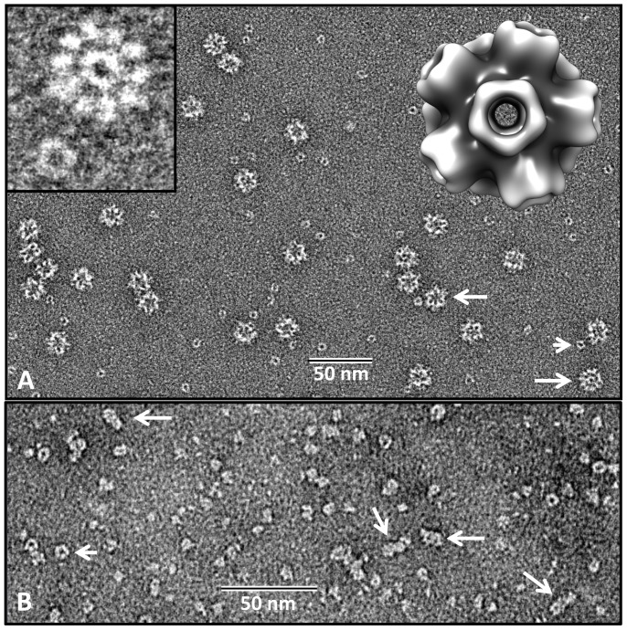
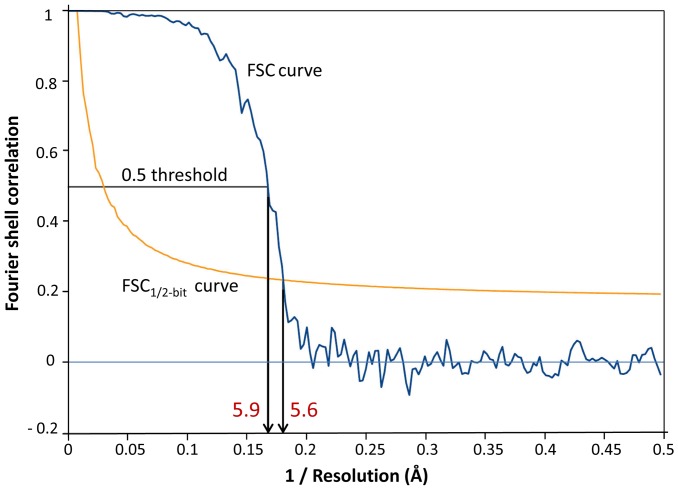
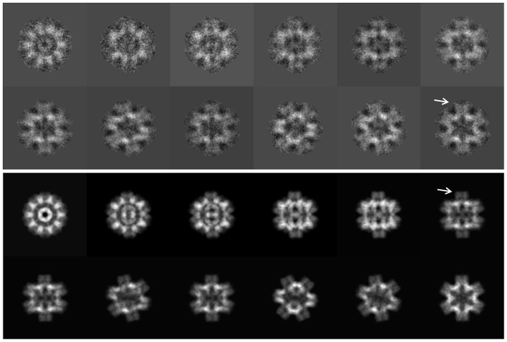
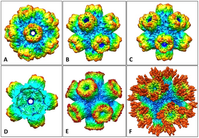
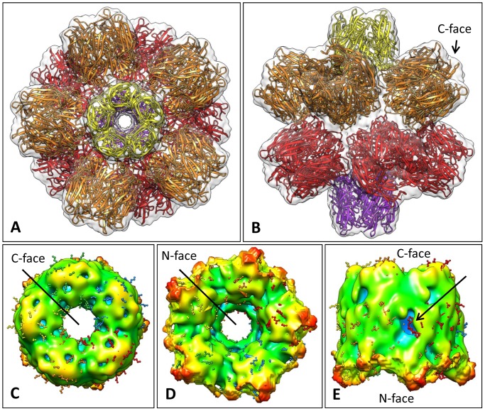
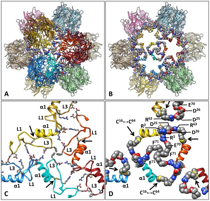
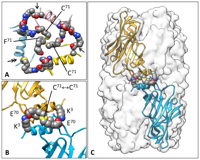
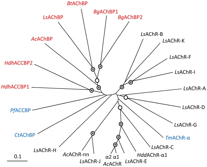
Nicotinic acetylcholine receptors (nAChR) play important neurophysiological roles and are of considerable medical relevance. They have been studied extensively, greatly facilitated by the gastropod acetylcholine-binding proteins (AChBP) which represent soluble structural and functional homologues of the ligand-binding domain of nAChR. All these proteins are ring-like pentamers. Here we report that AChBP exists in the hemolymph of the planorbid snail Biomphalaria glabrata (vector of the schistosomiasis parasite) as a regular pentagonal dodecahedron, 22 nm in diameter (12 pentamers, 60 active sites). We sequenced and recombinantly expressed two ∼25 kDa polypeptides (BgAChBP1 and BgAChBP2) with a specific active site, N-glycan site and disulfide bridge variation. We also provide the exon/intron structures. Recombinant BgAChBP1 formed pentamers and dodecahedra, recombinant BgAChBP2 formed pentamers and probably disulfide-bridged di-pentamers, but not dodecahedra. Three-dimensional electron cryo-microscopy (3D-EM) yielded a 3D reconstruction of the dodecahedron with a resolution of 6 Å. Homology models of the pentamers docked to the 6 Å structure revealed opportunities for chemical bonding at the inter-pentamer interfaces. Definition of the ligand-binding pocket and the gating C-loop in the 6 Å structure suggests that 3D-EM might lead to the identification of functional states in the BgAChBP dodecahedron.


Conflict of interest statement

Competing Interests: The authors have declared that no competing interests exist.

Figures

Figure 1
Figure 1. Electron microscopy and SDS-PAGE of BgAChBP.
(A) Negative staining EM of chromatographically purified rosette protein. 2% uranyl acetate was applied. Enlarged examples are also shown; note the peripheral protrusions (arrows). (B) Chromatographically enriched BgAChBP (R, from “rosette protein”) under reducing (R) and non-reducing (nR) conditions. Note that in the second case, some material migrates as subunit dimers, whereas other material remains in the monomeric state. M, marker proteins. (C) BgAChBP in glycosylated (R) and deglycosylated (dR) form, indicating that the 31 and 60 kDa bands represent the glycosylated and the 25 and 50 kDa bands the deglycosylated form (arrows). Asterisk, N-glycosidase F as deduced from controls; M, marker proteins. (D) BgAChBP material extracted through binding to amorphous CaCO3. R, BgAChBP starting material; S, supernatant after extraction; E, fraction eluted from CaCO3 by EDTA; W, wash buffer prior to EDTA extraction; M, marker proteins. Note that the wash buffer contains subunit dimers (arrow) and the slower migrating portion of the 31 kDa band, whereas the eluent contains its faster migrating portion. (E) Recombinant expression of BgAChBP1. S, bacterial cell supernatant; L, bacterial cell lysate; F, flow-through of Ni column; W, wash buffer of Ni column; E, eluent of Ni column, rich in recombinant BgAChBP1.
Figure 2
Figure 2. Sequence alignment of the BgAChBP subunits and LsAChBP (from Lymnaea stagnalis).
The red residues are addressed in the main text in the context of ligand binding (blue boxes), inter-pentamer linkage (red boxes), N-glycan binding (black boxes), or disulfide bridges (arrow symbols). The blue residues probably form salt bridges between adjacent subunits within the same pentamer (see Fig. 4E). Note the specific exchanges Y92→F92 in BgAChBP1 and Y193→F193 in BgAChBP2. Also note the strictly conserved disulfide bridges stabilizing the eponymous Cys-loop L7 and the gating C-loop L10, the putative additional disulfide bridge C16↔C64 in BgAChBP1, and the single cysteine C71 in BgAChBP2. (Chain-specific residue numbers are given.) The secondary structure elements predicted from the published crystal structures are also indicated (L, loop). The short helix following strand β2 and marked in blue is absent in the molecular models of the BgAChBP subunits. Genbank entries JQ814367, JQ814368, AAK64377.
Figure 3
Figure 3. Gene structure of BgAChBP1 and BgAChBP2.
Data retrieved from the preliminary B. glabrata genomic data (http://129.24.144.93/blast_bg/2index.html). Exon 1 and the first three amino acids encoded by exon 2 belong to the signal peptide, as deduced from evaluation in SignalP, and N-terminal protein sequencing (see Table 1). Genbank entries JQ814367, JQ814368.
Figure 4
Figure 4. Homology models of BgAChBP1.
(A) The modeled subunit showing the N-terminal helix α1, the 10-stranded β-sandwich, the connecting loops L1 to L10, the three disulfide bridges, and the potential attachment site for N-linked glycans. (B–D) The modeled pentamer in side view (B) and the two different top views (C, D). The C-face is defined by the five C-termini and eponymous Cys-loops L7, the N-face contains the five N-termini and α1 helices. (E) Two neighboring subunits extracted from the modeled pentamer, with amino acid residues in the principal side of the ligand-binding pocket highlighted. Note that instead of phenylalanine F92, other AChP-LBD/AChBP members possess a tyrosine. Putative salt bridges connecting both subunits are also shown. (F) The modeled subunit showing the three disulfide bridges and the amino acids presumably involved in inter-pentamer contacts. Red labels mark features that are specific for BgAChBP. (PDB-ID of the BbAChBP1 pentamer: 4AOD; PDB-ID of the BgAChBP2 pentamer: 4AOE).
Figure 5
Figure 5. Electron microscopy of recombinant BgAChBP1 and BgAChBP2 as expressed in E. coli.
(A) Recombinant BgAChBP1 pentamers (short arrow) and dodecahedra (large arrows). Left insert, enlarged view along the five-fold symmetry axis of a recombinant dodecahedron and a single pentamer, respectively. Right insert, 3D reconstruction (resolution ∼20 Å) from ∼3000 negatively stained particles of the recombinant BgAChBP1 dodecahedron. (B) Recombinant BgAChBP2 pentamers (short arrow) and presumed di-pentamers (large arrows). In several independent expression experiments, not a single dodecahedron was detected in the electron microscope.
Figure 6
Figure 6. Resolution determination of the final 3D reconstruction of the BgAChBP dodecahedron.
The results for the density map in Fig. 8A–D are shown. Compared to the 5.9 Å obtained by the FSC0.5 criterion, the 5.6 Å determined with the FSC1/2-bit criterion might be too optimistic. Therefore, this density map is further referred to as the “6-Å cryo-EM structure”.
Figure 7
Figure 7. 3D-EM processing of BgAChBP.
Characteristic class sum images (top), and the corresponding reprojections (bottom) of the density map shown in Fig. 8A–D. Note peripheral protrusions in class sum images (upper arrow) that are absent in the corresponding reprojections (lower arrow). This disappearance results from masking for avoiding noise bias (see Methods).
Figure 8
Figure 8. Cryo-EM structures of the BgAChBP dodecahedron.
(A) The final 6-Å cryo-EM structure viewed along one of six five-fold symmetry axes, exposing a central channel 2 nm in width. The overall diameter of the particle is 22 nm. (B) View along one of the 15 two-fold symmetry axes, exposing one of the 30 edges between two adjacent pentamers. (C) View along one of the 10 three-fold symmetry axes, exposing one of the 20 vertices at the junction between three neighboring pentamers. (D) Cut-open view to expose the central cavity (with the cut perpendicular to one of the five-fold axes of symmetry). (E) Unsharpened, unfiltered, unmasked version of the 6-Å cryo-EM structure to show the peripheral “fuzz” interpreted as glycans. (F) A 5.8-Å cryo-EM structure independently obtained from the same dataset. In this case, over-fitting of noise was accepted to avoid the loss of the putative carbohydrate side chains. (EM-DB ID of the 6-Å cryo-EM structure of the BgAChBP dodecahedron: EMD-2055).
Figure 9
Figure 9. Docking of the molecular model of BgAChBP1 into the 6-Å cryo-EM structure. (A)
The molecular model of the dodecahedron depicted along one of the five-fold symmetry axes. The 6-Å cryo-EM structure is shown in opaque to demonstrate the fitting. (B) The same model, viewed along one of the two-fold symmetry axes. (C) Top view of a pentamer extracted from the 6-Å cryo-EM structure, exposing the C-face; the docked molecular model is shown in ball & stick mode. (D) The same structure but rotated 180° to expose the N-face (the view from inside the central cavity). (E) The same structure in side view. Note the large cavity (blue) representing one of the five ligand-binding pockets, and the gating C-loop (arrow). Also note that in the cryo-EM structure the C-loop is too short to fully embed the molecular model (red) which might be due to its flexibility. (PDB-ID of the BbAChBP1 pentamer: 4AOD).
Figure 10
Figure 10. Putative inter-pentamer interfaces in a BgAChBP1 dodecahedron.
(A) Molecular model of the dodecahedron, viewed along one of the three-fold symmetry axes. The amino acid appositions providing opportunities for inter-pentamer bonding are highlighted. (B) The same view as in (A), with most of the three pentamers joined in the vertex removed. They are indicated by the yellow, light blue and orange red helices α1. In the center, the trigonal ring that makes the inter-pentamer contact is visible. (C) The residues that together with helix α1 form the trigonal ring, shown in ball & stick mode. In addition, loops L1 and L3 are indicated. Subunits of similar color stem from the same pentamer. Note the position of the C16↔C64 bridge (arrows). (D) Details of the central and three adjacent trigonal rings. Each ring connects three pentamers at their common vertex (via the F71 cluster, three salt bridges R3↔E70 and three salt bridges D25↔R63). Alternatively, two parallel salt bridges D25↔R63 can be considered as connection between two adjacent pentamers across their common edge. (PDB-ID of the BbAChBP1 pentamer: 4AOD).
Figure 11
Figure 11. Putative inter-pentamer interfaces with BgAChBP2.
(A) Combination of a disulfide-bridged BgAChBP2 subunit dimer and a BgAChBP1 monomer in the vertex of a hypothetical hetero-dodecahedron. Note that in the trigonal ring, the covalent intra-subunit C16↔C64 bridge (double arrow) is lacking at two positions (arrows), because it is absent in BgAChBP2 (that shows A16 and S64 instead). (B) Putative subunit dimer of BgAChBP2 stabilized by an inter-subunit disulfide bridge C71↔C71 and two flanking salt bridges K3↔E70. The same subunit model as in (A) was applied. (C) Density map of a speculative BgAChBP2 di-pentamer (opaque), simulated at 6 Å resolution from a molecular model of the di-pentamer. This model, of which one out of five subunit dimers is shown here, was constructed in silico by joining two BgAChBP2 pentamers at their free C71 residues. (PDB-ID of the BgAChBP2 pentamer: 4AOE).
Figure 12
Figure 12. Radial phylogenetic tree of AChBP, ACCBP and AChR-LBD.
Sequences of gastropod AChBP and ACCBP polypeptides (marked in red) are compared here to sequences of gastropod AChR-LBD polypeptides (marked in black). Sequences from the pearl oyster P. fucata, the polychaete annelid C. telata and the electric ray T. marmorata are also included (marked in blue). Nodes bootstrap-supported above 900 are indicated by a circle, those above 990 are additionally marked by an asterisk (1000 replicas were calculated). Note that the gastropod AChBP complex is clearly separated from the gastropod nAChR-LBD complex. Also note that BgAChBP1 and BgAChBP2 show a clear sister-group relationship, suggesting that they arose from a gene duplication event that occurred within the Planorbidae. The neighbor-joining method implemented in Clustal W was applied. A corresponding identity matrix is shown in Table 2. Ac, Aplysia californica (genbank entries AAL37250, AAL37251, AAL78648, AAL78649); Bg, Biomphalaria glabrata (JQ814367, JQ814368); Bt, Bulinus truncatus (PDB-ID 2BJ0); Ct, Capitella teleta (EY637248); Hdd, Haliotis discus discus (ABO26693); Hdh, Haliotis discus hanei (ABU51880, ABU62818); Ls, Lymnaea stagnalis (AAK64377, ABA60380 to ABA60390); Pf, Pinctada fucata (ABF13208); Tm, Torpedo marmorata (PDB-ID 2BG9); LBD, ligand binding domain.

References

    1. Brejc K, van Dijk WJ, Klaassen RV, Schuurmans M, van Der Oost J, et al. (2001) Crystal structure of an ACh-binding protein reveals the ligand-binding domain of nicotinic receptors. Nature 411: 269–276. - PubMed
    1. Hansen SB, Talley TT, Radic Z, Taylor P (2004) Structural and ligand recognition characteristics of an acetylcholine-binding protein from Aplysia californica . J Biol Chem 279: 24197–24202. - PMC - PubMed
    1. Celie PH, Klaassen RV, van Rossum-Fikkert SE, van Elk R, van Nierop P, et al. (2005) Crystal structure of acetylcholine-binding protein from Bulinus truncatus reveals the conserved structural scaffold and sites of variation in nicotinic acetylcholine receptors. J Biol Chem 280: 26457–26466. - PubMed
    1. McCormack T, Petrovich RM, Mercier KA, DeRose EF, Cuneo MJ, et al. (2010) Identification and functional characterization of a novel acetylcholine-binding protein from the marine annelid Capitella teleta . Biochemistry 49: 2279–2287. - PMC - PubMed
    1. Lester HA, Dibas MI, Dahan DS, Leite JF, Dougherty DA (2004) Cys-loop receptors: new twists and turns. Trends Neurosci 27: 329–336. - PubMed

Publication types