

Troglostrongylus brevior and Troglostrongylus subcrenatus (Strongylida: Crenosomatidae) as agents of broncho-pulmonary infestation in domestic cats
- PMID: 22916686
- PMCID: PMC3469345
- DOI: 10.1186/1756-3305-5-178
Troglostrongylus brevior and Troglostrongylus subcrenatus (Strongylida: Crenosomatidae) as agents of broncho-pulmonary infestation in domestic cats
Abstract
Background: Aelurostrongylus abstrusus is currently regarded as the main metastrongyloid infesting domestic cats, whereas the reports of Troglostrongylus spp. in domestic and wild felids largely remain anecdotic. This paper reports on pulmonary infestation caused by Troglostrongylus brevior and Troglostrongylus subcrenatus in two kittens and describes, for the first time, associated clinical presentations and pathological features. Morphometrical, molecular and phylogenetic analyses have also been conducted to differentiate here the examined Troglostrongylus species from A. abstrusus, towards a clearer delineation of metastrongyloids affecting cats.
Methods: Two kittens were referred for respiratory distress and hospitalized with a diagnosis of severe aelurostrongylosis, based on the presence of metastrongyloid larvae in the faeces. Despite prompt treatment, kittens died within 48 hours. Both kittens were submitted to necropsy to determine the cause of death.
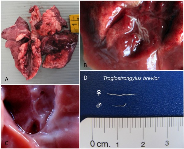
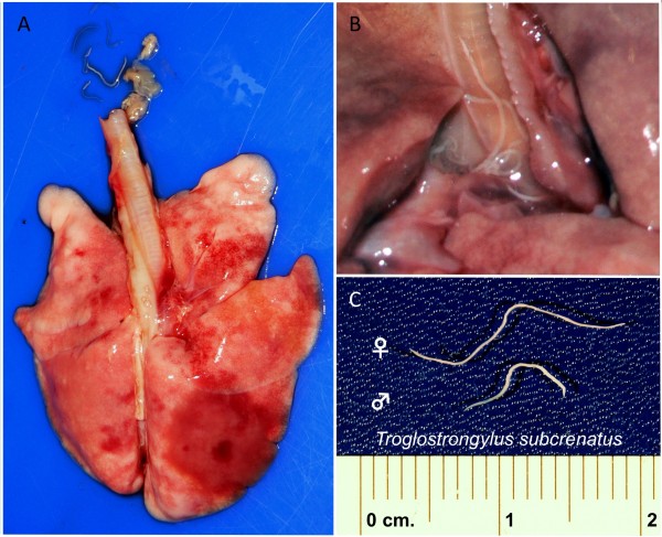
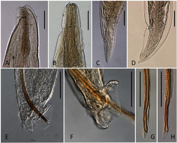
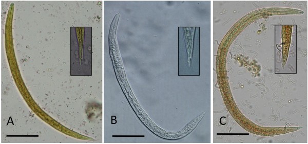
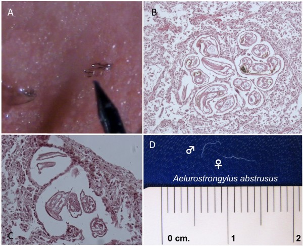
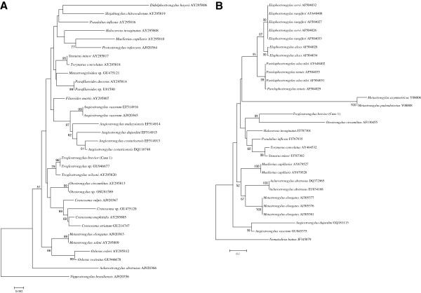
Results: At necropsy, nematode specimens were found in the trachea, bronchi and bronchioles and were associated with respiratory signs (i.e., dyspnoea, polypnea, severe coughing and nasal discharge). Morphology and measurements of adult parasites found allowed the unequivocal identification of T. brevior and T. subcrenatus, even if first stage larvae were rather similar to those of A. abstrusus. Briefly, T. brevior and T. subcrenatus larvae were shorter in length and lacking the typical knob-like terminal end of A. abstrusus. Molecular and phylogenetic analyses corroborated morphological identification and provided data on mitochondrial and ribosomal DNA genes of T. brevior.
Conclusions: Data presented here indicate that T. brevior and T. subcrenatus may cause major respiratory distress in domestic cats. Consequently, these two species should be included, along with A. abstrusus, in the differential diagnosis of cat bronchopulmonary affections and treatment protocols need to be evaluated. Through research on the biology, epidemiology and control of Troglostrongylus spp. infestations in domestic cats are advisable to implement current knowledge on these neglected metastrongyloids.
Figures
References
-
- Anderson RC. Their Development and Transmission. 2. New York: CABI Publishing; 2000. Nematode Parasites of Vertebrates.
-
- Bowman DD, Hendrix CM, Lindsay DS, Barr SC. Feline Clinical Parasitology. Ames: Iowa State University Press; 2002.
Publication types
MeSH terms
Substances
Associated data
- Actions
- Actions
- Actions
LinkOut - more resources
Full Text Sources
Medical
Molecular Biology Databases
Miscellaneous
