

Physiopathology of intratendinous calcific deposition
- PMID: 22917025
- PMCID: PMC3482552
- DOI: 10.1186/1741-7015-10-95
Physiopathology of intratendinous calcific deposition
Abstract
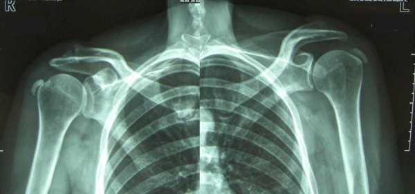
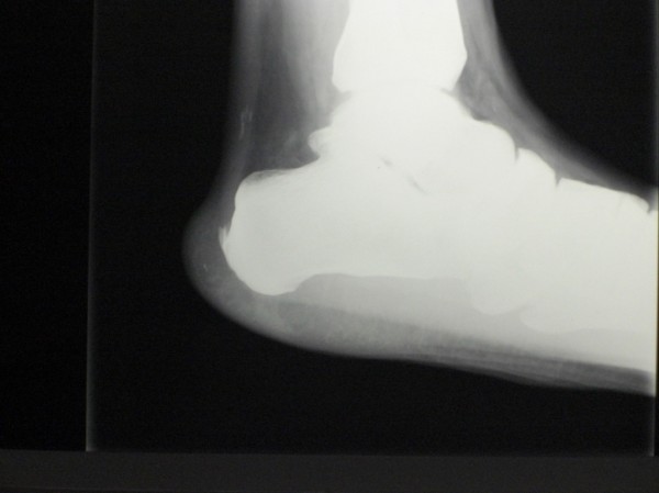
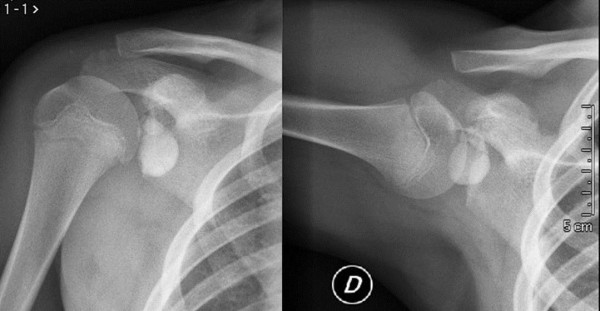
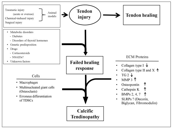
In calcific tendinopathy (CT), calcium deposits in the substance of the tendon, with chronic activity-related pain, tenderness, localized edema and various degrees of decreased range of motion. CT is particularly common in the rotator cuff, and supraspinatus, Achilles and patellar tendons. The presence of calcific deposits may worsen the clinical manifestations of tendinopathy with an increase in rupture rate, slower recovery times and a higher frequency of post-operative complications. The aetiopathogenesis of CT is still controversial, but seems to be the result of an active cell-mediated process and a localized attempt of the tendon to compensate the original decreased stiffness. Tendon healing includes many sequential processes, and disturbances at different stages of healing may lead to different combinations of histopathological changes, diverting the normal healing processes to an abnormal pathway. In this review, we discuss the theories of pathogenesis behind CT. Better understanding of the pathogenesis is essential for development of effective treatment modalities and for improvement of clinical outcomes.
Figures
References
-
- Coleman BD, Khan KM, Kiss ZS, Bartlett J, Young DA, Wark JD. Open and arthroscopic patellar tenotomy for chronic patellar tendinopathy. A retrospective outcome study. Am J Sports Med. 2000;28:183–190. - PubMed
Publication types
MeSH terms
LinkOut - more resources
Full Text Sources
Other Literature Sources
Medical
