. 2012 Nov;215(5):668-680.e1.
doi: 10.1016/j.jamcollsurg.2012.06.414. Epub 2012 Aug 21.

The role of extracellular signal-related kinase during abdominal aortic aneurysm formation

Affiliations

The role of extracellular signal-related kinase during abdominal aortic aneurysm formation

Abhijit Ghosh et al. J Am Coll Surg. 2012 Nov.

Abstract

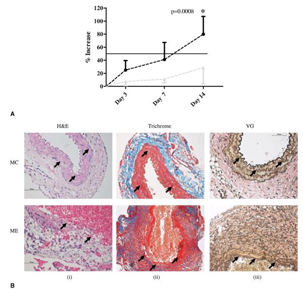
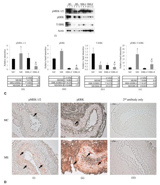
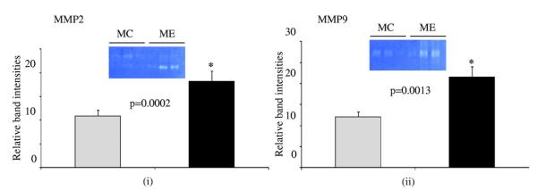
Background: It is hypothesized that activation of extracellular signal-related kinase (ERK) is critical in activating matrix metalloproteinases (MMPs) during abdominal aortic aneurysm (AAA) formation.

Study design: C57BL/6 male mice underwent either elastase or heat-inactivated elastase aortic perfusion (n = 9 per group). Mouse aortic smooth muscle cells were transfected with ERK-1 and 2 siRNA along with or without elastase treatment. Mouse and human aortic tissue were analyzed by Western blots, zymograms, and immunohistochemistry, and statistical analysis was done using Graphpad and Image J softwares.

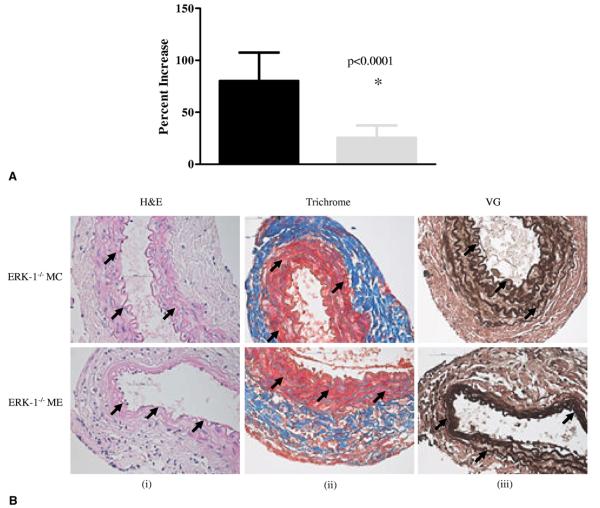
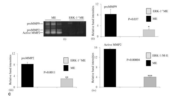
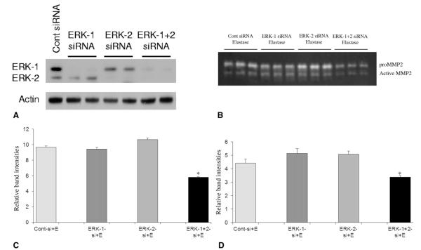
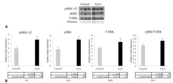
Results: Western blot and immunohistochemistry documented increased phospho-mitogen-activated protein kinase kinase-1/2 (pMEK-1/2; 153%, p = 0.270 by Western) and pERK (171%, p = 0.004 by Western blot) in the elastase perfused aortas. Male ERK-1(-/-) mice underwent elastase perfusion, and aortic diameter was determined at day 14. ERK-1(-/-) mice failed to develop AAA, and histologic analysis depicted intact collagen and elastin fibers in the aortas. Zymography of aortas of elastase-treated ERK-1(-/-) mice showed lower levels of proMMP2 (p < 0.005) and active MMP2 (p < 0.0001), as well as proMMP9 (p = 0.037) compared with C57BL/6 mice. siRNA transfection of ERK-1 and -2 significantly reduced formation of pro- and active MMP2 (p < 0.01 for both isoforms) in aortic smooth muscle cells treated with elastase in vitro. Human AAA tissue had significantly elevated levels of pMEK-1/2 (150%, p = 0.014) and pERK (159%, p = 0.013) compared with control tissues.

Conclusions: The MAPK (mitogen-activated protein kinase)/ERK pathway is an important modulator of MMPs during AAA formation. Targeting the ERK pathway by reagents that inhibit either the expression or phosphorylation of ERK isoforms could be a potential therapy to prevent AAA formation.


Figures

Figure 1
Figure 1
(A) Abdominal aortic aneurysm phenotype. Increase in aortic diameter in ME vs MC mice, p < 0.001 at day 14. Black line, ME mice; gray line, MC mice. (B) (i-iii) Histology of ME and MC mice. Arrows indicate aortic wall structure (hematoxylin and eosin), collagen (Trichrome), and elastin staining (Verhoff’s van Gieson, VG), magnification ×400. (C) (i-v) Western blot and densitometric analysis showing phosphorylation of MEK-1/2 (pMEK-1/2), and ERK (pERK) (ii, iii) and total ERK (T-ERK) (iv) in ME, MC, ERK-1−/− aorta perfused with elastase (ERK-E) and ERK-1−/− aorta perfused with heat-inactivated elastase (ERK-C) mice. The p values and percent change are given below the respective graphs. Each lane is a pool of 3 mice. Lane 3 was omitted for statistical calculations because it did not show any band. Densitometric analysis of the Western blot, p values, and percent change are tabulated below each graph. (D) (i-iii) Immunohistochemistry showing increased pMEK-1/2 and pERK in ME vs MC mice at day 14, magnification ×400. ERK, extracellular signal-related kinase; MC, male control mice; ME, male elastase infused mice; MEK, mitogen-activated protein kinase kinase.
Figure 1
Figure 1
(A) Abdominal aortic aneurysm phenotype. Increase in aortic diameter in ME vs MC mice, p < 0.001 at day 14. Black line, ME mice; gray line, MC mice. (B) (i-iii) Histology of ME and MC mice. Arrows indicate aortic wall structure (hematoxylin and eosin), collagen (Trichrome), and elastin staining (Verhoff’s van Gieson, VG), magnification ×400. (C) (i-v) Western blot and densitometric analysis showing phosphorylation of MEK-1/2 (pMEK-1/2), and ERK (pERK) (ii, iii) and total ERK (T-ERK) (iv) in ME, MC, ERK-1−/− aorta perfused with elastase (ERK-E) and ERK-1−/− aorta perfused with heat-inactivated elastase (ERK-C) mice. The p values and percent change are given below the respective graphs. Each lane is a pool of 3 mice. Lane 3 was omitted for statistical calculations because it did not show any band. Densitometric analysis of the Western blot, p values, and percent change are tabulated below each graph. (D) (i-iii) Immunohistochemistry showing increased pMEK-1/2 and pERK in ME vs MC mice at day 14, magnification ×400. ERK, extracellular signal-related kinase; MC, male control mice; ME, male elastase infused mice; MEK, mitogen-activated protein kinase kinase.
Figure 2
Figure 2
Zymogram and densitometric analysis showing increased Matrix metalloproteinase (MMP) (i) MMP2 and (ii) MMP9 activity levels in elastase infused (ME) compared with control (MC) mice, *p < 0.05. Gray bar, MC mice; black bar, ME mice.
Figure 3
Figure 3
(A) Percent aortic diameter increase showing that ERK-1−/− mice treated with elastase (ERK-1−/−ME) do not form abdominal aortic aneurysms compared to ME mice, *p < 0.0001. Black bar, ME mice; gray bar, ERK-1−/− ME. (B) (i-iii), Histology of ERK-1−/− mice treated with elastase (ERK-1−/−ME) or heat inactivated elastase (ERK-1−/−MC) at day 14. Arrows indicate aortic wall structure (hematoxylin and eosin), collagen (Trichrome), and elastin staining (Verhoff’s van Gieson, VG)), magnification ×400. (C) Zymography (i) and densitometric analysis (ii-iv) showing elastase treated ERK-1−/−ME had decreased proMMP9, proMMP2 and active MMP2 activity compared with ME mice, *p = 0.037, **p < 0.005 and ***p < 0.0001, respectively. ERK, extracellular signal-related kinase; MC, control mice; ME, elastase infused mice; MMP, matrix metalloproteinase.
Figure 3
Figure 3
(A) Percent aortic diameter increase showing that ERK-1−/− mice treated with elastase (ERK-1−/−ME) do not form abdominal aortic aneurysms compared to ME mice, *p < 0.0001. Black bar, ME mice; gray bar, ERK-1−/− ME. (B) (i-iii), Histology of ERK-1−/− mice treated with elastase (ERK-1−/−ME) or heat inactivated elastase (ERK-1−/−MC) at day 14. Arrows indicate aortic wall structure (hematoxylin and eosin), collagen (Trichrome), and elastin staining (Verhoff’s van Gieson, VG)), magnification ×400. (C) Zymography (i) and densitometric analysis (ii-iv) showing elastase treated ERK-1−/−ME had decreased proMMP9, proMMP2 and active MMP2 activity compared with ME mice, *p = 0.037, **p < 0.005 and ***p < 0.0001, respectively. ERK, extracellular signal-related kinase; MC, control mice; ME, elastase infused mice; MMP, matrix metalloproteinase.
Figure 4
Figure 4
(A) Western blot showing inhibition of ERK isoform expression in ERK siRNA transfected mouse smooth muscle cells (SMCs). Mouse SMCs were transfected with siRNA for either ERK-1, ERK-2, a combination of ERK-1 and 2 or control siRNA (Cont-siRNA) and the transfection efficiency was checked by Western blotting with total ERK antibody. Actin probing was used to check for equal loading of proteins in lanes. (B) Zymogram and its densitometric analysis (C, D) for cell culture media from mouse SMCs transfected with control siRNA (Cont siRNA) or siRNAs for ERK-1, ERK-2 or ERK-1 and ERK-2 combination and then treated with elastase for 24 h. For proMMP2 of (C) and for active MMP2 of (D), *p < 0.01 compared with cont-si+E. E, elastase; ERK, extracellular signal-related kinase; MMP, matrix metalloproteinase.
Figure 5
Figure 5
(A) Western blot showing levels for pMEK-1/2, pERK, and T-ERK in human AAA samples compared with control tissues. Ponceau staining was used for normalization of bands with respect to proteins. B. (i-iv) Densitometric analysis of the Western blot in (A), p values and percent change are tabulated below each graph. AAA, abdominal aortic aneurysm; ERK, extracellular signal-regulated kinase; MEK, mitogen-activated protein kinase kinase.

References

    1. Cho BS, Roelofs KJ, Ford JW, et al. Decreased collagen and increased matrix metalloproteinase-13 in experimental abdominal aortic aneurysms in males compared with females. Surgery. 2010;147:258–267. - PMC - PubMed
    1. Pearce WH, Shively VP. Abdominal aortic aneurysm as a complex multifactorial disease: Interactions of polymorphisms of inflammatory genes, features of autoimmunity, and current status of MMPs. Ann N Y Acad Sci. 2006;1085:117–132. - PubMed
    1. Petersen E, Gineitis A, Wagberg F, Angquist KA. Activity of matrix metalloproteinase-2 and -9 in abdominal aortic aneurysms. Relation to size and rupture. Eur J Vasc Endovasc Surg. 2000;20:457–461. - PubMed
    1. Eagleton MJ, Ballard N, Lynch E, et al. Early increased MT1-MMP expression and late MMP-2 and MMP-9 activity during Angiotensin II induced aneurysm formation. J Surg Res. 2006;135:345–351. - PubMed
    1. Wilson WR, Anderton M, Schwalbe EC, et al. Matrix metalloproteinase-8 and -9 are increased at the site of abdominal aortic aneurysm rupture. Circulation. 2006;113:438–445. - PubMed

Publication types

MeSH terms