Review
. 2012 Oct;26(10):2159-71.
doi: 10.1038/leu.2012.246. Epub 2012 Aug 24.

EuroClonality/BIOMED-2 guidelines for interpretation and reporting of Ig/TCR clonality testing in suspected lymphoproliferations

Affiliations
Free PMC article
Review

EuroClonality/BIOMED-2 guidelines for interpretation and reporting of Ig/TCR clonality testing in suspected lymphoproliferations

A W Langerak et al. Leukemia. 2012 Oct.
Free PMC article

Abstract

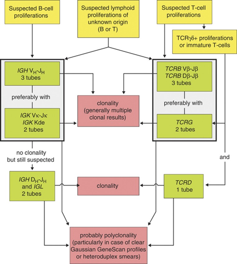
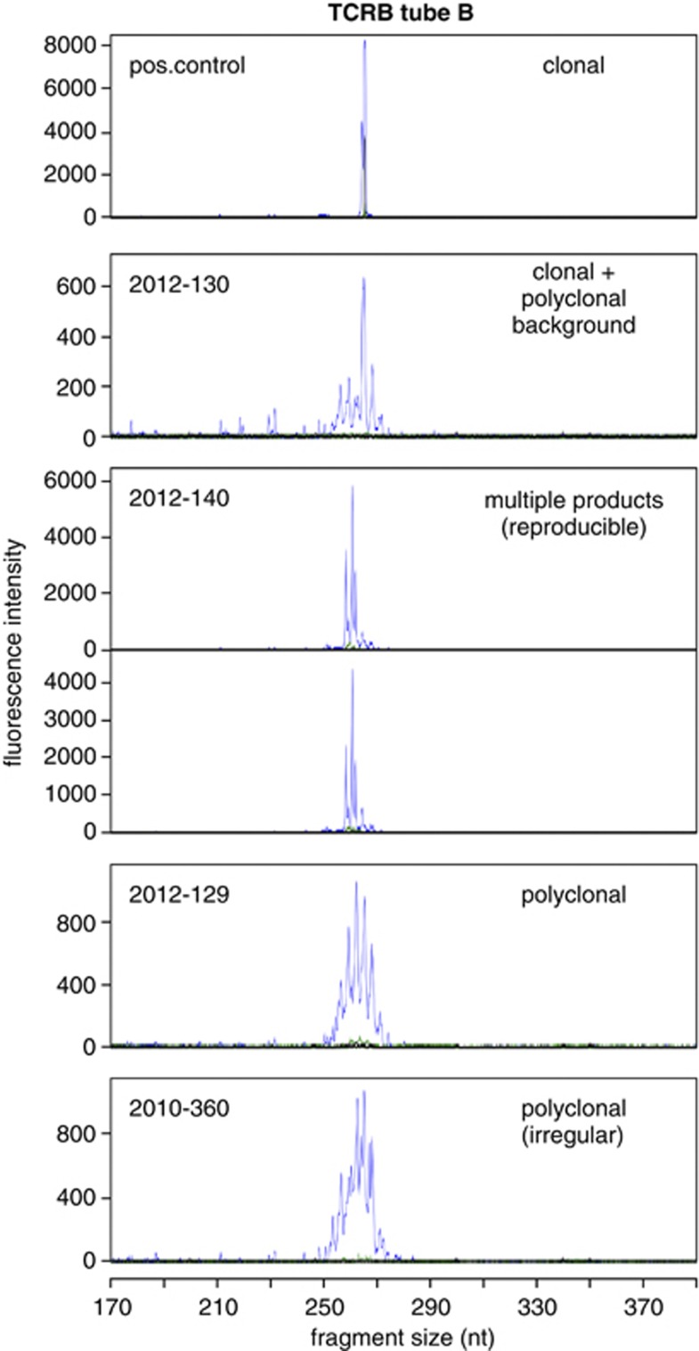
PCR-based immunoglobulin (Ig)/T-cell receptor (TCR) clonality testing in suspected lymphoproliferations has largely been standardized and has consequently become technically feasible in a routine diagnostic setting. Standardization of the pre-analytical and post-analytical phases is now essential to prevent misinterpretation and incorrect conclusions derived from clonality data. As clonality testing is not a quantitative assay, but rather concerns recognition of molecular patterns, guidelines for reliable interpretation and reporting are mandatory. Here, the EuroClonality (BIOMED-2) consortium summarizes important pre- and post-analytical aspects of clonality testing, provides guidelines for interpretation of clonality testing results, and presents a uniform way to report the results of the Ig/TCR assays. Starting from an immunobiological concept, two levels to report Ig/TCR profiles are discerned: the technical description of individual (multiplex) PCR reactions and the overall molecular conclusion for B and T cells. Collectively, the EuroClonality (BIOMED-2) guidelines and consensus reporting system should help to improve the general performance level of clonality assessment and interpretation, which will directly impact on routine clinical management (standardized best-practice) in patients with suspected lymphoproliferations.


Figures

Figure 1
Figure 1
Strategy for PCR-based clonality diagnostics in suspected lymphoproliferations with an inconclusive diagnosis or with unusual histology, immunophenotype or clinical presentation, using the EuroClonality/BIOMED-2 multiplex PCR protocols. In case of a suspected B-cell proliferation, IGH VH–JH multiplex PCR analysis is best performed first. As a second step, IGK PCR analysis (Vκ–Jκ and Kde rearrangements) can be chosen. Preferably, these two steps are combined to avoid delay in the diagnostic process. Finally, IGH DH1-6–JH PCR analysis (potentially combined with IGL analyses) can be reserved for remaining suspected cases, in which the preceding PCR assays have failed to detect monoclonality and have not shown clear signs of polyclonality either. For suspected T-cell proliferations, TCRB multiplex PCR is generally slightly more informative than TCRG PCR, but the order of analysis of these two loci can be changed as they provide complementary information; preferably both targets should be used in parallel. Only in case of suspected TCRγδ+ T-cell proliferations and immature T-cell proliferations (suspicion of lymphoblastic malignancies), combined TCRG and TCRD PCR analysis is preferred. In case of suspected lymphoproliferations of unknown origin, both Ig and TCR genes should be used as PCR targets. It should be noted that in such cases the clonal Ig/TCR results cannot be used straightforwardly for B-/T-lineage assignment. A full-proof diagnosis of polyclonality remains difficult, but a high probability of polyclonality is supported by clear Gaussian GS curves or HD smears in the absence of clonal results.
Figure 2
Figure 2
Example highlighting the difficulty to correlate the number of bands in HD analysis to the number of rearrangements. Using TCRG multiplex PCR tube A, two clonal peaks are observed (biallelic rearrangements) in GS analysis (a). In contrast, in HD analysis (b) four bands are seen, two representing the homoduplexes of ∼144 and ∼216 bp, and the other two representing clonal HDs between the two clonal rearrangements (indicated by arrows).
Figure 3
Figure 3
Examples of technical descriptions of different IGH GS profiles. Representative examples of profiles are shown for IGH multiplex tube A. All assays have been performed in duplicate, but owing to space constraints duplicates are only shown for some technical descriptions in which the reproducibility of the pattern is crucial for proper choice of the term.
Figure 4
Figure 4
Examples of technical descriptions of different IGK GS profiles. Representative examples of profiles are shown for IGK multiplex tube A. All assays have been performed in duplicate, but owing to space constraints duplicates are only shown for some technical descriptions in which the reproducibility of the pattern is crucial for proper choice of the term.
Figure 5
Figure 5
Examples of technical descriptions of different TCRB (V–J) GS profiles. Representative examples of profiles are shown for TCRB multiplex tube B. All assays have been performed in duplicate, but owing to space constraints duplicates are only shown for some technical descriptions in which the reproducibility of the pattern is crucial for proper choice of the term.
Figure 6
Figure 6
Examples of technical descriptions of different TCRB (D–J) GS profiles. Representative examples of profiles are shown for TCRB multiplex tube C. All assays have been performed in duplicate, but owing to space constraints duplicates are only shown for some technical descriptions in which the reproducibility of the pattern is crucial for proper choice of the term.
Figure 7
Figure 7
Examples of technical descriptions of different TCRG GS profiles. Representative examples of profiles are shown for TCRG multiplex tube A. All assays have been performed in duplicate, but owing to space constraints duplicates are only shown for some technical descriptions in which the reproducibility of the pattern is crucial for proper choice of the term.
Figure 8
Figure 8
Examples of technical descriptions of different TCRD GS profiles. Representative examples of profiles are shown for TCRD multiplex tube. All assays have been performed in duplicate, but owing to space constraints duplicates are only shown for some technical descriptions in which the reproducibility of the pattern is crucial for proper choice of the term.

References

    1. Van Dongen JJM, Wolvers-Tettero IL. Analysis of Ig and TCR genes, part II. Clin Chim Acta. 1991;198:93–174. - PubMed
    1. Van Krieken JHJM, Langerak AW, Macintyre EA, Kneba M, Smith JL, Garcia Sanz R, et al. Improved reliability of lymphoma diagnostics via PCR-based clonality testing. Report of the BIOMED-2 Concerted Action BHM4-CT98-3936. Leukemia. 2007;21:201–206. - PubMed
    1. Dik WA, Pike-Overzet K, Weerkamp F, De Ridder D, De Haas EF, Baert MRM, et al. New insights on human T-cell development by quantitative T-cell receptor gene rearrangement studies and gene expression profiling. J Exp Med. 2005;201:1715–1723. - PMC - PubMed
    1. Van Zelm MC, van der Burg M, de Ridder D, Barendregt BH, de Haas EF, Reinders MJ, et al. Ig gene rearrangement steps are initiated in early human precursor B cell subsets and correlate with specific transcription factor expression. J Immunol. 2005;175:5912–5922. - PubMed
    1. Van Dongen JJM, Hooijkaas H, Michiels JJ, Grosveld G, de Klein A, van der Kwast TH, et al. Richter's syndrome with different immunoglobulin light chains and different heavy chain gene rearrangements. Blood. 1984;64:571–575. - PubMed

MeSH terms