. 2012 Sep 15;11(18):3513-7.
doi: 10.4161/cc.21803. Epub 2012 Aug 23.

Multipotent nestin-expressing stem cells capable of forming neurons are located in the upper, middle and lower part of the vibrissa hair follicle

Affiliations

Multipotent nestin-expressing stem cells capable of forming neurons are located in the upper, middle and lower part of the vibrissa hair follicle

Yasuyuki Amoh et al. Cell Cycle. .

Abstract

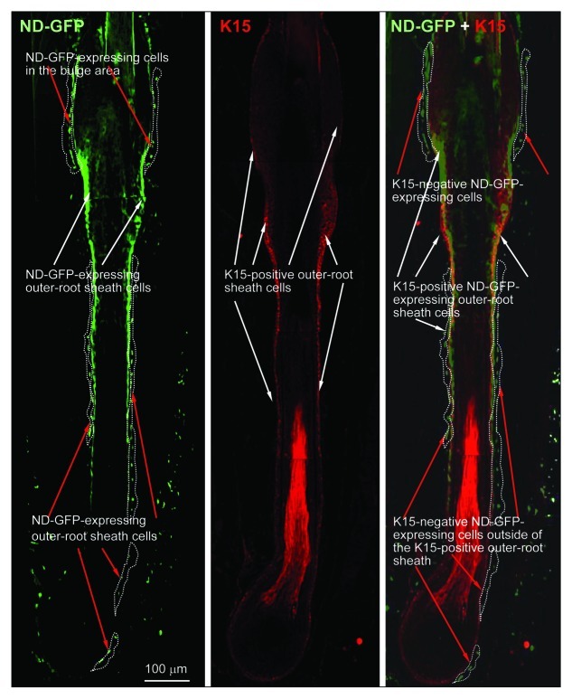
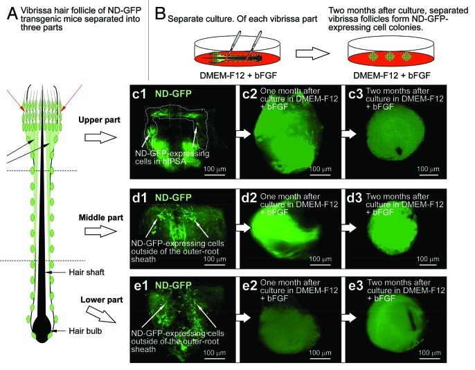
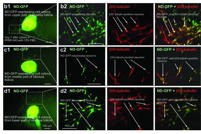
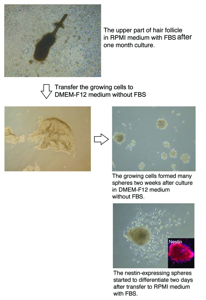
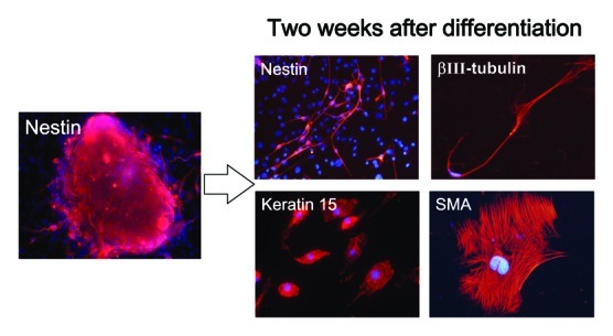
We have previously demonstrated that the neural stem-cell marker nestin is expressed in hair follicle stem cells. Nestin-expressing cells were initially identified in the hair follicle bulge area (BA) using a transgenic mouse model in which the nestin promoter drives the green fluorescent protein (ND-GFP). The hair-follicle ND-GFP-expressing cells are keratin 15-negative and CD34-positive and could differentiate to neurons, glia, keratinocytes, smooth muscle cells and melanocytes in vitro. Subsequently, we showed that the nestin-expressing stem cells could affect nerve and spinal cord regeneration after injection in mouse models. In the present study, we separated the mouse vibrissa hair follicle into three parts (upper, middle and lower). Each part of the follicle was cultured separately in DMEM-F12 containing B-27 and 1% methylcellulose supplemented with basic FGF. After 2 mo, the nestin-expressing cells from each of the separated parts of the hair follicle proliferated and formed spheres. Upon transfer of the spheres to RPMI 1640 medium containing 10% FBS, the nestin-expressing cells in the spheres differentiated to neurons, as well as glia, keratinocytes, smooth muscle cells and melanocytes. The differentiated cells were produced by spheres which formed from nestin-expressing cells from all segments of the hair follicle. However, the differentiation potential is greatest in the upper part of the follicle. This result is consistent with trafficking of nestin-expressing cells throughout the hair follicle from the bulge area to the dermal papilla that we previously observed. The nestin-expressing cells from the upper part of the follicle produced spheres in very large amounts, which in turn differentiated to neurons and other cell types. The results of the present study demonstrate that multipotent, nestin-expressing stem cells are present throughout the hair follicle and that the upper part of the follicle can produce the stem cells in large amounts that could be used for nerve and spinal cord repair.


Figures

None
Figure 1. Frozen sections with IF staining showing presence of nestin- and keratin-15-expressing cells in various parts of the whisker hair follicle (see Materials and Methods for details).
None
Figure 2. Vibrissa hair follicles from nestin-expressing green fluorescent protein (ND-GFP) transgenic mice were divided into three parts and were suspended in DMEM-F12 containing B-27 and 1% methylcellulose supplemented with bFGF every 2 d. After 2 mo, the divided ND-GFP-expressing vibrissa hair follicle fragments formed ND-GFP-expressing spherical colonies.
None
Figure 3. Neurons differentiating from ND-GFP cells in spherical colonies formed from various parts of the whisker follicle. Seven days after switching to RPMI 1640 containing 10% FBS, the ND-GFP-expressing cells differentiated to βIII-tubulin-positive neurons. (b2, c2, d2) are higher magnification of (b1, c1, d1), indicated by the white-dashed box (see Materials and Methods for details).
None
Figure 4. The number of neurons differentiating from ND-GFP-expressing spheres from the upper part of the whisker follicle was significantly higher compared with the middle and lower parts of the follicle. * p < 0.001 vs. control (see Materials and Methods for details).
None
Figure 5. The upper part of the hair follicle was cultured in RPMI medium with FBS and produced numerous ND-GFP-expressing spheres (see Materials and Methods for details).
None
Figure 6. Cell types differentiating from spheres formed from ND-GFP cells in culture of the upper part of the whisker follicle (see Materials and Methods for details).

References

    1. Li L, Mignone J, Yang M, Matic M, Penman S, Enikolopov G, et al. Nestin expression in hair follicle sheath progenitor cells. Proc Natl Acad Sci USA. 2003;100:9958–61. doi: 10.1073/pnas.1733025100. - DOI - PMC - PubMed
    1. Sieber-Blum M, Grim M, Hu YF, Szeder V. Pluripotent neural crest stem cells in the adult hair follicle. Dev Dyn. 2004;231:258–69. doi: 10.1002/dvdy.20129. - DOI - PubMed
    1. Sieber-Blum M, Schnell L, Grim M, Hu YF, Schneider R, Schwab ME. Characterization of epidermal neural crest stem cell (EPI-NCSC) grafts in the lesioned spinal cord. Mol Cell Neurosci. 2006;32:67–81. doi: 10.1016/j.mcn.2006.02.003. - DOI - PubMed
    1. Hoffman RM. The pluripotency of hair follicle stem cells. Cell Cycle. 2006;5:232–3. doi: 10.4161/cc.5.3.2397. - DOI - PubMed
    1. Biernaskie J, Paris M, Morozova O, Fagan BM, Marra M, Pevny L, et al. SKPs derive from hair follicle precursors and exhibit properties of adult dermal stem cells. Cell Stem Cell. 2009;5:610–23. doi: 10.1016/j.stem.2009.10.019. - DOI - PMC - PubMed

Publication types

LinkOut - more resources