. 2012 Nov-Dec;6(5):477-88.
doi: 10.4161/pri.21914. Epub 2012 Aug 23.

Induction of ligand-specific PrP (C) signaling in human neuronal cells

Affiliations

Induction of ligand-specific PrP (C) signaling in human neuronal cells

Ryan J Arsenault et al. Prion. 2012 Nov-Dec.

Abstract

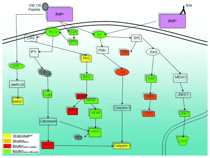
Cellular prion protein (PrP (C) ) has attracted considerable attention for its role in transmissible spongiform encephalopathies (TSEs). In spite of being a point of intense research effort critical questions still remain regarding the physiological function of PrP (C) and how these functions may change with the conversion of the protein into the infectious and pathological conformation (PrP (Sc) ). While emerging evidence suggests PrP (C/Sc) are involved in signal transduction there is little consensus on the signaling pathways associated with the normal and diseased states. The purported involvement of PrP (C) in signal transduction, and the association of TSEs with neural pathology, makes kinome analysis of human neurons an interesting and appropriate model to characterize patterns of signal transduction following activation of PrP (C) by two commonly employed experimental ligands; antibody-induced dimerization by 6H4 and the amino acids 106-126 PrP peptide fragment (PrP 106-126). Analysis of the induced kinome responses reveals distinct patterns of signaling activity following each treatment. Specifically, stimulation of human neurons with the 6H4 antibody results in alterations in mitogen activated protein kinase (MAPK) signaling pathways while the 106-126 peptide activates growth factor related signaling pathways including vascular endothelial growth factor (VEGF) signaling and the phosphoinositide-3 kinase (PI3K) pathway. These pathways were validated through independent functional assays. Collectively these results indicate that stimulation of PrP (C) with distinct ligands, even within the same cell type, results in unique patterns of signaling. While this investigation highlights the apparent functional versatility of PrP (C) as a signaling molecule and may offer insight into cellular mechanisms of TSE pathology it also emphasizes the potential dangers associated with attributing activation of specific intracellular events to particular receptors through artificial models of receptor activation.


Figures

Figure 1.
Figure 1.
PrP surface expression. Shown are flow cytometic analyses plots of neuronal cells labeled with antibody 6H4. A secondary antibody, GAM-FITC, was used to visualize the cells. The plots to the left shows non-specific GAM-FITC binding following incubation with an irrelevant isotype control IgG antibody. The plot to the right depicts cell fluorescence following incubation with the PrP-specific antibody 6H4 and the FITC-conjugated GAM. The % shows the percent of cells which have shifted, indicating fluorescence. Media control cell autofluorescence (no antibody added) resulted in 1.05% of cells indicating fluorescence (data not shown).
Figure 2.
Figure 2.
Peptide array pseudo-image. Shown is a pseudo-image of a peptide array block combining the prion protein stimulation data. Transformed data for PrP 106-126 is compared with scramble control peptide data, and a significance value is determined based on a significant increase or decrease in phosphorylation between the two. This process is also performed for 6H4 antibody stimulated and isotype control antibody. The relative intensity of each spot indicates the level of significance of the phosphorylation of that spot. The left side of each spot is the response to PrP 106-126 stimulation. The right side of each spot indicates 6H4 stimulation response. Grey spots represent inconsistent peptides between array peptide replicates as determine by Χ2 test
Figure 3.
Figure 3.
Phosphorylation Heat Map and Clustering. (A) The background-corrected raw data collected from the peptide arrays were VSN-transformed, and a heatmap/clustering of the data was produced using the Complete Linkage + Euclidian Distance method. The lines at the top of the heatmap indicate the relative similarity between the stimulants indicated at the bottom of the heatmap. The shorter the lines, the more similar the two connected stimulants. The lines on the left side of the heatmap indicate the relative similarity in signal between the 300 individual peptides on the array. The colored lines indicate the relative degree of phosphorylation of each peptide from strongly phosphorylated (red) to non-phosphorylated (green) as indicated by a Z-score. (B) Shown here is the 3D Principal Component Analysis of the five treatments. Relative distance on the three axes indicates level of similarity or difference among the treatments. PrP refers to PrP 106-126 peptide. Scram refers to scramble control peptide. 6H4 refers to the PrP-specific antibody. Iso refers to the IgG1 isotype control antibody. Media refers to media control.
Figure 4.
Figure 4.
Stimulation comparison of peptide phosphorylation. Peptides displaying differential phosphorylation following the two stimulations are compared. The majority of the significantly differentially phosphorylated peptides are unique to one stimulant or the other. A minority of the peptides are common to both PrP 106-126 and 6H4. Common Direction of Phosphorylation refers to peptides displaying either increased or decreased phosphorylation following both stimulations. Opposite Direction of Phosphorylation refers to one stimulation resulting in an increased phosphorylation, while the other stimulation results in a decrease.
Figure 5.
Figure 5.
Signaling pathways linked to PrPC stimulation. Shown is a selection of proteins implicated by peptide array data that is organized into interconnected pathways. Yellow indicates unique PrP 106-126-related phosphorylation. Orange indicates unique 6H4-related phosphorylation. Red indicates that both stimulants affect the peptide in the same direction of phosphorylation. Green indicates that both stimulants affect the peptide in opposite directions. Clear indicates insignificant signal or that the peptide is not present on the array.
Figure 6.
Figure 6.
Cell viability. Stimulated human neuronal cells were assayed for viability using trypan blue dye exclusion and a hemocytometer. Camptothecin refers to the inducer of apoptosis. PrP 106-126 refers to the prion peptide fragment. 6H4 refers to the 6H4 antibody. PrP 106-126 + VEGF refers to PrP 106-126 peptide plus the addition of recombinant VEGF. 6H4 + VEGF refers to 6H4 antibody plus recombinant VEGF. Each stimulated sample was compared with its respective control sample to keep analyses as similar to peptide array analyses as possible. Camptothecin and VEGF counts were compared with unstimulated control counts. 6H4 counts were compared with IgG1 isotype control counts. 6H4 + VEGF counts were compared with 6H4-stimulated counts. PrP 106-126 counts were compared with scramble peptide control counts. PrP 106-126 + VEGF counts were compared with PrP 106–126-stimulated counts. Assays were performed in triplicate with fold change and standard error presented. *Indicates a statistical confidence of p ≤ 0.05.
Figure 7.
Figure 7.
Intracellular calcium. Stimulated human neuronal cells were assayed for intracellular calcium as measured by fluorescence. The assay was performed in triplicate. The means of the replicates are presented along with standard error of the means. Analysis of variance indicated a p-value of 0.032 between groups. The greater the fluorescence signal, the more intracellular calcium. Pairs which showed a statistically significant difference in signal of at least p ≤ 0.05 are indicated.

References

    1. Prusiner SB. Novel proteinaceous infectious particles cause scrapie. Science. 1982;216:136–44. doi: 10.1126/science.6801762. - DOI - PubMed
    1. Büeler H, Fischer M, Lang Y, Bluethmann H, Lipp HP, DeArmond SJ, et al. Normal development and behaviour of mice lacking the neuronal cell-surface PrP protein. Nature. 1992;356:577–82. doi: 10.1038/356577a0. - DOI - PubMed
    1. Roucou X, LeBlanc AC. Cellular prion protein neuroprotective function: implications in prion diseases. J Mol Med (Berl) 2005;83:3–11. doi: 10.1007/s00109-004-0605-5. - DOI - PubMed
    1. Brown DR, Qin K, Herms JW, Madlung A, Manson J, Strome R, et al. The cellular prion protein binds copper in vivo. Nature. 1997;390:684–7. doi: 10.1038/37733. - DOI - PubMed
    1. Shorter J, Lindquist S. Prions as adaptive conduits of memory and inheritance. Nat Rev Genet. 2005;6:435–50. doi: 10.1038/nrg1616. - DOI - PubMed

Publication types