Review
. 2012 Sep;12(9):669-77.
doi: 10.1038/nri3279.

Why must T cells be cross-reactive?

Affiliations
Review

Why must T cells be cross-reactive?

Andrew K Sewell. Nat Rev Immunol. 2012 Sep.

Abstract

Clonal selection theory proposed that individual T cells are specific for a single peptide-MHC antigen. However, the repertoire of αβ T cell receptors (TCRs) is dwarfed by the vast array of potential foreign peptide-MHC complexes, and a comprehensive system requires each T cell to recognize numerous peptides and thus be cross-reactive. This compromise on specificity has profound implications because the chance of any natural peptide-MHC ligand being an optimal fit for its cognate TCR is small, as there will almost always be more-potent agonists. Furthermore, any TCR raised against a specific peptide-MHC complex in vivo can only be the best available solution from the naive T cell pool and is unlikely to be the best possible solution from the substantially greater number of TCRs that could theoretically be produced. This 'systems view' of TCR recognition provides a plausible cause for autoimmune disease and substantial scope for multiple therapeutic interventions.


Conflict of interest statement

The author declares no competing financial interests.

Figures

Figure 1
Figure 1. TCR and peptide–MHC structures.
a | Depicted is a ribbon model of an αβ T cell receptor (TCR) showing the positions of the six variable complementarity-determining region (CDR) loops. b,c | MHC class I and class II molecules can accommodate antigenic peptides of different lengths. The closed ends of the MHC class I binding groove cause long peptides to 'bulge' out of the binding groove, and this bulging increases with each additional amino acid in the peptide. By contrast, the ends of the MHC class II binding cleft are open, which allows the accommodation of much longer peptides without the need for peptide kinking. d,e | The images show HLA-A*0201 (in grey) presenting the immunodominant GLCTLVAML peptide (stick model) from Epstein–Barr virus and HLA-DR4 (in grey) presenting a peptide from myelin basic protein (MBP). TCRs dock on a peptide–MHC complex in a diagonal mode that is conserved for binding to MHC class I and class II molecules. The colours indicate the docking footprints of the AS01 TCR96 and MSC-2C8 TCR97 on their cognate peptide–MHC complexes and show the 'footprints' on the MHC complex of the six CDR loops. In general, the germline-encoded CDR1 and CDR2 loops interact mainly with the MHC molecule itself, whereas the hypervariable CDR3 loops sit over the peptide. However, the small structural database that has been compiled to date already contains examples in which CDR1 and CDR2 make substantial interactions with the peptide and in which CDR3 has an important role in contacting the MHC molecule,.
Figure 2
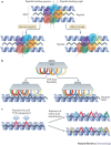
Figure 2. The TCR uses multiple mechanisms to engage numerous peptide–MHC molecules.
a | Macro-level changes enable the T cell receptor (TCR) to bind to peptide–MHC complexes with an altered peptide binding angle (red dotted line) and/or peptide binding register (black dotted line) within a roughly diagonal binding mode. The cartoon shows 'footprints' of the TCR complementarity-determining region (CDR) loops projected down onto the peptide–MHC platform. b | Micro-level CDR loop flexibility enables the accommodation of different peptide–MHC 'landscapes'. The cartoon shows a side view of a TCR engaging a peptide–MHC complex. c | Structural studies show that most TCRs focus on two to four upward-facing peptide residues. In this example, the TCR is focused on the two peptide residues shown in red. Such residue-focused interaction allows the TCR to tolerate multiple amino acid substitutions at other positions in the peptide (indicated by different colours). The above examples are not mutually exclusive and represent only some of the possibilities. MHC-binding motifs often allow for different residues at primary MHC anchors. It should also be noted that TCRs can change the conformation of the peptide–MHC complex following engagement,,.
Figure 3
Figure 3. T cell cross-reactivity causes autoimmunity.
T cells expressing autoreactive T cell receptors (TCRs) are able to bypass system 'safety checks' and populate the periphery. Such T cells generally remain harmless. However, if such T cells become activated in response to a pathogen-derived peptide and become effector T cells, they may then cross-recognize a self-derived peptide to cause autoimmune disease. APC, antigen-presenting cell.
Figure 4
Figure 4. Enhanced TCRs as soluble therapies.
a | The MHC class I presentation pathway presents peptides at the cell surface from intracellular proteins. This potentially allows soluble high-affinity 'monoclonal' T cell receptors (TCRs) to target any cell based on its expression of any protein. 'Monoclonal' TCRs are able to use the MHC class I presentation pathway to 'see inside' cells and scan them for internal anomalies. This 'X-ray vision' opens up access to a far greater range of disease-relevant antigens than are available for monoclonal antibodies. TCRs can be engineered to deliver a variety of molecules that stimulate or suppress the immune system. Potential 'payloads' include antibody Fab fragments that then deliver a signal to immune cells. As MHC-bound peptides are often present at low copy numbers (<50 copies per cell), the payloads delivered by TCRs must act at very low concentrations. b | High-affinity tumour-specific TCRs that are manufactured as bispecific T cell-engaging molecules by linking them to CD3-specific Fab fragments can direct the lysis of tumour cells by CD8+ T cells and thereby induce the regression of established tumours. These molecules do not activate T cells as monomers at the concentrations used. T cell-engaging TCRs bind to the cognate antigen on the tumour cell surface with long half-lives and 'present' the linked CD3-specific Fab fragments. These Fab fragments then crosslink TCRs on the surface of antigen-experienced CD8+ T cells, resulting in cellular activation and elimination of the target cell. The delivery of toxins with soluble TCRs is not recommended, as the soluble TCR constructs are taken up by scavenging cells such as macrophages. Thus, molecules that deliver a particular signal to a specific effector cell are preferable. For example high-affinity TCRs could be used to downregulate immune responses by signalling through inhibitory receptors such as cytotoxic T lymphocyte antigen 4 (CTLA4) (not shown).
Figure 5
Figure 5. TCR-optimized peptide skewing of the repertoire of T cells.
Clonotypic T cell receptors (TCRs) that recognize the same antigen are not all equal, and one TCR may provide the most effective immunity. In the case of HIV for example, one TCR may be more difficult for the virus to escape from than other TCRs. If the required TCR is public (that is, it occurs in all individuals with the restricting HLA molecule) or has a public-type motif, then a TCR-optimized peptide for this clonotype could be used to skew the response towards the most effective clonotype(s). There are no known rules that enable the prediction of which TCRs a particular ligand will stimulate. Thus, this process requires pre-testing using in vitro priming assays to ensure that it induces the required clonotype(s) while minimizing the induction of suboptimal clonotypes.

References

    1. Davis MM, et al. Ligand recognition by αβ T cell receptors. Annu. Rev. Immunol. 1998;16:523–544. - PubMed
    1. Davis MM. T cell receptor gene diversity and selection. Annu. Rev. Biochem. 1990;59:475–496. - PubMed
    1. Davis MM, Bjorkman PJ. T-cell antigen receptor genes and T-cell recognition. Nature. 1988;334:395–402. - PubMed
    1. Lefranc MP, et al. IMGT, the international ImMunoGeneTics information system. Nucleic Acids Res. 2009;37:D1006–D1012. - PMC - PubMed
    1. Rudolph MG, Stanfield RL, Wilson IA. How TCRs bind MHCs, peptides, and coreceptors. Annu. Rev. Immunol. 2006;24:419–466. - PubMed

Publication types

MeSH terms