Review
. 2013 Jun;70(11):1875-95.
doi: 10.1007/s00018-012-1119-x. Epub 2012 Aug 24.

RNA recognition by double-stranded RNA binding domains: a matter of shape and sequence

Affiliations
Review

RNA recognition by double-stranded RNA binding domains: a matter of shape and sequence

Grégoire Masliah et al. Cell Mol Life Sci. 2013 Jun.

Abstract

The double-stranded RNA binding domain (dsRBD) is a small protein domain of 65-70 amino acids adopting an αβββα fold, whose central property is to bind to double-stranded RNA (dsRNA). This domain is present in proteins implicated in many aspects of cellular life, including antiviral response, RNA editing, RNA processing, RNA transport and, last but not least, RNA silencing. Even though proteins containing dsRBDs can bind to very specific dsRNA targets in vivo, the binding of dsRBDs to dsRNA is commonly believed to be shape-dependent rather than sequence-specific. Interestingly, recent structural information on dsRNA recognition by dsRBDs opens the possibility that this domain performs a direct readout of RNA sequence in the minor groove, allowing a global reconsideration of the principles describing dsRNA recognition by dsRBDs. We review in this article the current structural and molecular knowledge on dsRBDs, emphasizing the intricate relationship between the amino acid sequence, the structure of the domain and its RNA recognition capacity. We especially focus on the molecular determinants of dsRNA recognition and describe how sequence discrimination can be achieved by this type of domain.


Figures

Fig. 1
Fig. 1
Sequence alignment of various double-stranded RNA binding domains. Multiple sequence alignment of various dsRBD from human (Homo sapiens, Hs), fruit fly (Drosophila melanogaster, Dm), baker’s yeast (Saccharomyces cerevisiae, Sc), frogs (Xenopus leavis, Xl), plants (Arabidopsis thaliana, At) and bacteria (Escherichia coli, Ec and Aquifex aeolicus, Aa). Alignment was done with Multalin [117, 118] and manually optimized using 3D structural information. For each sequence, the name of the protein and the dsRBD number are given in the first item. The second item corresponds to the accession code in the UniProt database (http://www.uniprot.org). The third item corresponds to the range of amino-acid composing the dsRBD in the numbering of the full-length protein. The alignment is coloured by amino-acid conservation (>40 %) and properties. The sequence consensus (>40 %), the residues conserved for the fold and/or dsRNA binding and the canonical secondary structured elements are shown below the alignment. The three regions of interaction with dsRNA are also indicated
Fig. 2
Fig. 2
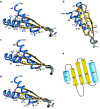
Structure of the double-stranded RNA binding domain. a, b Three-dimensional structure of a dsRBD shown in two different orientations. The structure of XlRBPA-ds2 (PDB code 1DI2) is shown as a cartoon representation with α-helices (blue) and β-strands (yellow). The secondary structure elements composing the αβββα fold are labelled on the structure. Conserved residues matching the sequence consensus of Fig. 1 are shown as sticks and labelled with the residue number corresponding to the sequence alignment of Fig. 1. c Conserved residues important for the fold of the domain. d Conserved residues important for dsRNA binding. Note that Y21 and F35 are important for both the fold and dsRNA binding by orienting the long lysine side-chains of K59 and K55, respectively. e Topology of a dsRBD. Residue numbers corresponding to the sequence alignment of Fig. 1 are indicated
Fig. 3
Fig. 3
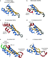
Variations and extensions to the canonical dsRBD fold. Various dsRBDs structures are shown as cartoons with α-helices (blue) and β-strands (yellow). Variations and extensions to the fold are shown in red or green. a Xenopus leavis RNA binding protein A (Xlrbpa) dsRBD2 constitute the archetype of a canonical dsRBD (PDB code 1DI2). b Mammalian ADAR2 dsRBD2 with a shorter helix α1 (PDB code 2L2K). c Budding yeast RNase III (S. cerevisiae Rnt1p) with a shorter helix α1 and an α-helical C-terminal extension (helix α3) (PDB code 2LBS). d Budding yeast Dicer (K. polysporus Dcr1) with a short α-helical C-terminal extension (helix α3) (PDB code 3RV0). e Fission yeast Dicer (S. pombe Dcr1) with a C-terminal extension composed of a short helix α3 and a CHCC zinc-binding motif (green). It also bears a long insertion in loop 2 (PDB code 2L6M). f Plant small RNA methyltransferase (A. thaliana HEN1) with a long insertion in loop 2 (PDB code 3HTX)
Fig. 4
Fig. 4
Sequence discrimination in the minor groove. a, b Differences in the topology of the A-form dsRNA helix and the B-form dsDNA helix. Note especially the differences in the dimension and accessibility of the minor and major grooves. Chemical groups of an A–U pair (c) and a G–C pair (d) lying in the minor and major grooves. Discrimination in the minor groove relies on the appreciation of the chemical group in position 2 of purine rings
Fig. 5
Fig. 5
Global view of the canonical RNA binding surface of dsRBDs. Region 1 (helix α1) and region 2 (loop 2) insert in two successive RNA minor grooves, region 3 (loop 4) contacts the phosphodiester backbone of the intervening RNA major groove. The residues shown in red sticks correspond to a the canonical KKxAK motif of the ADAR2 dsRBD1-RNA stem–loop complex (PDB code 2L3C) and b to the bipartite motif found in the RNase III–RNA complex (PDB code 2NUE) where one of the lysine is coming from helix α1. The numbering of the lysines corresponds to the sequences alignment of Fig. 1. See text for details
Fig. 6
Fig. 6
Sequence specific interactions in the dsRNA minor grooves. Amino acids side chains important for RNA binding are represented as sticks. a Recognition of the minor groove by helix α1 (region 1), showing the sequence specific hydrogen bond between the Gln 11 side chain (Gln 161 in the wild-type sequence) and the atom N3 and the exocyclic amino group of a guanine in the minor groove. (A. aeolicus RNase III dsRBD, PDB code 2EZ6). b Recognition of the dsRNA minor groove by helix α1 (region 1), showing the sequence specific van der Waals contact between the methyl group of Met 4 (Met 238 in the wild-type sequence) and the H2 atom of an adenine in the minor groove. (ADAR2 dsRBD2, PDB code 2L2K). c Recognition of the major and minor grooves by region 3 and 2. Lys 55, Lys 56 and Lys 59 (Lys 127, Lys 128 and Lys 131 in the wild-type sequence) interact with the phosphodiester backbone on opposite strands, across the major groove. The conformation of Lys 55 and Lys 59 side chains is stabilised by hydrophobic contacts with Val 3, Tyr 21, Leu 23, Phe 35 and Met 37. The main chain carbonyl group of Val 30 makes a sequence specific hydrogen bond to the amino group of a guanine in the minor groove (ADAR2 dsRBD2, PDB code 2L2K). d Recognition of the minor groove by loop 2 (Xlrbpa dsRBD2, PDB code 1DI2). The two RNA strands are ‘bridged’ by hydrogen bonds involving the peptide backbone carbonyl and the Nδ atom of the imidazole ring of His 31 (His 141 in the wild-type sequence) and one hydroxyl group on each strand. The peptide backbone carbonyl group of P30 makes a sequence-specific hydrogen bond with the amino group of a guanine
Fig. 7
Fig. 7
a Schematic representation of helix α1 showing the position of the residues involved in dsRNA binding. The numbers inside the circles correspond to the positions in the sequence alignment of Figs. 1, 7b. b Alignment of helix α1 sequences emphasizing the variability of the residues at positions 3, 4, 7 and 11, involved in RNA binding. c Schematic representation of helix α1 of ADAR2 dsRBD2 showing the location of the methionine making a sequence-specific contact to the H2 proton of an adenine. d Schematic representation of helix α1 of the dsRBD of Aa RNase III showing the location of the glutamine making a pair of sequence specific contact to the N3 and to the exocyclic amino group of a guanine
Fig. 8
Fig. 8
Interaction of dsRBDs with RNA apical loops. a Structure of Rnt1p dsRBD with helix α1 interacting with the RNA AGAA apical loop (PDB code 1T4L). The surface of the RNA hairpin is shown for the RNA stem (white) and for the RNA loop (red). The protein is shown as a cartoon, with α helices represented as blue cylinders. b Structure of ADAR2 dsRBD2 showing no interaction between the protein and the RNA apical loop (PDB code 2L2K)
Fig. 9
Fig. 9
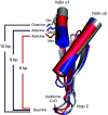
Register variability between sequence specific contacts from helix α1 and loop 2. Structure superposition of ADAR2 dsRBD1 (blue) and dsRBD2 (red) and of the dsRBD of Aa RNase III (grey) (PDB codes 2L3C, 2L2K and 2EZ6). The glutamine side chain (Aa RNase III dsRBD), the methionine side chain (ADAR2 dsRBD1 and 2) from helix α1 and the peptide backbone carbonyl of loop 2, which make sequence-specific contacts with the RNA bases in the minor groove are represented as sticks. The variety of registers observed in these three structures mainly results from the different position of the glutamine (position 11) and the two methionines (position 4) on helix α1 (Fig. 7c, d)

References

    1. Gatignol A, Buckler-White A, Berkhout B, Jeang KT. Characterization of a human TAR RNA-binding protein that activates the HIV-1 LTR. Science. 1991;251(5001):1597–1600. doi: 10.1126/science.2011739. - DOI - PubMed
    1. Ferrandon D, Elphick L, Nüsslein-Volhard C, St Johnston D. Staufen protein associates with the 3′UTR of bicoid mRNA to form particles that move in a microtubule-dependent manner. Cell. 1994;79(7):1221–1232. doi: 10.1016/0092-8674(94)90013-2. - DOI - PubMed
    1. Higuchi M, Single FN, Köhler M, Sommer B, Sprengel R, Seeburg PH. RNA editing of AMPA receptor subunit GluR-B: a base-paired intron-exon structure determines position and efficiency. Cell. 1993;75(7):1361–1370. doi: 10.1016/0092-8674(93)90622-W. - DOI - PubMed
    1. Nicholson AW. Function, mechanism and regulation of bacterial ribonucleases. FEMS Microbiol Rev. 1999;23(3):371–390. doi: 10.1111/j.1574-6976.1999.tb00405.x. - DOI - PubMed
    1. Bernstein E, Caudy AA, Hammond SM, Hannon GJ. Role for a bidentate ribonuclease in the initiation step of RNA interference. Nature. 2001;409(6818):363–366. doi: 10.1038/35053110. - DOI - PubMed

Publication types