Review
. 2012:62:301-26.
doi: 10.1007/978-94-007-4572-8_16.

The wonders of flap endonucleases: structure, function, mechanism and regulation

Affiliations
Review

The wonders of flap endonucleases: structure, function, mechanism and regulation

L David Finger et al. Subcell Biochem. 2012.

Abstract

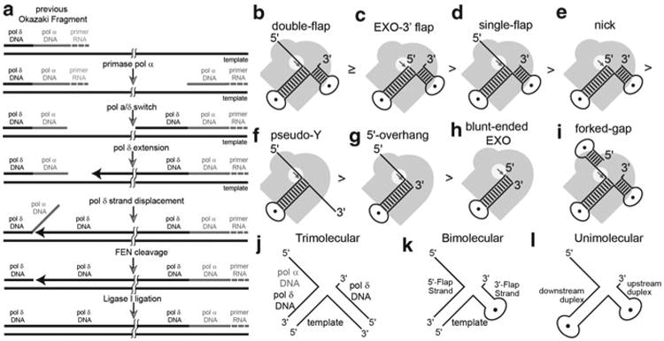
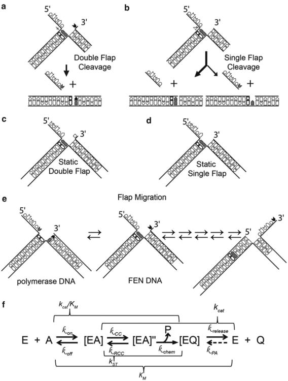
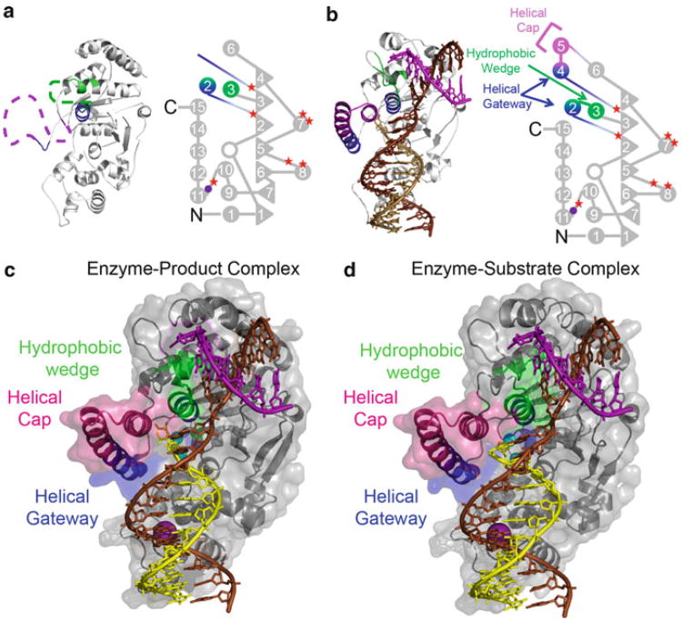
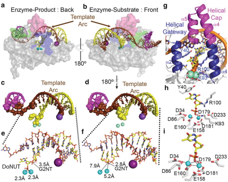
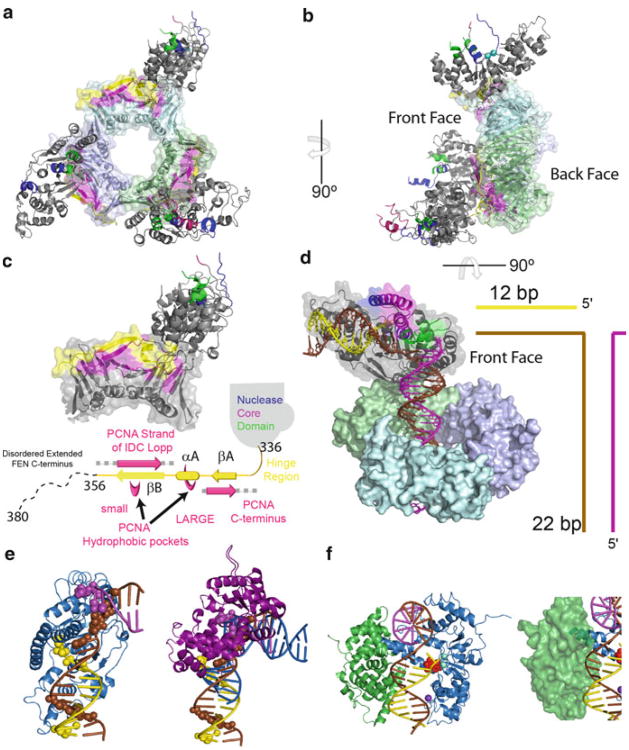
Processing of Okazaki fragments to complete lagging strand DNA synthesis requires coordination among several proteins. RNA primers and DNA synthesised by DNA polymerase α are displaced by DNA polymerase δ to create bifurcated nucleic acid structures known as 5'-flaps. These 5'-flaps are removed by Flap Endonuclease 1 (FEN), a structure-specific nuclease whose divalent metal ion-dependent phosphodiesterase activity cleaves 5'-flaps with exquisite specificity. FENs are paradigms for the 5' nuclease superfamily, whose members perform a wide variety of roles in nucleic acid metabolism using a similar nuclease core domain that displays common biochemical properties and structural features. A detailed review of FEN structure is undertaken to show how DNA substrate recognition occurs and how FEN achieves cleavage at a single phosphate diester. A proposed double nucleotide unpairing trap (DoNUT) is discussed with regards to FEN and has relevance to the wider 5' nuclease superfamily. The homotrimeric proliferating cell nuclear antigen protein (PCNA) coordinates the actions of DNA polymerase, FEN and DNA ligase by facilitating the hand-off intermediates between each protein during Okazaki fragment maturation to maximise through-put and minimise consequences of intermediates being released into the wider cellular environment. FEN has numerous partner proteins that modulate and control its action during DNA replication and is also controlled by several post-translational modification events, all acting in concert to maintain precise and appropriate cleavage of Okazaki fragment intermediates during DNA replication.


Figures

Fig. 16.1
Fig. 16.1
The role of FENs in lagging-strand DNA replication and various FEN substrates in vitro. (a) Simplified diagram of the Okazaki fragment maturation (bi) Various activities that can be observed with FENs and model substrates in vitro. The grey image represents the protein with the active site highlighted by the white circle to show how these activities are achieved. (bh) The strength of each activity is indicated by the order they are placed as illustrated by the symbols between each. (i) Example of a forked-gap substrate. (jl) Examples of double flap constructs used in biochemical studies. Illustrations are labelled to (j) show how in vivo Okazaki fragments correspond to in vitro substrates and (k) each region or (l) component of the substrates are referred to as herein
Fig. 16.2
Fig. 16.2
The importance of the 3′-flap in vitro and in vivo. Reactions on (a) double flaps result in all dsDNA product being a ligatable nick, whereas (b) single flaps produce a minor product that is a 1 nt gap. Examples of static (i.e., single conformation) double-(c) and single-(d) flaps that are commonly used in vitro. (e) Flaps generated in replication are potentially migrating flaps (equilibrating flaps) and can theoretically form multiple structures. Those labelled here represent the two conformations important for polymerase and FENs. (f) Diagram of the FEN reaction pathway on double-flap substrates deduced from studies of FENs to date. Note this is only a model, and there may be more steps in the reaction pathway than illustrated. The FEN enzyme (E) binds its substrate (S) to form an enzyme-substrate complex ([ES]). To be able to cleave the substrate, the protein, DNA, or both have to change conformation to create a cleavage competent complex ([ES]″). Upon cleavage, ssDNA (P) and dsDNA (Q) products are created. The ssDNA (P) likely dissociates form the complex immediately upon cleavage resulting in the enzyme-dsDNA product complex ([EQ]). The dissociation of [EQ] results in nicked dsDNA and enzyme turnover. The rates (k) associated with each steps are listed above or below the corresponding arrow: kon – bimolecular association (i.e., diffusion), koff – dissociation, kCC – conformational change, kRCC – reverse conformational change, kchem – chemical catalysis, krelease – product dissociation, kPA – product association. Note, kPA can be ignored when measuring initial rates of reaction. The macroscopic rate constants commonly measured kinetically are above or below a bracket that encompasses the rates that can in fluence the measured parameter. Because reactions with FEN have intermediates after initial [ES] formation, the KM is an overall dissociation constant for all enzyme-bound species ([ES] + [ES]″ + [EQ]). The turnover number (kcat) in WT FENs is mainly a reflection of the slowest step (enzyme product release), but can be affected by other first order rates in the reaction pathway. The second order rate constant (kcat/KM) for WT is mainly a measure of diffusion (kon), but mutants of FENs can sometimes change and represent anyone or some combination of steps within the bracket. The rate measured under single turnover conditions can measure any rates after initial [ES] complex formation and before [EQ] release and is a measure of some physical limitation such as conformational change in WT FENs
Fig. 16.3
Fig. 16.3
Comparison of DNA-free hFEN1 and DNA-bound hFEN1. Structures of (a) hFEN1 without DNA and (b) hFEN1-product dsDNA with domain maps to highlight the ordering of the helical gateway and cap. Filled circles and triangles represent α-helices and β-sheets, respectively, and are numbered accordingly. The open circle represents a single helical turn of 4 amino acids. The approximate locations of the seven acidic residues for divalent metal sequestration and of the three amino acid residues responsible for coordination of the K+ ion are represented by filled red stars and purple circle, respectively. Structures of the enzyme-product (c) and enzymesubstrate (d) complexes with the protein illustrated as ribbon diagrams with translucent surface representations. The DNA template, 5′-flap and 3′-flap strand are shown in brown, yellow and purple, respectively
Fig. 16.4
Fig. 16.4
FENs effectively utilize the helical properties of DNA to deliver the scissile phosphate to the active site. Due to the spacing of the dsDNA binding regions and the bend induced on the template strand of the two-way DNA junction, the template arc directs the 5′-flap strand towards the active in both the enzyme product (a) and enzyme substrate (b) complexes. The DNA strands and translucent surface representation of the protein are coloured as in Fig. 16.3. The two trivalent samarium ions (Sm3+) and the K+ ions are represented as cyan and purple spheres, respectively. Looking at the DNA alone and representing sites of protein contact to DNA by spheres, the product (c) and substrate (d) DNA are almost the same except for important changes near the scissile phosphate. Note the lack of direct contacts in the template arc region. Focussing on the downstream duplex region, comparison of the product (e) and substrate (f) DNA near the active site shows that the two nucleotides have unpaired for the scissile phosphate to interact with the catalytically important metal ions. In addition, the Gly2 N-terminus (G2NT) interacts initially with the scissile phosphate diester in the enzyme substrate complex (f), but interacts with the phosphate diester 3′ to the scissile phosphate in the enzyme-product complex (e). (g) View of the active site (coloured as above) from the back of the helical gateway and helical cap to highlight some of the catalytically important residues. (h) View of the complete hFEN1 active site that includes the seven highly conserved carboxylates, two basic residues from helix four (Lys93 and Arg100), and an aromatic stacking partner (Tyr40) from helix two. (i) View of the four active site acidic residues (D86, E160, D179, and D181) and product complex unpaired base phosphate monoester directly coordinating the Sm3+ metals (grey dashed lines)
Fig. 16.5
Fig. 16.5
How FENs work with other proteins. (a) Structure of homotrimeric hPCNA in complex with three subunits of hFEN1. The nuclease core domain is coloured as in Fig. 16.3. PCNA subunits are shown as combined ribbon and transparent surface representation. The portion of the extended C-terminus of hFEN1 that was observed in the crystal (residues 336–356) and the regions important for interaction in PCNA are shown in yellow and magenta, respectively. (b) Turning the structure 90° shows that the protein is positioned on one face of the protein. (c) Closer view of the βA-αA-βB motif with schematic illustration below it coloured as above. Alpha helices are represented as cylinders and arrows indicate the position of the β-sheets. (d) Model of hFEN1 interacting with PCNA and DNA simultaneously and coloured as above. (e) Comparison of the hFEN1 (blue) DNA (coloured as above) structure with the pol β-DNA (Purple) structures shows that the only regions available for FENs to initially contact in a handoff model is the downstream dsDNA binding region. (f) Comparison of the DBD domain of ligase (green) and its known interaction site (minor groove) in comparison to hFEN1 (blue) DNA product complex (coloured as above). The groove necessary for DBD interaction is accessible. Below is a translucent surface representation of the ligase DBD showing a steric clash between the DNA and the helical cap of hFEN1. This steric interference may be the initial manner in which ligase facilitates hFEN1 release of its product

References

    1. Balakrishnan L, Stewart J, Polaczek P, Campbell JL, Bambara RA. Acetylation of Dna2 endonuclease/helicase and flap endonuclease 1 by p300 promotes DNA stability by creating long flap intermediates. J Biol Chem. 2009;285:4398–4404. - PMC - PubMed
    1. Beattie TR, Bell SD. The role of the DNA sliding clamp in Okazaki fragment maturation in archaea and eukaryotes. Biochem Soc Trans. 2011;39:70–76. - PubMed
    1. Berg OG, von Hippel PH. Diffusion-controlled macromolecular interactions. Annu Rev Biophys Biophys Chem. 1985;14:131–160. - PubMed
    1. Bochman ML, Sabouri N, Zakian VA. Unwinding the functions of the Pif1 family helicases. DNA Repair. 2010;9:237–249. - PMC - PubMed
    1. Bornarth CJ, Ranalli TA, Henricksen LA, Wahl AF, Bambara RA. Effect of flap modifications on human FEN1 cleavage. Biochemistry. 1999;38:13347–13354. - PubMed

Publication types

MeSH terms