Review
. 2012:62:327-41.
doi: 10.1007/978-94-007-4572-8_17.

DNA ligase I, the replicative DNA ligase

Affiliations
Review

DNA ligase I, the replicative DNA ligase

Timothy R L Howes et al. Subcell Biochem. 2012.

Abstract

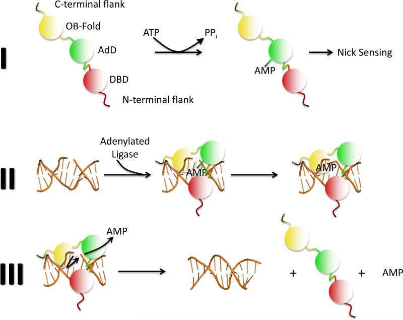
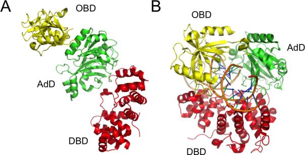
Multiple DNA ligation events are required to join the Okazaki fragments generated during lagging strand DNA synthesis. In eukaryotes, this is primarily carried out by members of the DNA ligase I family. The C-terminal catalytic region of these enzymes is composed of three domains: a DNA binding domain, an adenylation domain and an OB-fold domain. In the absence of DNA, these domains adopt an extended structure but transition into a compact ring structure when they engage a DNA nick, with each of the domains contacting the DNA. The non-catalytic N-terminal region of eukaryotic DNA ligase I is responsible for the specific participation of these enzymes in DNA replication. This proline-rich unstructured region contains the nuclear localization signal and a PCNA interaction motif that is critical for localization to replication foci and efficient joining of Okazaki fragments. DNA ligase I initially engages the PCNA trimer via this interaction motif which is located at the extreme N-terminus of this flexible region. It is likely that this facilitates an additional interaction between the DNA binding domain and the PCNA ring. The similar size and shape of the rings formed by the PCNA trimer and the DNA ligase I catalytic region when it engages a DNA nick suggest that these proteins interact to form a double-ring structure during the joining of Okazaki fragments. DNA ligase I also interacts with replication factor C, the factor that loads the PCNA trimeric ring onto DNA. This interaction, which is regulated by phosphorylation of the non-catalytic N-terminus of DNA ligase I, also appears to be critical for DNA replication.


Figures

Figure 17.1
Figure 17.1. Three-step mechanism of DNA ligation
(1) DNA ligase I binds and hydrolyses adenosine triphosphate (ATP), releasing pyrophosphate (PPi) and a covalent ligase-adenosine monophosphate (AMP) intermediate. (2) The AMP group is subsequently transferred from the ligase polypeptide to the 5’ phosphate termini of a nick in duplex DNA. (3) The non-adenylated ligase catalyzes phosphodiester bond formation in a reaction involving nucleophilic attack by the 3’HO group and release of AMP.
Figure 17.2
Figure 17.2. Alignment of the polypeptides encoded by the three human LIG genes
The conserved catalytic regions of the human DNA ligases each contain a DNA binding (DBD, red), adenylation (AdD, green) and OB-fold (OBD, yellow) domain. The AdD and OBD, which make up the catalytic core of the nucleotidyl transferase family that also includes RNA ligases and mRNA capping enzymes, contain six highly conserved motifs (I, III, IIIa, IV, V and VI). The position of the active site lysine residue within motif I that forms the covalent bond with AMP is indicated for each DNA ligase. The non-catalytic regions that flank the DNA ligase catalytic region determine the subcellular distribution and cellular functions of the DNA ligases. The positions of the nuclear localization signals (NLS, blue) of DNA ligase I and DNA ligase IIIβ, and the mitochondrial leader sequence (MLS, cyan) of DNA ligase IIIα are shown. The replication factory targeting sequence (RFTS), which is also a PCNA interacting peptide (PIP) box that targets DNA ligase I replication and interacts with PCNA, is indicated in grey. Sites of phosphorylation on serine residues within the non-catalytic N-terminal region of DNA ligase I are indicated. Amino acids, glutamine 566 and arginine 771, are replaced with lysine and tryptophan, respectively in the polypeptides encoded by the mutant ligI alleles of the only known DNA ligase I deficient human identified to date. All the DNA ligases encoded by the LIG3 gene have an N-terminal zinc finger (orange) that is involved in DNA binding. Both DNA ligase IIIα and DNA ligase IV contain a breast and ovarian cancer susceptibility protein-1 C-terminal motifs (BRCT, dark green) domain that are involved in protein-protein interactions.
Figure 17.3
Figure 17.3. DNA binding induces a large conformational change in the DNA ligase catalytic region
(A) In the crystal structure of Sulfolobus solfataricus DNA ligase obtained in the absence of DNA (Pascal, et al., 2006), the DNA binding (DBD, shown in red), adenylation domain (AdD, green), and OB-fold (OBD, yellow) domains are in an extended conformation with relatively few contacts between the domains. (B) In the crystal structure of human DNA ligase I in complex with nicked DNA (Pascal, et al., 2004), the DNA binding (DBD, red), adenylation domain (AdD, green) and OB-fold (OBD, yellow) domains encircle the nicked DNA forming a ring structure that is stabilized by each domains interacting with the DNA and by contacts between the DNA binding and OB-fold domains.
Figure 17.4
Figure 17.4. Models for the interaction between PCNA and DNA ligase I on nicked DNA
(A) DNA ligase I initially engages a PCNA trimer (orange) by an interaction between the DNA ligase I PIP box (gray) and the interdomain connector loop (IDCL) region of the one of PCNA subunits. This docking facilitates a lower affinity interaction between the DNA ligase I DNA binding domain (red) and the PCNA trimer. At this stage, the DNA ligase I catalytic region remains in an extended conformation. This complex of PCNA and DNA ligase I slides freely along the duplex until it encounters a nick. The catalytic region of DNA ligase I then encircles the DNA nick. Interactions with the face of the PCNA trimer are likely to facilitate the transition of the catalytic region from the extended conformation into the compact ring structure. Domains of DNA ligase I are coloured as Figure 17.3. (B) A theoretical space-filling model of the double ring structure formed by PCNA (shown predominantly in orange but with the IDCL highlighted in blue; PDB: 1AXC) and the catalytic region of DNA ligase I (PDB: 1X9N) on nicked duplex DNA.

References

    1. Barnes DE, Johnston LH, Kodama K, Tomkinson AE, Lasko DD, Lindahl T. Human DNA ligase I cDNA: cloning and functional expression in Saccharomyces cerevisiae. Proc Natl Acad Sci U S A. 1990;87:6679–6683. - PMC - PubMed
    1. Barnes DE, Tomkinson AE, Lehmann AR, Webster AD, Lindahl T. Mutations in the DNA ligase I gene of an individual with immunodeficiencies and cellular hypersensitivity to DNA-damaging agents. Cell. 1992;69:495–503. - PubMed
    1. Bentley D, Selfridge J, Millar JK, Samuel K, Hole N, Ansell JD, Melton DW. DNA ligase I is required for fetal liver erythropoiesis but is not essential for mammalian cell viability. Nat Genet. 1996;13:489–491. - PubMed
    1. Bentley DJ, Harrison C, Ketchen AM, Redhead NJ, Samuel K, Waterfall M, Ansell JD, Melton DW. DNA ligase I null mouse cells show normal DNA repair activity but altered DNA replication and reduced genome stability. J Cell Sci. 2002;115:1551–1561. - PubMed
    1. Cardoso MC, Joseph C, Rahn HP, Reusch R, Nadal-Ginard B, Leonhardt H. Mapping and use of a sequence that targets DNA ligase I to sites of DNA replication in vivo. J Cell Biol. 1997;139:579–587. - PMC - PubMed

MeSH terms