Cerebral coenurosis in a cat caused by Taenia serialis: neurological, magnetic resonance imaging and pathological features
- PMID: 22918847
- PMCID: PMC10822227
- DOI: 10.1177/1098612X12458211
Cerebral coenurosis in a cat caused by Taenia serialis: neurological, magnetic resonance imaging and pathological features
Abstract
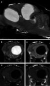
CLINICAL SUMMARY: A 4-year-old Birman cat was presented with marked obtundation and non-ambulatory tetraparesis. Two well-demarcated, intra-axial T2-hyperintense, T1-hypointense structures, which did not contrast enhance, were evident on magnetic resonance imaging (MRI). Histopathology of the structures revealed metacestodes that were morphologically indicative of larval stages of Taenia species. Polymerase chain reaction amplification of a fragment within the 12S rRNA gene confirmed the subspecies as Taenia serialis. PRACTICAL SIGNIFICANCE: This is the first report of MRI findings of cerebral coenurosis caused by T serialis in a cat. Early MRI should be considered an important part of the diagnostic work-up for this rare clinical disease, as it will help guide subsequent treatment and may improve the prognosis.
Conflict of interest statement
The authors do not have any potential conflicts of interest to declare.
Figures


Similar articles
-
A Case of Coenurosis in a Wild Rabbit (Lepus sinensis) Caused by Taenia serialis Metacestode in Qinghai Tibetan Plateau Area, China.Korean J Parasitol. 2018 Apr;56(2):195-198. doi: 10.3347/kjp.2018.56.2.195. Epub 2018 Apr 30. Korean J Parasitol. 2018. PMID: 29742875 Free PMC article.
-
Cerebral coenurosis in a cat.J Am Vet Med Assoc. 1988 Jan 1;192(1):82-4. J Am Vet Med Assoc. 1988. PMID: 3343187
-
Fatal cerebral coenurosis in a cat.J Am Vet Med Assoc. 1994 Jul 1;205(1):69-71. J Am Vet Med Assoc. 1994. PMID: 7928552
-
Cerebral and non-cerebral coenurosis in small ruminants.Trop Biomed. 2014 Mar;31(1):1-16. Trop Biomed. 2014. PMID: 24862039 Review.
-
Taenia multiceps coenurosis: a review.Parasit Vectors. 2022 Mar 12;15(1):84. doi: 10.1186/s13071-022-05210-0. Parasit Vectors. 2022. PMID: 35279199 Free PMC article. Review.
Cited by
-
A Case of Coenurosis in a Wild Rabbit (Lepus sinensis) Caused by Taenia serialis Metacestode in Qinghai Tibetan Plateau Area, China.Korean J Parasitol. 2018 Apr;56(2):195-198. doi: 10.3347/kjp.2018.56.2.195. Epub 2018 Apr 30. Korean J Parasitol. 2018. PMID: 29742875 Free PMC article.
-
Symptomatic lateral ventricular cystic lesion in a young cat.JFMS Open Rep. 2020 Jun 18;6(1):2055116920930181. doi: 10.1177/2055116920930181. eCollection 2020 Jan-Jun. JFMS Open Rep. 2020. PMID: 32595977 Free PMC article.
-
Magnetic Resonance Imaging in 50 Captive Non-domestic Felids - Technique and Imaging Diagnoses.Front Vet Sci. 2022 Feb 8;9:827870. doi: 10.3389/fvets.2022.827870. eCollection 2022. Front Vet Sci. 2022. PMID: 35211543 Free PMC article.
-
Supratentorial arachnoid cyst management by cystoperitoneal shunt in a 1-year-old European cat.JFMS Open Rep. 2015 Jul 14;1(2):2055116915593970. doi: 10.1177/2055116915593970. eCollection 2015 Jul-Dec. JFMS Open Rep. 2015. PMID: 28491374 Free PMC article.
-
Identifying wildlife reservoirs of neglected taeniid tapeworms: Non-invasive diagnosis of endemic Taenia serialis infection in a wild primate population.PLoS Negl Trop Dis. 2017 Jul 13;11(7):e0005709. doi: 10.1371/journal.pntd.0005709. eCollection 2017 Jul. PLoS Negl Trop Dis. 2017. PMID: 28704366 Free PMC article.
References
-
- von Nickisch-Rosenegk M, Silva-Gonzalez R, Lucius R. Modification of universal 12S rDNA primers for specific amplification of contaminated Taenia species (Cestoda) gDNA enabling phylogenetic studies. Parasitol Res 1999; 85: 819–825. - PubMed
-
- Slocombe RF, Arundel JH, Labuc R, Doyle MK. Cerebral coenuriasis in a domestic cat. Aust Vet J 1989; 66: 92–93. - PubMed
-
- Georgi JR, De Lahunta A, Percy DH. Cerebral coenurosis in a cat. Report of a case. Cornell Vet 1969; 59: 127–134. - PubMed
Publication types
MeSH terms
LinkOut - more resources
Full Text Sources
Medical
Miscellaneous

