. 2012 Oct;23(20):3970-81.
doi: 10.1091/mbc.E12-05-0352. Epub 2012 Aug 23.

APC(FZR1) prevents nondisjunction in mouse oocytes by controlling meiotic spindle assembly timing

Affiliations

APC(FZR1) prevents nondisjunction in mouse oocytes by controlling meiotic spindle assembly timing

Janet E Holt et al. Mol Biol Cell. 2012 Oct.

Abstract

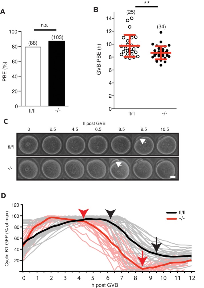
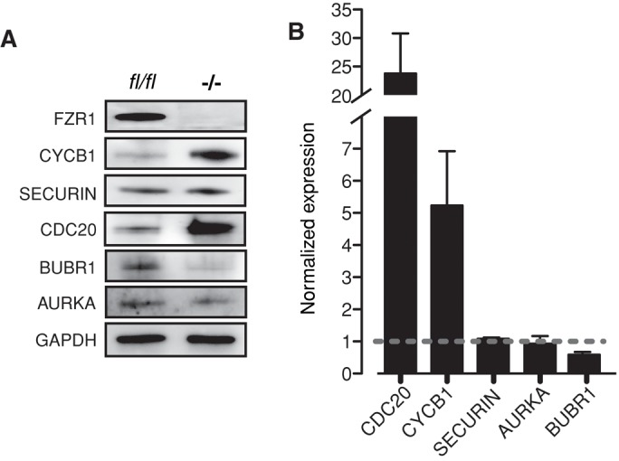
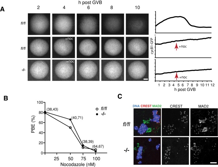
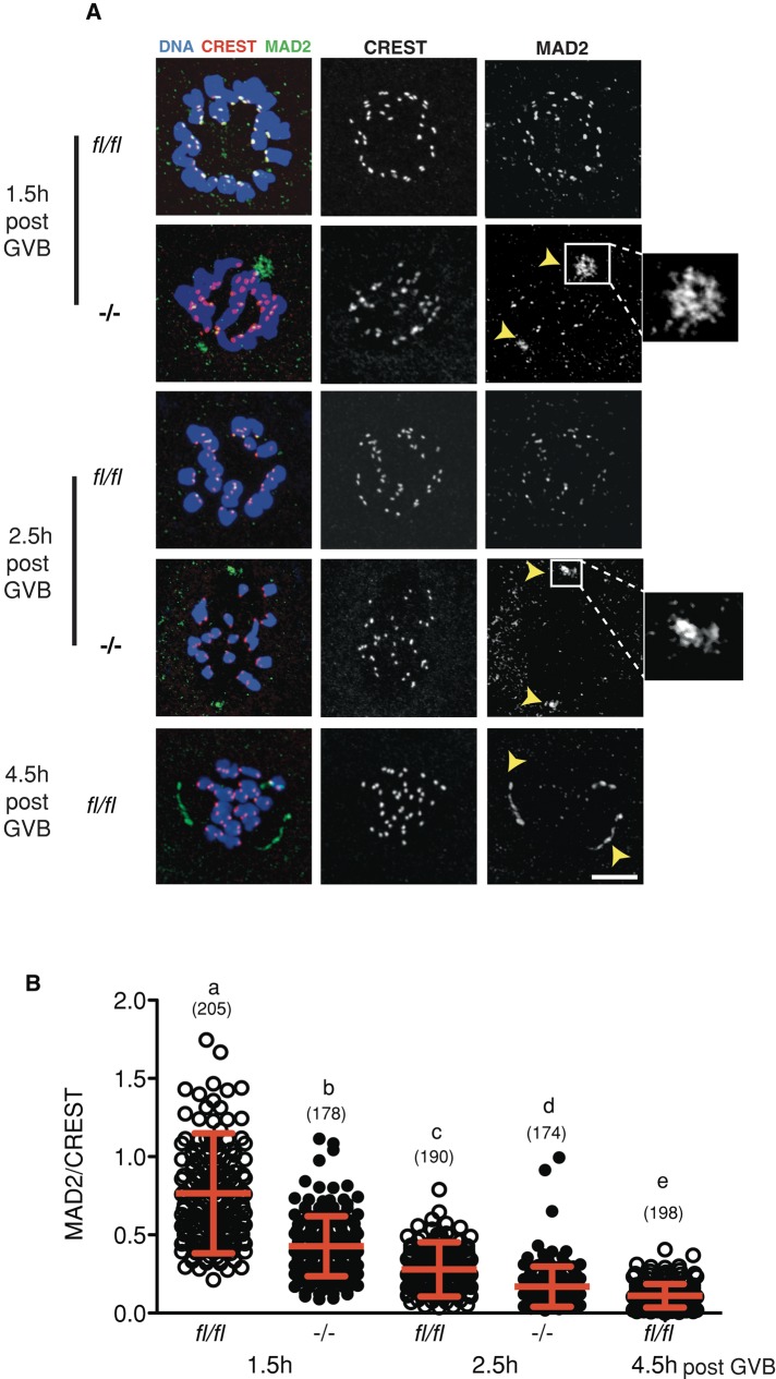
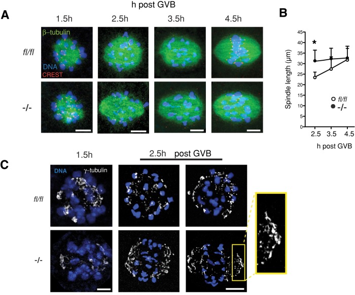
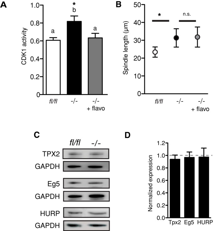
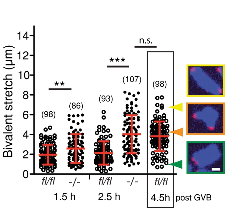
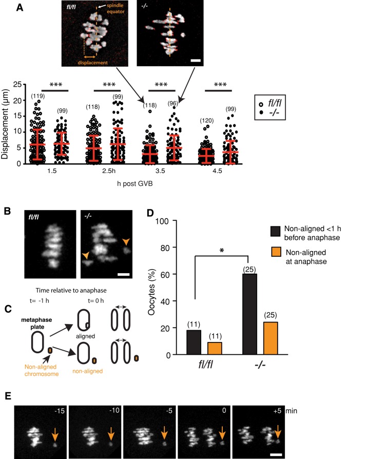
FZR1 is an anaphase-promoting complex (APC) activator best known for its role in the mitotic cell cycle at M-phase exit, in G1, and in maintaining genome integrity. Previous studies also established that it prevents meiotic resumption, equivalent to the G2/M transition. Here we report that mouse oocytes lacking FZR1 undergo passage through meiosis I that is accelerated by ~1 h, and this is due to an earlier onset of spindle assembly checkpoint (SAC) satisfaction and APC(CDC20) activity. However, loss of FZR1 did not compromise SAC functionality; instead, earlier SAC satisfaction was achieved because the bipolar meiotic spindle was assembled more quickly in the absence of FZR1. This novel regulation of spindle assembly by FZR1 led to premature bivalent attachment to microtubules and loss of kinetochore-bound MAD2. Bivalents, however, were observed to congress poorly, leading to nondisjunction rates of 25%. We conclude that in mouse oocytes FZR1 controls the timing of assembly of the bipolar spindle and in so doing the timing of SAC satisfaction and APC(CDC20) activity. This study implicates FZR1 as a major regulator of prometaphase whose activity helps to prevent chromosome nondisjunction.


Figures

FIGURE 1:
FIGURE 1:
Accelerated passage through meiosis I in the absence of FZR1 is associated with earlier degradation of cyclin B1. (A) Polar body extrusion rates for fl/fl and Fzr1−/− oocytes matured in vitro (n.s., p = 0.2; χ2). (B) Time between GVB and PBE for oocytes matured in vitro (**p = 0.004; t test) (C) Representative brightfield time-lapse images used to determine the GVB–PBE interval calculated in B. Arrows indicate the first appearance of PBE. (D) Cyclin B1-GFP degradation profile for fl/fl and Fzr1−/− oocytes (n = 27 and 24, respectively). Arrows indicate mean time of polar body extrusion for microinjected oocytes; arrowheads indicate onset of cyclin B1 degradation. In A and B, parentheses give numbers of oocytes analyzed; error bars in B show SD. Scale bar in C, 20 μm.
FIGURE 2:
FIGURE 2:
FZR1 regulates protein levels of CDC20, cyclin B1, and BUBR1. (A) Representative immunoblots of GV-arrested oocytes collected from Fzr1−/− mice or control littermates. At least three separate runs were performed, with n = 50 oocytes per lane. (B) Densitometric analysis of those immunoblots from A. Levels of the APCFZR1 substrates cyclin B1 and CDC20 were raised, whereas BUBR1 levels were less in Fzr1−/− oocytes. Error bars show SD.
FIGURE 3:
FIGURE 3:
The microtubule poison nocodazole induces a robust SAC response in Fzr1−/− oocytes. (A) Cyclin B1-GFP expression and degradation profiles for fl/fl and Fzr1−/− oocytes in the absence or presence of nocodazole. Representative oocytes and traces are shown. Drug addition 4 h after GVB prevented cyclin B1 degradation in both fl/fl and Fzr1−/− oocytes. (B) Dose response of oocytes to nocodazole assessed by the ability of oocytes to undergo PBE within 12 h of GVB. A 100 nM dose of nocodazole blocked >95% of PBE in control fl/fl and Fzr1−/− oocytes (n.s., 50 nM, p = 0.41; 75 nM, p = 0.77; 100 nM, p = 0.56; χ2). (C) MAD2 immunolocalization to kinetochores of fl/fl and Fzr1−/− oocytes after 100 nM nocodazole treatment 4 h post GVB. In B, parentheses give numbers of fl/fl and Fzr1−/− oocytes analyzed. Scale bar, (A) 20 μm, (B) 10 μm.
FIGURE 4:
FIGURE 4:
Earlier loss of MAD2 from kinetochores during prometaphase in Fzr1−/− oocytes. (A) Representative confocal z-stack images of MAD2/CREST immunostained in fl/fl and Fzr1−/− oocytes fixed during prometaphase. Arrowheads and insets indicate sites of intense MAD2 accumulation at predicted spindle poles for Fzr1−/− oocytes, similar to fl/fl oocytes at 4.5 h post GVB. (B) Kinetochore MAD2/CREST intensity ratios from at 1.5 and 2.5 h post GVB (p < 0.001; Kruskal–Wallis test with Dunn's post hoc test). Error bars (B) show SD; parentheses show numbers of kinetochores analyzed. Scale bar in A, 10 μm.
FIGURE 5:
FIGURE 5:
Accelerated meiosis I spindle elongation in Fzr1−/− oocytes. (A) Representative confocal z-stacks of meiosis I spindles in tubulin-immunostained oocytes at 1.5–4.5 h post GVB. Elongating bipolar spindles are present by 2.5 h in Fzr1−/− but not control fl/fl oocytes. (B) Spindle length measurements for Fzr1−/- and fl/fl oocytes fixed and immunostained at the indicated times post GVB (*p = 0.012; 3.5 h, p = 0.16; 4.5 h, p = 0.8; Students t test; n = 5–7 oocytes per time point). (C) γ-Tubulin–immunostained oocytes 1.5 and 2.5 h post GVB. Inset highlights accumulation of γ-tubulin at the developing spindle pole caps. Error bars in B indicate SD. Scale bars in A and C, 10 μm.
FIGURE 6:
FIGURE 6:
Premature spindle elongation is independent of CDK1 activity or TPX2, HURP, and Eg5 protein level in Fzr1−/− oocytes. (A) CDK1 activity measured using an enzyme-linked immunosorbent assay in Fzr1−/− and control fl/fl oocytes at 2.5 h after GVB. Activity levels were significantly higher in Fzr1−/− oocytes (*p = 0.037, one-way ANOVA with Tukey's test). Treatment with 2.5 μM flavopiridol reduced CDK1 activity to levels that were not significantly different from control oocytes. (B) Spindle length for control and Fzr1−/− oocytes, with and without flavopiridol treatment, measured 2.5 h after GVB. Flavopiridol treatment of Fzr1−/− oocytes did not significantly alter spindle length. (C) Representative immunoblots for TPX2, HURP, and Eg5 on protein from GV-arrested oocytes collected from Fzr1−/− mice or controls, with n = 50–100 oocytes per lane and at least two separate experiments performed. (D) Densitometric analysis of immunoblots in C. Error bars show SD.
FIGURE 7:
FIGURE 7:
Earlier onset of bivalent stretching in the absence of FZR1. Bivalent stretch was measured as the distance between the pairs of sister kinetochores. Mean distances were increased for Fzr1−/− oocytes compared with fl/fl at 1.5 h and 2.5 h post GVB (**p = 0.008, ***p < 0.001; Mann–Whitney test). Images show representative homologue bivalent conformation types with corresponding distances. Error bars indicate SD; parentheses show the numbers of kinetochores analyzed. Scale bar, 2 μm.
FIGURE 8:
FIGURE 8:
Congression and metaphase alignment failure in Fzr1−/− oocytes. (A) Displacement values for Fzr1−/− and fl/fl bivalents from the spindle equator during prometaphase. The spread of displacement values was greater for Fzr1−/− oocytes at all time points measured, indicating poor congression of bivalents compared with controls (***p < 0.001; Levene's test for variance). Representative confocal z-stack images of fl/fl and Fzr1−/− H2BmCherry/CENPC-labeled chromosomes at 3.5 h after GVB are shown. Error bars indicate SD, and the line indicates the mean. Parentheses indicate the numbers of bivalents examined from a total of five to six oocytes per group. (B) Representative confocal z-stack images of fl/fl and Fzr1−/− H2BmCherry-labeled chromosomes 1 h before anaphase I, showing failure of congression in the Fzr1−/− oocyte. Arrowheads indicate nonaligned bivalents. (C) Schematic showing fate of nonaligned bivalents in the 1 h before anaphase. Nonaligned bivalents either returned to the metaphase plate or remained separate and were consequently nonaligned at anaphase onset. (D) Percentage of fl/fl and Fzr1−/− oocytes with nonaligned bivalents 1 h before anaphase and at the onset of anaphase (*p = 0.03; Fisher's exact test). (E) Representative time series of Fzr1−/− bivalents in the minutes preceding anaphase I. Arrows indicate a nonaligned bivalent that fails to return to the metaphase plate before anaphase onset. Parentheses in C indicate the numbers of eggs analyzed. Scale bars, (A) 5 μm, (D) 15 μm.
FIGURE 9:
FIGURE 9:
Increased rates of homologue nondisjunction in Fzr1−/− oocytes. (A) Representative confocal image of a monastrol-induced spread from a meiosis II (MII) egg used for aneuploidy analysis. (B) Aneuploidy rates in fl/fl and Fzr1−/− oocytes matured in vitro. *p = 0.045; χ2. (C) Kinetochore counts from individual eggs scored in B. Rates of hyperploidy matched rates of hypoploidy in Fzr1−/− eggs (n.s., p = 0.41; χ2). (D) Nondisjunction prevailed over PSSC in aneuploid Fzr1−/− eggs (*p = 0.02; χ2). Parentheses in B and D indicate numbers of eggs analyzed. Scale bar in A, 2 μm.

Similar articles

Cited by

References

    1. Breuer M, Kolano A, Kwon M, Li CC, Tsai T, Pellman D, Brunet S, Verlhac MH. HURP permits MTOC sorting for robust meiotic spindle bipolarity, similar to extra centrosome clustering in cancer cells. J Cell Biol. 2010;191:1251–1260. - PMC - PubMed
    1. Brunet S, Dumont J, Lee KW, Kinoshita K, Hikal P, Gruss OJ, Maro B, Verlhac MH. Meiotic regulation of TPX2 protein levels governs cell cycle progression in mouse oocytes. PloS One. 2008;3:e3338. - PMC - PubMed
    1. Brunet S, Pahlavan G, Taylor S, Maro B. Functionality of the spindle checkpoint during the first meiotic division of mammalian oocytes. Reproduction. 2003;126:443–450. - PubMed
    1. Cahu J, Olichon A, Hentrich C, Schek H, Drinjakovic J, Zhang C, Doherty-Kirby A, Lajoie G, Surrey T. Phosphorylation by Cdk1 increases the binding of Eg5 to microtubules in vitro and in Xenopus egg extract spindles. PloS One. 2008;3:e3936. - PMC - PubMed
    1. Chiang T, Duncan FE, Schindler K, Schultz RM, Lampson MA. Evidence that weakened centromere cohesion is a leading cause of age-related aneuploidy in oocytes. Curr Biol. 2010;20:1522–1528. - PMC - PubMed

Publication types

MeSH terms