

Acute stress, but not corticosterone, disrupts short- and long-term synaptic plasticity in rat dorsal subiculum via glucocorticoid receptor activation
- PMID: 22918985
- PMCID: PMC4457523
- DOI: 10.1093/cercor/bhs247
Acute stress, but not corticosterone, disrupts short- and long-term synaptic plasticity in rat dorsal subiculum via glucocorticoid receptor activation
Abstract
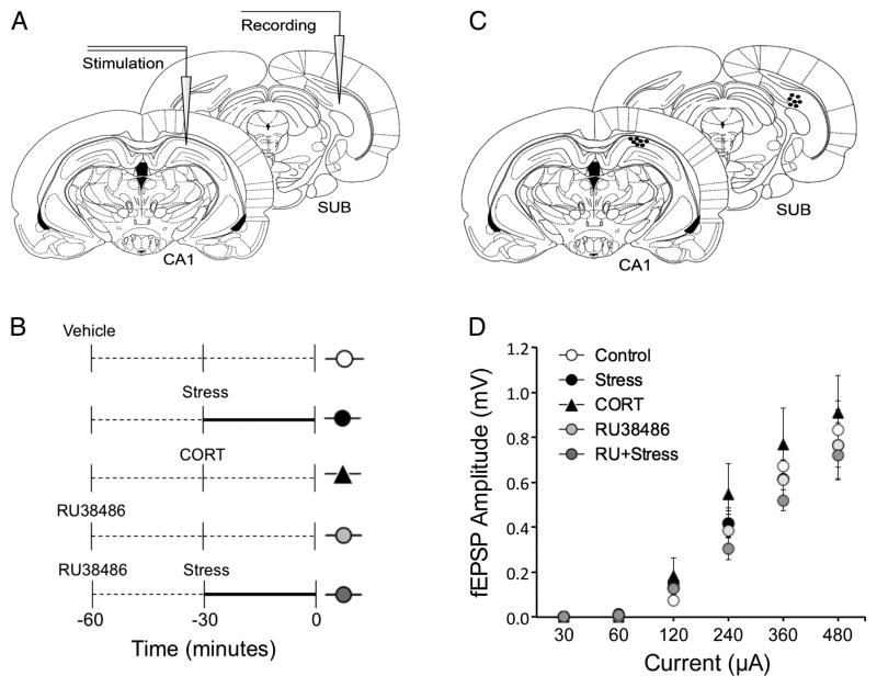
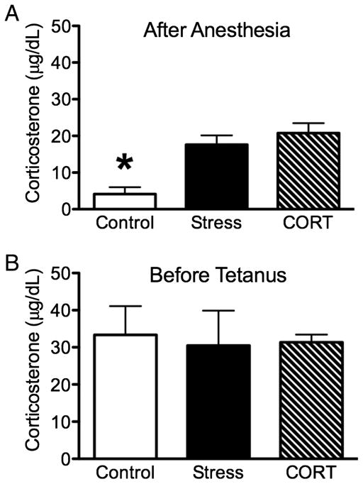
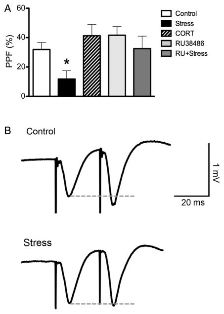
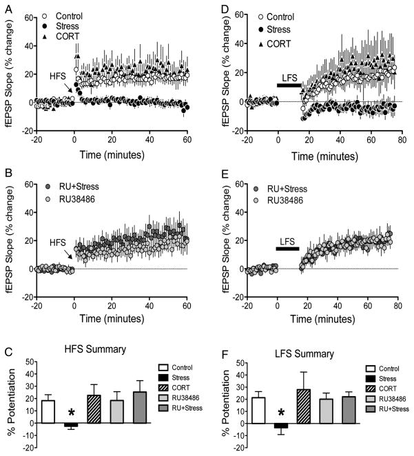
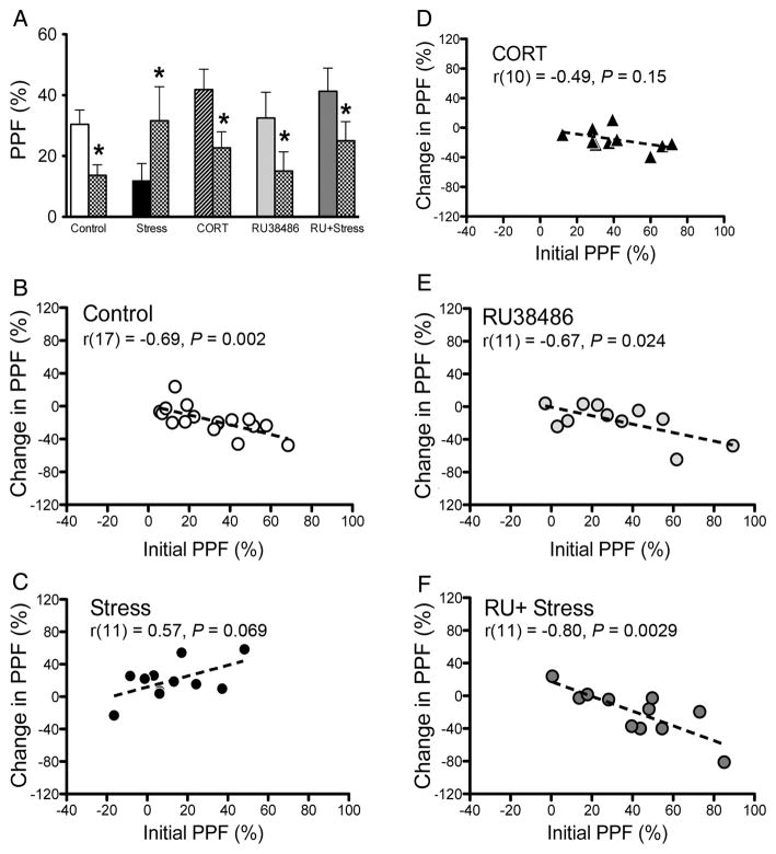
The subiculum (SUB) serves as the major output structure of the hippocampus; therefore, exploring synaptic plasticity within this region is of great importance for understanding the dynamics of hippocampal circuitry and hippocampal-cortical interactions. Previous research has shown exposure to acute stress dramatically alters synaptic plasticity within the hippocampus proper. Using in vivo electrophysiological recordings in urethane-anesthetized adult male Sprague-Dawley rats, we tested the effects of either acute restraint stress (30 min) or corticosterone (CORT) injections (3 mg/kg; s.c.) on short- and long-term forms of synaptic plasticity in the Cornu Ammonis 1-SUB pathway. Paired-pulse facilitation and two forms of long-term plasticity (long-term potentiation and late-developing potentiation) were significantly reduced after exposure to acute stress but not CORT treatment. Measurements of plasma CORT confirmed similar levels of circulating hormone in animals exposed to either acute stress or CORT treatment. The disruptive effects of acute stress on both short- and long-term forms of synaptic plasticity are mediated by glucocorticoid receptor (GR) activation as these disruptions were blocked by pre-treatment with the selective GR antagonist RU38486 (10 mg/kg; s.c.). The present results highlight the susceptibility of subicular plasticity to acute stress and provide evidence that GR activation is necessary but not sufficient for mediating these alterations.
Keywords: hippocampus; in vivo electrophysiology; late-developing potentiation; long-term potentiation; paired-pulse facilitation.
Conflict of interest statement
Figures
References
-
- Andersen P, Morris R, Bliss T, Amaral D, O’Keefe J. The Hippocampus Book. New York: Oxford University Press US; 2006.
-
- Anderson M, Commins S, O’Mara SM. The effects of low frequency and two-pulse stimulation protocols on synaptic transmission in the CA1–subiculum pathway in the anaesthetized rat. Neurosci Lett. 2000;279:181–184. - PubMed
-
- Behr J, Wozny C, Fidzinski P, Schmitz D. Synaptic plasticity in the subiculum. Prog Neurobiol. 2009;89:334–342. - PubMed
-
- Boeijinga PH, Boddeke HWGM. Activation of 5-HT1B receptors suppresses low but not high frequency synaptic transmission in the rat subicular cortex in vitro. Brain Res. 1996;721:59–65. - PubMed
-
- Cazakoff BN, Howland JG. Acute stress disrupts paired pulse facilitation and long-term potentiation in rat dorsal hippocampus through activation of glucocorticoid receptors. Hippocampus. 2010;20:1327–1331. - PubMed
Publication types
MeSH terms
Substances
Grants and funding
LinkOut - more resources
Full Text Sources
Molecular Biology Databases
